This website uses cookies to ensure you get the best experience on our website.


- Table of Contents
Plan CNN1 staining in paraffin sections using cytoplasmic smooth muscle staining as the principal readout (HPA tissue IHC). This guide covers controls, antigen retrieval, chromogenic detection and interpretation of myoepithelial staining (HPA tissue IHC).

Expected localisation, validated protocols, controls and antibodies — the at-a-glance facts below, then the full design guide.
| Expected localisation | Cytoplasm of smooth muscle and myoepithelial cells (HPA tissue IHC) | |
| Staining pattern | Selective cytoplasmic staining in smooth muscle and myoepithelial cells (HPA tissue IHC) | |
| Antigen retrieval | Citrate pH 6 HIER, heat-mediated (datasheet PA2224) | |
| Positive control | Endometrium+2 more · see all | |
| Negative control | Adipose tissue+4 more · see all |
| Fixation | Keep fixation consistent across sections. (standard IHC practice; not target-specific) | |
| Caveat | Smooth muscle in mixed tissue may dominate the signal (HPA tissue IHC) | |
| Regulation | Expression is enriched in smooth muscle (UniProt) | |
| Isoform / epitope | 2 isoforms; epitope coverage is unspecified (UniProt) |
The catalog antibody protocol is followed by published CNN1 IHC methods from two PMC articles: tumor tissue staining and staining of clinical HGSC and normal fimbriae (PMC13221474; PMC5617412).
| Sample | Tissue sections; selected-image fixative not specified (standard IHC workflow) |
| Fixation | Image fixative and duration unreported (datasheet PA2224); verify before use. |
| Sectioning | 4–5 µm sections on charged slides (standard) |
| Deparaffinisation | Xylene, graded ethanol series to water (standard) |
| Antigen retrieval | Heat retrieval: Citrate pH 6, 20 min (datasheet PA2224) |
| Peroxidase block | 3% H2O2, 10 min, room temperature (standard) |
| Blocking | 10% goat serum (datasheet PA2224) |
| Primary antibody | Rabbit anti-CNN1, 0.5-1μg/ml (datasheet PA2224) |
| Primary incubation | Overnight at 4 °C (datasheet PA2224) |
| Detection | Streptavidin-biotin complex (SABC), DAB chromogen (datasheet PA2224) |
| Counterstain | Hematoxylin, blue, dehydrate and mount (standard) |
| Expected result | CNN1-positive staining in smooth muscle cells of endometrium (HPA tissue IHC: High). HPA tissue profile: Highly selective cytoplasmic expression in smooth muscle and myoepithelial cells. No signal in the no-primary control. |
In paraffin section IHC, expect CNN1 in the cytoplasm of smooth muscle cells and breast myoepithelial cells (HPA tissue IHC: Enhanced; highly selective cytoplasmic expression). ICC-IF places it on actin filaments, while UniProt gives no subcellular annotation and reports no transmembrane segment (HPA ICC-IF: approved actin filament location; UniProt P51911: topology).
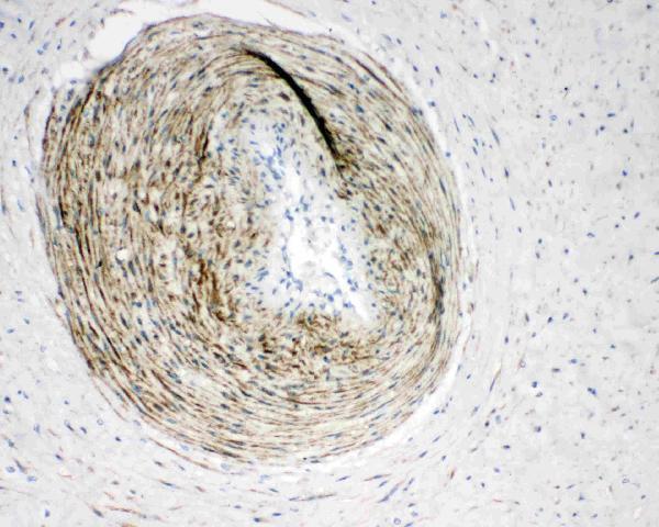
| Strong cytoplasmic staining outlines smooth muscle cells in a vessel wall or smooth muscle tissue. | This matches the reported smooth muscle pattern; confirm the stained cells by section morphology rather than judging color alone (HPA tissue IHC: high in smooth muscle cells; general IHC practice). |
| Moderate cytoplasmic staining follows myoepithelial cells in breast tissue. | This is an expected positive pattern at a lower reported level than smooth muscle; assess the cell layer and surrounding tissue separately (HPA tissue IHC: medium in breast myoepithelial cells; high in smooth muscle cells). |
| The dominant signal is nuclear or restricted to cell membranes. | Treat this as discordant with the reported cytoplasmic IHC pattern; check morphology, counterstain and detection controls before calling CNN1 positive (HPA tissue IHC: cytoplasmic; UniProt P51911: no transmembrane segment; general IHC practice). |
| Adipocytes or respiratory epithelial cells stain while expected smooth muscle cells do not. | Possible nonspecific antibody binding or endogenous detection activity needs investigation; the named cell types were reported as undetected, though nearby smooth muscle may still stain (HPA tissue IHC: adipocytes and respiratory epithelial cells not detected; general IHC practice). |
| A broad haze obscures cell borders, or a known-positive smooth muscle area is blank. | Neither appearance supports a reliable negative or positive call. Compare a reagent control and an intact positive area before interpreting the specimen (HPA tissue IHC: high in smooth muscle cells; general IHC practice). |
| Cell type and tissue context | Score the stained cell, not the whole section: smooth muscle can be present beside a cell type reported as undetected (HPA tissue IHC: high in smooth muscle cells; adipocytes not detected). |
| Antibody validation | Two listed antibodies have Enhanced IHC status; this supports the tissue pattern but does not validate every staining condition (HPA antibodies: HPA014263 and CAB000007, IHC Enhanced; general IHC practice). |
| Antigen retrieval | No CNN1-specific retrieval setting is supplied. Compare standard retrieval conditions using a positive tissue control and consistent detection settings (general IHC practice). |
| Isoforms and phosphorylation | UniProt lists 2 isoforms and 5 modified residues; the record does not say whether either changes IHC staining or antibody recognition (UniProt P51911: isoforms and modified residues). |
| Processing and topology | The recorded chain spans residues 1–297, with no signal peptide, propeptide or transmembrane segment; these facts do not establish an epitope location (UniProt P51911: processing and topology). |
| Situation | Likely cause | Next action |
|---|---|---|
| No stain in a known-positive smooth muscle area. | A missing target-rich area, weak detection or a failed run is possible; the image alone cannot distinguish these (HPA tissue IHC: high in smooth muscle cells; general IHC practice). | Verify smooth muscle morphology, then review the run control, antibody dilution, retrieval and chromogen development under the same conditions (general IHC practice). |
| Diffuse brown signal covers many unrelated cells. | Background may arise from excess antibody, incomplete blocking or endogenous detection activity (general IHC practice). | Compare reagent controls; review blocking, antibody dilution, washes and detection chemistry before scoring any cytoplasmic signal (general IHC practice). |
| Nuclei or cell membranes carry the strongest signal. | That distribution conflicts with the reported cytoplasmic pattern; staining alone cannot identify its source (HPA tissue IHC: cytoplasmic; general IHC practice). | Check counterstain and detection controls, then compare the same run with positive smooth muscle morphology (general IHC practice; HPA tissue IHC: high in smooth muscle cells). |
| Adipocytes or respiratory epithelial cells appear positive. | Those cell types were reported as undetected; nearby smooth muscle or background can complicate visual assignment (HPA tissue IHC: both cell types not detected; general IHC practice). | Trace cell boundaries and adjacent smooth muscle, then use a reagent control to assess nonspecific or endogenous signal (general IHC practice). |
| Breast myoepithelial staining seems weaker than smooth muscle. | The reported levels differ: medium in breast myoepithelial cells and high in smooth muscle cells (HPA tissue IHC: respective levels). | Score each cell population against its expected pattern; do not require identical intensity across the two contexts (HPA tissue IHC: respective levels; general IHC practice). |
| What should an IF/ICC image show? | HPA reports an approved actin filament location in ICC-IF images; that finding does not define paraffin section resolution (HPA ICC-IF: actin filaments; general microscopy practice). | Use the separate IF/ICC guide for that application; interpret this paraffin IHC result by cytoplasmic signal in the expected cells (HPA tissue IHC: smooth muscle and myoepithelial cells). |


![HPA ICC-IF BJ [Human fibroblast] — CNN1 (HPA014263)](https://images.proteinatlas.org/14263/1446_H4_3_blue_red_green.jpg)
![HPA ICC-IF BJ [Human fibroblast] — CNN1 (HPA014263)](https://images.proteinatlas.org/14263/1446_H4_6_blue_red_green.jpg)
Comprehensive Human Protein Atlas IHC scoring per tissue (reliability: Enhanced — High consistency between antibody staining and RNA expression data.). Rows are taken directly from the HPA tissue chart — click any row's HPA link to view the source.
| Tissue | Cell type | Level | Evidence | Source |
|---|---|---|---|---|
| Endometrium | Smooth muscle cells | High | Protein (IHC) | HPA → |
| Smooth muscle | Smooth muscle cells | High | Protein (IHC) | HPA → |
| Breast | Myoepithelial cells | Medium | Protein (IHC) | HPA → |
| Tissue | Cell type | Level | Evidence | Source |
|---|---|---|---|---|
| Adipose tissue | Adipocytes | Not detected | Protein (IHC) | HPA → |
| Adrenal gland | Glandular cells | Not detected | Protein (IHC) | HPA → |
| Appendix | Endocrine cells | Not detected | Protein (IHC) | HPA → |
| Bone marrow | Hematopoietic cells | Not detected | Protein (IHC) | HPA → |
| Bronchus | Respiratory epithelial cells | Not detected | Protein (IHC) | HPA → |
Troubleshoot CNN1 staining in paraffin sections by checking retrieval, cell identity, cytoplasmic localisation and detection controls (datasheet PA2224; HPA tissue IHC).
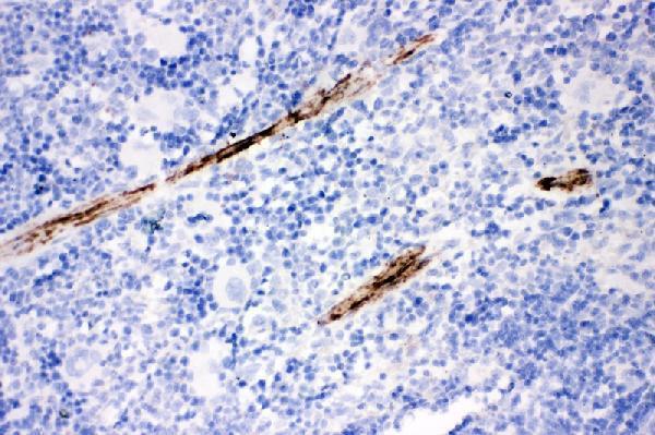
Anti-CNN1 antibodies with real IHC images cover paraffin sections of human placenta and appendicitis tissue, and mouse and rat spleen (catalog IHC captions).
PA2224 is listed for IHC and shows human placenta and mouse and rat spleen in paraffin sections (PA2224 applications and IHC captions). M08065-1 is listed for IHC, IF and ICC, with a paraffin-section IHC image from human appendicitis tissue (M08065-1 applications and IHC caption).
Which to pick: For tissue IHC across human, mouse and rat, choose PA2224: all three species are listed as reactive and represented in its IHC captions (PA2224 reactivity and IHC captions). For IF/ICC, choose the rabbit monoclonal M08065-1 because those applications are listed, with a stated IF dilution of 1:50; its supplied image documents IHC, not IF (M08065-1 catalog applications, clone, IF dilution and IHC caption). Both IHC captions describe paraffin sections but do not report the fixative (PA2224 and M08065-1 IHC captions).