This website uses cookies to ensure you get the best experience on our website.


- Table of Contents
Plan CNR1 paraffin IHC with the catalog antibody at 0.5–1 µg/mL (datasheet A01291-1). Use adipocytes as a positive reference and score cytoplasmic and membranous staining by cell type (HPA tissue IHC).

Expected localisation, validated protocols, controls and antibodies — the at-a-glance facts below, then the full design guide.
| Expected localisation | Cytoplasmic and membranous tissue staining (HPA tissue IHC) | |
| Staining pattern | High in adipocytes; cytoplasmic/membranous (HPA tissue IHC) | |
| Antigen retrieval | Citrate pH 6 HIER, heat-mediated (datasheet A01291-1) | |
| Positive control | Adipose tissue+4 more · see all | |
| Negative control | Adrenal gland+4 more · see all |
| Fixation | Keep fixation consistent across sections (standard IHC practice; not target-specific); Matched tissue-IHC evidence does not establish the fixation claim. Validate the specimen-specific method before use. (selected-SKU IHC image A01291-1) | |
| Caveat | CNS staining varies by cell type; hippocampal glia are undetected (HPA tissue IHC) | |
| Regulation | Tissue dependent; brain enriched (UniProt) | |
| Isoform / epitope | 3 isoforms; check extracellular versus cytoplasmic epitope coverage (UniProt) |
The catalog antibody’s IHC-P protocol is paired with published chromogenic IHC protocols for placental tissue and monkey cerebrum (PMC12984115; PMC9496684).
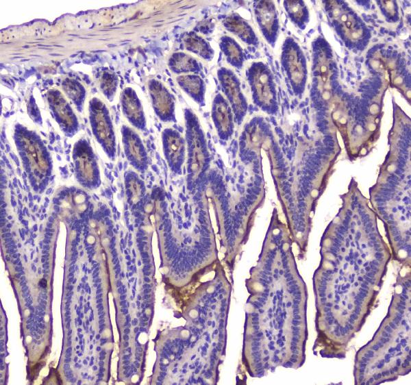
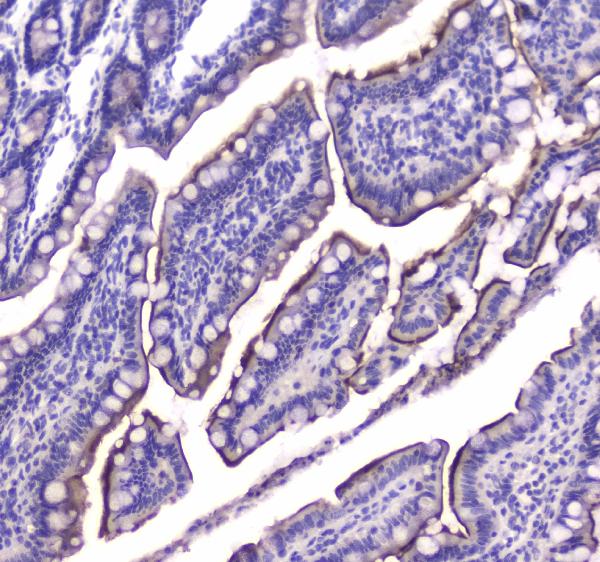
| Sample | Paraffin-embedded mouse small intestine tissue; fixative not specified (datasheet A01291-1) |
| Fixation | Image fixative and duration unreported (datasheet A01291-1); verify before use. |
| Sectioning | 4–5 µm sections on charged slides (standard) |
| Deparaffinisation | Xylene, graded ethanol series to water (standard) |
| Antigen retrieval | Heat retrieval: Citrate pH 6, 20 min (datasheet A01291-1) |
| Peroxidase block | 3% H2O2, 10 min, room temperature (standard) |
| Blocking | 10% goat serum (datasheet A01291-1) |
| Primary antibody | Rabbit anti-CNR1, 0.5-1μg/ml (datasheet A01291-1) |
| Primary incubation | Overnight at 4 °C (datasheet A01291-1) |
| Detection | Streptavidin-biotin complex (SABC), DAB chromogen (datasheet A01291-1) |
| Counterstain | Hematoxylin, blue, dehydrate and mount (standard) |
| Expected result | CNR1-positive staining in adipocytes of adipose tissue (HPA tissue IHC: High). HPA tissue profile: Cytoplasmic and membranous expression in several different tissue types, most abundant in adipocytes, pituitary gland and CNS. No signal in the no-primary control. |
CNR1 is a seven-transmembrane receptor associated with the cell membrane, axons and presynapses; UniProt also annotates the mitochondrial outer membrane (UniProt P21554 topology; UniProt P21554 subcellular). In tissue IHC, HPA reports cytoplasmic and membranous staining, including high staining in adipocytes and anterior pituitary cells (HPA tissue IHC). HPA rates the tissue pattern Enhanced while reporting medium consistency with RNA data (HPA tissue IHC).
| Membranous staining, with some cytoplasmic staining, in adipocytes or anterior pituitary cells. | This matches HPA's tissue pattern and its high staining levels in those cells (HPA tissue IHC). Score the named cell population and compartment together; intensity alone does not establish specificity, especially where the tissue contains several cell types (general IHC practice). |
| Predominantly nuclear staining, without the expected membranous or cytoplasmic pattern. | Treat this as a possible artefact: HPA describes cytoplasmic and membranous tissue staining, while UniProt annotates membrane-associated locations (HPA tissue IHC; UniProt P21554 subcellular). Review the control and antibody conditions before calling it CNR1 (general IHC practice). |
| Strong staining in glomerular cells, cardiomyocytes or esophageal squamous cells. | HPA reports CNR1 as not detected in these specific cell populations (HPA tissue IHC). Check for cross-reactivity or endogenous detection activity before accepting the signal (general IHC practice). A negative annotation for those cells does not classify every cell in the same organ. |
| Diffuse color across tissue and empty slide areas, obscuring cell boundaries. | This distribution is difficult to reconcile with HPA's named positive cell populations and membranous or cytoplasmic pattern (HPA tissue IHC). Evaluate nonspecific reagent binding, detection background and washing with suitable controls (general IHC practice). Do not score diffuse color as cellular positivity. |
| No staining in an adipocyte-rich area or anterior pituitary cells. | HPA reports high staining in adipocytes and anterior pituitary cells, so an absent result needs technical review (HPA tissue IHC). Confirm that the expected cells are present, then check the antibody, detection reagents and run controls (general IHC practice). |
| Compartment and epitope position | CNR1 has seven transmembrane segments and distinct extracellular and cytoplasmic regions (UniProt P21554 topology). Interpret staining against the antibody's stated epitope if available; the supplied record gives no epitope for a catalog antibody. HPA's tissue IHC pattern is both membranous and cytoplasmic (HPA tissue IHC). |
| Tissue and cell selection | HPA reports high staining in adipocytes, anterior pituitary cells, placental cytotrophoblasts and testicular Sertoli cells, but medium staining in caudate neuronal cells (HPA tissue IHC). Record the cell population examined; a whole-organ positive or negative label would hide these differences. |
| Isoform coverage | UniProt lists three CNR1 isoforms and reports much lower expression of isoforms 2 and 3 than isoform 1 (UniProt P21554). The supplied evidence does not identify an antibody epitope or establish which isoforms an IHC signal detects; avoid assigning an isoform from staining alone. |
| Strength of tissue evidence | HPA labels tissue IHC reliability Enhanced and notes medium consistency between staining and RNA expression (HPA tissue IHC). Its negative calls apply to specified cell populations, including hippocampal glial cells, rather than every cell in those tissues (HPA tissue IHC). |
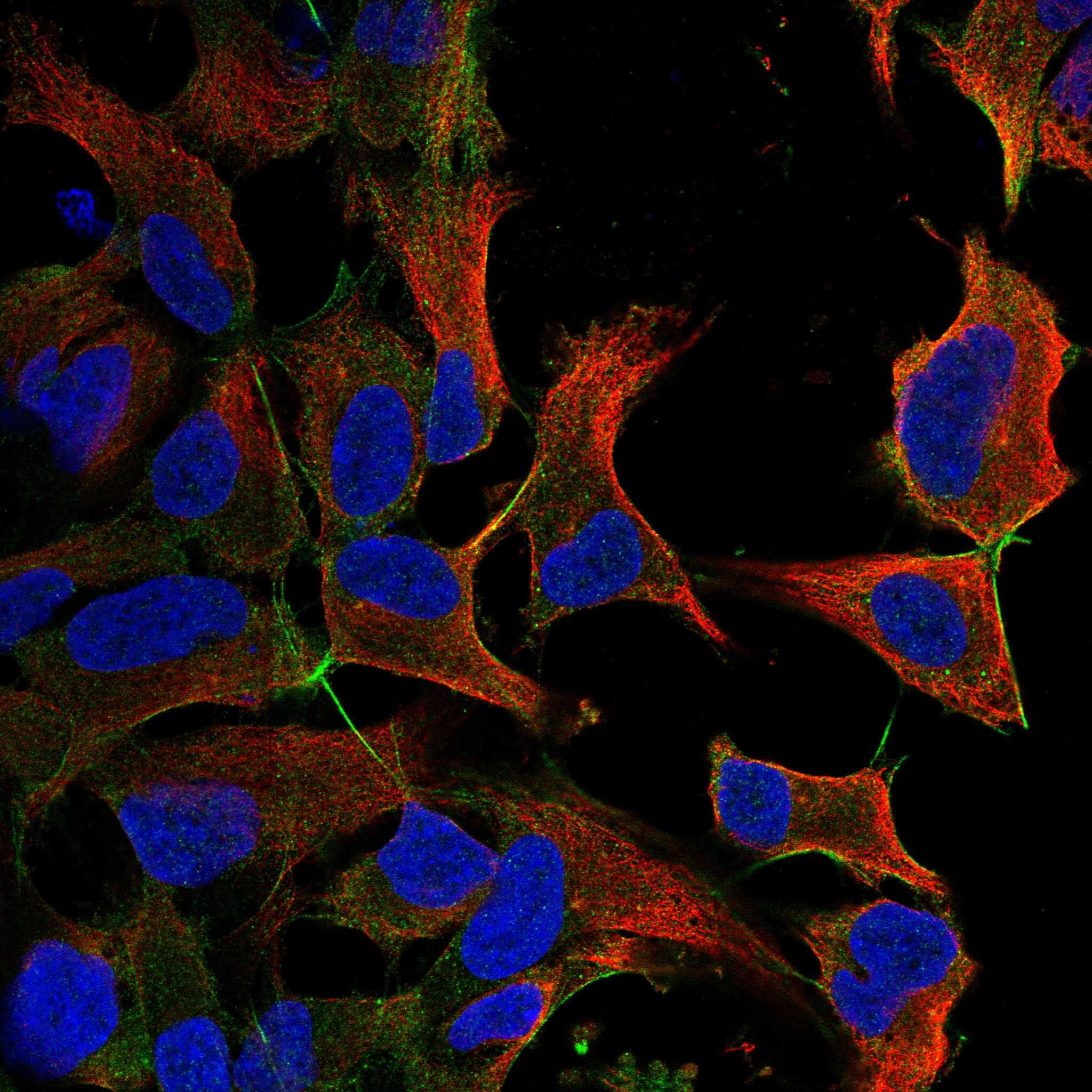
| IF/ICC Q: What localisation is reported? | A: HPA reports plasma membrane as the supported main ICC-IF location and actin filaments as an uncertain additional location (HPA subcellular ICC-IF). This is an IF interpretation cue; it does not supply an IF protocol or replace the tissue IHC pattern. |
| Situation | Likely cause | Next action |
|---|---|---|
| Expected positive cells are unstained. | The sampled field may lack the named positive population (HPA tissue IHC), or the staining run may have failed (general IHC practice). | Locate identifiable adipocytes or anterior pituitary cells and compare them with a positive run control; then check antibody and detection steps (HPA tissue IHC; general IHC practice). |
| Signal is weak in a purported negative control. | The control may contain a different CNR1-staining cell population; HPA negative calls are cell-specific (HPA tissue IHC). | Score the annotated cells, such as cardiomyocytes in heart muscle, and document the exact population examined (HPA tissue IHC). |
| Color appears after omitting the primary antibody. | Primary-independent signal can arise from endogenous detection activity or other detection reagents (general IHC practice). | Review the detection-only control and use the blocking or detection controls appropriate to the chromogenic system (general IHC practice). |
| Color spreads across many cell types and the slide background. | Diffuse signal can reflect nonspecific binding or excess detection background (general IHC practice). | Compare with a primary-omission control, review blocking and washing, and reassess antibody concentration using the assay instructions (general IHC practice). |
| A predominantly nuclear pattern appears in otherwise positive tissue. | The pattern conflicts with HPA's membranous and cytoplasmic tissue description and UniProt's membrane-associated locations (HPA tissue IHC; UniProt P21554 subcellular). | Check whether the nuclear color persists in controls; withhold a specific CNR1 call until the expected cellular pattern is resolved (general IHC practice). |
| An antigen retrieval change gives a different result. | Retrieval conditions can alter IHC staining generally; CNR1-specific fixation or retrieval sensitivity is unreported in the supplied sources (general IHC practice). | Compare conditions on the same known-positive cell population with matched controls; do not infer a CNR1-specific fixation effect from this change alone (HPA tissue IHC; general IHC practice). |


Comprehensive Human Protein Atlas IHC scoring per tissue (reliability: Enhanced — Medium consistency between antibody staining and RNA expression data. Antibody staining in cells/structures not annotated, view images.). Rows are taken directly from the HPA tissue chart — click any row's HPA link to view the source.
| Tissue | Cell type | Level | Evidence | Source |
|---|---|---|---|---|
| Adipose tissue | Adipocytes | High | Protein (IHC) | HPA → |
| Breast | Adipocytes | High | Protein (IHC) | HPA → |
| Bronchus | Ciliated cells (tip of cilia) | High | Protein (IHC) | HPA → |
| Pituitary gland | Cells in anterior | High | Protein (IHC) | HPA → |
| Placenta | Cytotrophoblasts | High | Protein (IHC) | HPA → |
| Tissue | Cell type | Level | Evidence | Source |
|---|---|---|---|---|
| Adrenal gland | Glandular cells | Not detected | Protein (IHC) | HPA → |
| Cervix | Glandular cells | Not detected | Protein (IHC) | HPA → |
| Esophagus | Squamous epithelial cells | Not detected | Protein (IHC) | HPA → |
| Heart muscle | Cardiomyocytes | Not detected | Protein (IHC) | HPA → |
| Hippocampus | Glial cells | Not detected | Protein (IHC) | HPA → |
Troubleshoot CNR1 chromogenic IHC in paraffin sections by checking retrieval, staining controls, expected localisation, and scoring before interpreting regional differences.
A01291-1 is listed for IHC and ICC in human, mouse and rat; its IHC images show paraffin sections of mouse and rat small intestine (catalog applications/reactivity; IHC image captions).
A01291-1 has IHC images from paraffin sections of mouse and rat small intestine (A01291-1 IHC image captions). It lists human, mouse and rat reactivity and ICC as an application, but supplies no IF image (catalog reactivity/applications; IF image alts).
Which to pick: Choose A01291-1 for paraffin-section tissue IHC: its mouse and rat captions document citrate retrieval at pH 6 for 20 minutes and 1 μg/ml primary antibody; the fixative is unreported (A01291-1 IHC image captions). For IF/ICC, A01291-1 lists ICC at 0.5–1 μg/ml, but supplies no IF image; clonality is unreported (catalog applications/dilution; IF image alts; clone field). For cross-species work, A01291-1 lists human, mouse and rat reactivity, with tissue IHC images for mouse and rat (catalog reactivity; A01291-1 IHC image captions).