CNR2 / Cannabinoid receptor 2 · IHC design guide

Design Immunohistochemistry for CNR2

Plan chromogenic IHC-P for CNR2 using the catalog antibody at 1:10–1:50 (datasheet: IHC-P, 1:10–1:50). Choose tissue controls and interpret variable cytoplasmic staining in light of HPA’s uncertain reliability (HPA tissue IHC).

Evidence assembled Oct 2026 · For research use; verify linked source records and product datasheet before use
Immunohistochemistry protocol sheet for CNR2 (IHC for CNR2): expected localisation Variable cytoplasmic staining in most tissues (HPA tissue IHC), antibody A01307-3, validated IHC image, and IHC protocol steps
Printable CNR2 IHC protocol sheet — expected localisation Variable cytoplasmic staining in most tissues (HPA tissue IHC), antibody A01307-3, controls and protocol steps. Open the full CNR2 IHC guide →

CNR2 Immunohistochemistry Experimental Design Guide

Expected localisation, validated protocols, controls and antibodies — the at-a-glance facts below, then the full design guide.

Must know before staining
Expected localisation Variable cytoplasmic staining in most tissues (HPA tissue IHC)
Staining pattern High glandular-cell staining; variable cytoplasmic pattern (HPA tissue IHC)
Antigen retrieval Citrate pH 6.0 HIER, 95–98 °C, 20 min (rule: cytoplasmic / membrane antigen)
Positive control ⓘ Appendix+4 more · see all
Negative control ⓘ Liver
Important caveats
Reasons your staining may differ from the expected pattern.
Fixation Keep formalin fixation consistent across compared sections. (selected-SKU IHC image A01307-3)
Caveat Antibody staining has low consistency with RNA data (HPA tissue IHC)
Regulation Higher expression in B cells and NK cells (UniProt)
Isoform / epitope CB2A and CB2B differ only in the 5′ UTR; epitope map unchanged (UniProt)
Section 1

Recommended CNR2 IHC & IF Protocols

The catalog antibody’s IHC-P protocol is paired with published CNR2 staining protocols for mouse asthma sections (PMC8267324) and human placental sections (PMC12984115).

Recommended immunohistochemistry (IHC-P) protocol parameters
SampleParaffin-embedded tissue sections; fixative not specified (datasheet A01307-3; sample unspecified)
FixationImage fixative and duration unreported (datasheet A01307-3); verify before use.
Sectioning4–5 µm sections on charged slides (standard)
DeparaffinisationXylene, graded ethanol series to water (standard)
Antigen retrievalHeat-induced epitope retrieval in citrate buffer, pH 6.0, 20 min at 95–98 °C (standard rule: cytoplasmic / membrane antigen)
Peroxidase block3% H2O2, 10 min, room temperature (standard)
Blocking10% normal serum of the secondary host, 30 min, room temperature (standard)
Primary antibodyRabbit anti-CNR2, 1:10-1:50 (datasheet A01307-3)
Primary incubationOvernight at 4 °C (standard)
DetectionHRP-polymer secondary, DAB chromogen 5–10 min (standard)
CounterstainHematoxylin, blue, dehydrate and mount (standard)
Expected resultCNR2-positive staining in glandular cells of appendix (HPA tissue IHC: High). HPA tissue profile: Cytoplasmic expression at variable levels in most tissues. No signal in the no-primary control.
💡Decision noteStart with citrate pH 6.0 retrieval at 95–98 °C for 20 min (page antigen retrieval); the placental protocol used EDTA pH 8.0 (PMC12984115).
Section 2

What Is the Expected CNR2 Staining Pattern?

CNR2 is a seven-pass cell-membrane receptor, with reported localisation in dendrites and perikarya (UniProt P34972 topology and subcellular location). In tissue sections, examine lymph-node germinal-center cells and selected glandular cells for staining (HPA: High in lymph-node germinal-center cells; High in colon glandular cells). HPA describes variable cytoplasmic staining across tissues, but rates its tissue IHC pattern Uncertain because staining and RNA data show low consistency (HPA: tissue IHC reliability).

What am I looking at on my slide?
Membrane-associated staining in germinal-center cells or colon glands.These are plausible positive cells (HPA: High in both). CNR2 is a membrane receptor (UniProt P34972 topology). Assess the cell pattern alongside a control because HPA rates tissue IHC Uncertain (HPA: reliability).
Cytoplasmic staining accompanies a plausible cellular pattern.HPA reports variable cytoplasmic expression in most tissues (HPA: tissue IHC profile). Cytoplasmic signal alone does not establish receptor localisation; compare its distribution with controls and the membrane expectation (UniProt P34972 subcellular location; general IHC practice).
Strong nuclear staining dominates the section.The nucleus is outside the reported CNR2 locations (UniProt P34972 subcellular location). Treat a nuclear-only pattern as suspect and check background and antibody controls before scoring it as CNR2 (general IHC practice).
Liver cholangiocytes stain strongly.HPA reports CNR2 as Not detected in these cells (HPA: liver cholangiocytes). Unexpected signal may reflect cross-reactivity or endogenous detection activity; assess adjacent morphology and detection controls (general IHC practice).
No staining appears in lymph-node germinal centers.These cells are reported High by HPA (HPA: lymph-node germinal-center cells). A blank section raises a staining-run question, but HPA's Uncertain IHC reliability means one negative result cannot settle CNR2 expression (HPA: reliability; general IHC practice).
💡Expected CNR2 appearanceA credible positive is cell-associated signal in lymph-node germinal-center cells or reported High glandular cells, with a membrane-compatible distribution; strong nuclear-only or cholangiocyte signal is suspect (HPA: tissue IHC; UniProt P34972 subcellular location).
How each factor affects the staining
Membrane topologyCNR2 has seven transmembrane segments and extracellular and cytoplasmic regions (UniProt P34972 topology). The antibody epitope is unspecified, so topology alone cannot prescribe antigen retrieval or permeabilisation.
Tissue and cell selectionHPA reports High staining in lymph-node germinal-center cells and colon, appendix and rectum glandular cells; liver cholangiocytes are Not detected (HPA: tissue IHC). Compare named cell populations within preserved morphology.
Strength of validationThe listed antibody CAB009719 has Uncertain IHC status, and the tissue pattern has low consistency with RNA data (HPA: antibody validation; tissue IHC reliability). Interpret isolated strong staining cautiously.
Promoter-derived transcriptsCB2A and CB2B differ in their 5′ untranslated regions (UniProt P34972 tissue specificity). A protein-staining pattern cannot assign signal to either transcript on that basis.
IF/ICC Q: What localisation is supported?HPA summarises the subcellular pattern as Membrane, but lists no main location or ICC-IF image cell lines (HPA: subcellular record). UniProt also reports membrane, dendrite and perikaryon localisation (UniProt P34972 subcellular location).
Retrieval and fixation evidenceNo target-specific fixation sensitivity or retrieval condition is supplied (HPA: tissue IHC record; UniProt P34972 record). Optimise these as general IHC workflow variables without treating a changed signal as proof of CNR2 identity (general IHC practice).
Why is my staining missing, weak or wrong?
SituationLikely causeNext action
Known-positive tissue has no signal.The staining run may have failed, or the selected cells may have been missed (general IHC practice).Confirm germinal-center cells or reported High glandular cells are present; check the staining-run positive control before interpreting absence (HPA: tissue IHC; general IHC practice).
Signal covers cells and empty areas diffusely.Background from detection or incomplete blocking can obscure cellular localisation (general IHC practice).Review a no-primary control and blocking and wash steps; score only signal that follows identifiable cells (general IHC practice).
Nuclei are the dominant positive compartment.That pattern conflicts with reported membrane, dendrite and perikaryon localisation (UniProt P34972 subcellular location).Check counterstain and no-primary control, then repeat with a verified staining run before assigning nuclear signal to CNR2 (general IHC practice).
Cholangiocytes appear strongly positive.HPA reports them as Not detected; cross-reactivity or endogenous detection activity is possible (HPA: liver cholangiocytes; general IHC practice).Inspect a detection-only control and compare with a reported High cell population in the same run (HPA: tissue IHC; general IHC practice).
Staining is weak or patchy across a tissue.HPA reports variable cytoplasmic levels and Uncertain tissue IHC reliability (HPA: tissue IHC profile and reliability).Score the named cell type and compartment rather than the whole section; compare with controls and avoid inferring expression from intensity alone (general IHC practice).
Membrane and cytoplasmic signals disagree.UniProt assigns CNR2 to the membrane, while HPA describes cytoplasmic tissue staining with Uncertain reliability (UniProt P34972 subcellular location; HPA: tissue IHC).Record each compartment separately and compare reproducibility across reported positive cells and controls before calling a specific pattern (HPA: tissue IHC; general IHC practice).

Sample controls for CNR2 IHC & IF

🧪Run lymph node first: germinal center cells should stain (HPA: High in lymph node germinal center cells). Use liver cholangiocytes as the negative tissue (HPA: Not detected); on the positive slide, use any neighboring cells without specific staining as internal negatives, showing counterstain only, without assuming a particular cell type is negative.
Positive control tissue: Appendix (Glandular cells, HPA High)
Negative control tissue: Liver (HPA Not detected)
ICC-IF cell lines (HPA subcellular resource): HPA carries no ICC-IF cell line for CNR2; derive a cell-line control from the positive tissue's cell type (Glandular cells) and confirm it by RNA or western blot first.
Technical controls: Include a no-primary (secondary-only) control, a host- and class-matched isotype control, and a CNR2 knockout tissue control where available (standard IHC practice). For lymph node DAB staining, quench endogenous peroxidase and check for background from immune cells (standard IHC practice).
⚠️Feasibility: The selected A01307-3 paraffin-section caption gives a 1:25 primary dilution but no fixative; a target-specific fixation window or fixation effect is unreported (selected tissue-IHC caption). Retrieval dependence is also unreported, so compare antigen retrieval conditions during validation (standard IHC practice). The supplied evidence does not establish that frozen sections or IF/ICC are easier, and HPA lists no ICC-IF image cell lines for CNR2 (HPA: subcellular record); immune-cell background is a practical concern in lymph node staining (standard IHC practice).

HPA tissue IHC evidence for CNR2

Comprehensive Human Protein Atlas IHC scoring per tissue (reliability: Uncertain — Low consistency between antibody staining and RNA expression data.). Rows are taken directly from the HPA tissue chart — click any row's HPA link to view the source.

Positive expression · recommended positive controls

TissueCell typeLevelEvidenceSource
Appendix Glandular cells High Protein (IHC) HPA →
Breast Glandular cells High Protein (IHC) HPA →
Colon Glandular cells High Protein (IHC) HPA →
Duodenum Glandular cells High Protein (IHC) HPA →
Lymph node Germinal center cells High Protein (IHC) HPA →

Undetected expression · recommended negative controls

TissueCell typeLevelEvidenceSource
Liver Cholangiocytes Not detected Protein (IHC) HPA →
Section 3

Advanced CNR2 IHC Tips

Troubleshoot CNR2 staining in paraffin sections by checking retrieval, antibody specificity, cellular localisation and controls before interpreting chromogenic signal.

How should I retrieve CNR2 antigen when staining is weak?
Start with citrate buffer at pH 6.0, heated to 95–98 °C for 20 min (page retrieval specification). Allow sections to cool consistently, then compare staining with a matched section processed without retrieval; differences can reveal whether heat exposure improves signal or increases background (standard IHC practice). Keep section thickness, antibody incubation and DAB development constant across the comparison so retrieval is the main variable (standard IHC practice). If staining remains weak, optimise retrieval cautiously on serial sections, because this 7-transmembrane receptor has extracellular and cytoplasmic regions that may differ in antibody accessibility (UniProt P34972 topology).
Can I infer the best fixative from the published CNR2 staining pattern?
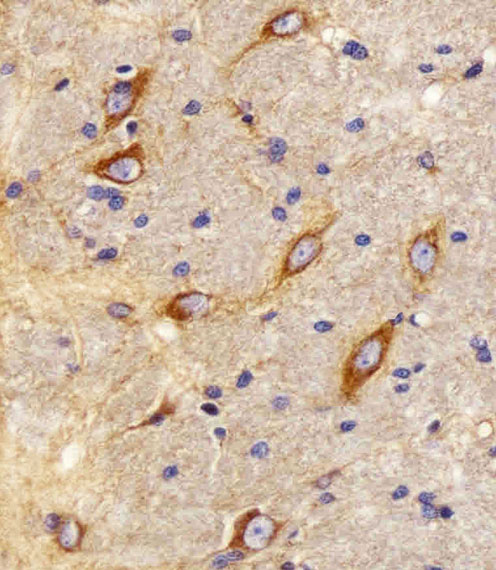
No target-specific fixation sensitivity is supplied for CNR2, so treat the effect of fixative type and fixation time as unknown (provided evidence). The selected antibody image documents a paraffin-embedded R. brain section, but its caption does not state the fixative (A01307-3 tissue-IHC caption). For a controlled comparison, process matched samples with recorded fixation and handling conditions, then stain serial sections in the same run (standard IHC practice). Compare signal, morphology and background together, keeping retrieval at citrate pH 6.0, 95–98 °C for 20 min while assessing fixation as the variable (page retrieval specification; standard IHC practice).
Should CNR2 staining look membranous or cytoplasmic in paraffin sections?
CNR2 is annotated at the cell membrane, dendrites and perikaryon, including the apical dendrite of pyramidal neurons (UniProt P34972 subcellular location). HPA instead describes variable cytoplasmic tissue staining and rates its tissue-IHC reliability uncertain because staining has low consistency with RNA expression (HPA tissue IHC). Examine membrane outlines and cell-associated cytoplasm at high magnification, recording each compartment separately rather than treating diffuse colour as equivalent to a membrane signal (standard IHC practice). Check whether staining follows intact cells in the relevant tissue region, and interpret neuronal staining cautiously because normal-brain expression has also been reported as undetectable in one study (UniProt P34972 tissue specificity).
Can this C-terminal antibody distinguish CB2A from CB2B?
CB2A and CB2B arise from alternative promoter usage and differ only in their 5′ UTR, so a protein stain cannot assign signal to either transcript on that basis (UniProt P34972 tissue specificity). The selected product is described as a C-terminal antibody, but its caption does not specify a precise immunising peptide or residue range (A01307-3 tissue-IHC caption). The annotated CNR2 cytoplasmic tail spans residues 302–360 and includes phosphorylation sites at 335, 336, 338 and 352 (UniProt P34972 topology and modified residues). Confirm the antibody’s documented epitope before attributing a staining difference to tail accessibility or phosphorylation (standard IHC practice).
How can IF help verify which cells carry CNR2 signal?
On a separate IF/ICC validation workflow, multiplex CNR2 with a marker for the cell type being evaluated, such as a B-cell marker when assessing a lymphoid region (UniProt P34972 tissue specificity; standard IF practice). Choose fluorophores after inspecting unstained tissue autofluorescence, and place the weaker expected signal in a channel with lower background (standard IF practice). If the antibody recognises the cytoplasmic C-terminal region, test controlled permeabilisation; an extracellular epitope calls for a different accessibility assessment (UniProt P34972 topology; standard IF practice). Include single-label and secondary-only controls before interpreting overlap, because the paraffin-section chromogenic caption does not establish IF/ICC performance (A01307-3 tissue-IHC caption; standard IF practice).
How do I reduce diffuse or widespread CNR2 DAB staining?
First compare an antibody-omission control with the stained section to identify signal from the detection system or endogenous peroxidase (standard IHC practice). Apply a peroxidase block before the peroxidase-conjugated secondary, then control secondary incubation, washes and DAB development across sections (standard IHC practice; A01307-3 tissue-IHC caption). The selected paraffin-section image used A01307-3 at 1:25, a peroxidase-conjugated goat anti-rabbit IgG at 1:400, and DAB, but those image conditions do not establish an optimal dilution for every specimen (A01307-3 tissue-IHC caption). Titrate the primary while comparing a plausible positive region and a low-signal region, mindful that HPA reports variable cytoplasmic staining and uncertain reliability (HPA tissue IHC; standard IHC practice).
What should I measure when comparing CNR2 staining between sections? ⚠ ANSWER MARKED FOR VERIFICATION
Define the tissue region, eligible cell type and scoring compartment before examining group labels, because CNR2 has membrane and perikaryal annotations while HPA reports variable cytoplasmic staining (UniProt P34972 subcellular location; HPA tissue IHC; standard IHC practice). For cell-based scoring, report the percentage of positive eligible cells and an H-score using intensity categories 0–3, with the same threshold across slides (standard IHC practice). For sparse positive cells, report density per mm² of viable tissue rather than raw counts (standard IHC practice). Normalise each result to the eligible cell population or measured viable area, and document excluded folds, edges and necrosis (standard IHC practice).
Which findings make a CNR2-positive cell convincing rather than artefactual?
A convincing result follows intact cell boundaries or a defined cell-associated compartment and recurs in the expected cell population across comparable sections (UniProt P34972 subcellular location; standard IHC practice). Evaluate immune-cell-rich regions alongside morphology, since CNR2 is preferentially expressed in immune cells, with higher protein expression in B cells and NK cells (UniProt P34972 tissue specificity). Treat staining confined to section edges, damaged or necrotic areas, or an antibody-omission control as suspect; check for endogenous peroxidase before assigning DAB colour to CNR2 (standard IHC practice). Interpret diffuse cytoplasmic staining cautiously because HPA calls its tissue-IHC evidence uncertain owing to low consistency with RNA expression (HPA tissue IHC).
Boster reagents

Best CNR2 / Cannabinoid receptor 2 IHC Antibodies

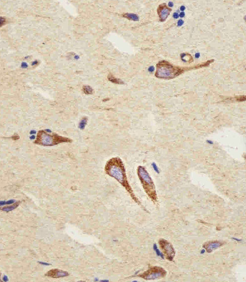
A01307-3 has IHC-P images of paraffin-embedded brain sections and paraffin-section, paraffin-embedded human skin carcinoma (catalog image captions). Human reactivity is listed; IF/ICC data are absent (catalog reactivity/applications).

Real IHC data Immunohistochemical analysis of paraffin-embedded R. brain section using CB2 Antibody(C-term). A01307-3 was diluted at 1:25 dilution. A peroxidase-conjugated goat anti-rabbit IgG at 1:400 dilution was used as the secondary antibody, followed by DAB staining.
Anti-CB2 Antibody (C-term)
Cat # A01307-3

A01307-3 is listed for human IHC-P and has DAB images of paraffin-embedded R. and H. brain sections (catalog applications/reactivity; image captions). A separate image shows paraffin-section, paraffin-embedded human skin carcinoma; fixation is unreported for the brain sections (catalog image captions).

Which to pick: Choose A01307-3 for human tissue IHC-P: it is a rabbit polyclonal with listed human reactivity and an IHC image of paraffin-section, paraffin-embedded human skin carcinoma (catalog host/dilution_raw/reactivity/applications; image caption). No SKU in the payload is listed for IF/ICC or cross-species reactivity; the R. brain image does not establish another species as reactive (catalog applications/reactivity; image caption). The selected A01307-3 tissue-IHC caption documents paraffin sections, but does not specify the fixative (selected-SKU IHC image A01307-3).

Each figure is that product's own IHC / IF validation image from its datasheet.

References

  1. UniProt Consortium. UniProt entry P34972 (CNR2_HUMAN, Cannabinoid receptor 2).
  2. Human Protein Atlas. CNR2 tissue IHC expression (reliability: Uncertain).
  3. Human Protein Atlas. CNR2 subcellular location (ICC-IF): Membrane.
  4. Human Protein Atlas. CNR2 antibody validation summary (1 antibodies).
  5. Cannabinoid type 2 receptors in dopamine neurons inhibits psychomotor behaviors, alters anxiety, depression and alcohol preference. Scientific reports 2017 — PMC5727179.
  6. Selective activation of cannabinoid receptor 2 regulates Treg/Th17 balance to ameliorate neutrophilic asthma in mice. Annals of translational medicine 2021 — PMC8267324.
  7. Altered Endocannabinoid Signaling in Placentas from SARS-CoV-2-Infected Pregnancies. Diagnostics (Basel, Switzerland) 2026 — PMC12984115.
  8. Cannabinoid CB2 receptor potentiates obesity-associated inflammation, insulin resistance and hepatic steatosis. PloS one 2009 — PMC2688760.
  9. PubMed PMID:7689702 — UniProt-cited evidence.
  10. PubMed PMID:19496827 — UniProt-cited evidence.
  11. PubMed PMID:16710414 — UniProt-cited evidence.