This website uses cookies to ensure you get the best experience on our website.


- Table of Contents
Plan COL14A1 paraffin-section IHC using reported cytoplasmic staining, including high signal in appendix glandular cells (HPA tissue IHC). Start the catalog antibody at 1:50 and optimize with controls (datasheet: 1:50); interpret staining cautiously because the tissue IHC evidence is uncertain (HPA tissue IHC).

Expected localisation, validated protocols, controls and antibodies — the at-a-glance facts below, then the full design guide.
| Expected localisation | Cytoplasmic staining in most tissues (HPA tissue IHC) | |
| Staining pattern | Cytoplasmic signal; high in glandular and squamous cells (HPA tissue IHC) | |
| Antigen retrieval | Citrate pH 6.0 HIER, 95–98 °C, 20 min (rule: cytoplasmic / membrane antigen) | |
| Positive control | Appendix+4 more · see all | |
| Negative control | Adipose tissue+4 more · see all |
| Fixation | Keep fixation consistent across paraffin sections (standard IHC practice; not target-specific) | |
| Caveat | Tissue staining is uncertain for this secreted protein (HPA tissue IHC) | |
| Regulation | Tissue-enhanced RNA in blood vessels (HPA tissue RNA) | |
| Isoform / epitope | 3 isoforms; signal peptide removed; epitope unknown (UniProt) |
The catalog antibody's IHC-P protocol is accompanied by 3 published COL14A1 paraffin IHC protocols (PMC9977988; PMC10401928; PMC12372714).
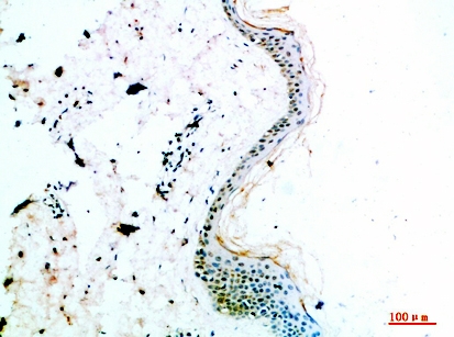
| Sample | Paraffin-embedded human skin tissue; fixative not specified (datasheet A06685) |
| Fixation | Image fixative and duration unreported (datasheet A06685); verify before use. |
| Sectioning | 4–5 µm sections on charged slides (standard) |
| Deparaffinisation | Xylene, graded ethanol series to water (standard) |
| Antigen retrieval | Heat-induced epitope retrieval in citrate buffer, pH 6.0, 20 min at 95–98 °C (standard rule: cytoplasmic / membrane antigen) |
| Peroxidase block | 3% H2O2, 10 min, room temperature (standard) |
| Blocking | 10% normal serum of the secondary host, 30 min, room temperature (standard) |
| Primary antibody | Rabbit anti-COL14A1, 1:50 recommended; image 1:200 (datasheet A06685) |
| Primary incubation | Overnight at 4 °C (standard) |
| Detection | HRP-polymer secondary, DAB chromogen 5–10 min (standard) |
| Counterstain | Hematoxylin, blue, dehydrate and mount (standard) |
| Expected result | COL14A1-positive staining in glandular cells of appendix (HPA tissue IHC: High). HPA tissue profile: Cytoplasmic expression in most tissues. No signal in the no-primary control. |
COL14A1 is a secreted, extracellular matrix protein with no transmembrane segment (UniProt Q05707 topology). For paraffin-section IHC, assess matrix-associated staining alongside the cytoplasmic staining reported in glandular cells of appendix, colon and rectum and squamous epithelial cells of esophagus (HPA tissue IHC). Treat either pattern cautiously: HPA rates its tissue IHC reliability Uncertain, noting that secreted protein can complicate the match between RNA and protein location (HPA tissue IHC).
| Signal outlines interstitial structures, with staining near appendix or colon glands (UniProt Q05707 subcellular location; HPA tissue IHC). | An extracellular distribution is consistent with COL14A1 secretion and its proposed association with collagen fibrils (UniProt Q05707 function). Judge it alongside the documented glandular-cell signal; HPA has not verified a definitive tissue pattern (HPA tissue IHC: Uncertain). |
| The strongest signal is confined to nuclei, without a comparable extracellular or reported cytoplasmic pattern. | Nuclear-only staining conflicts with the extracellular location assigned by UniProt and the cytoplasmic tissue profile reported by HPA (UniProt Q05707 subcellular location; HPA tissue IHC). Check counterstain overlap and repeat with appropriate controls before calling it COL14A1-specific signal (standard IHC practice). |
| Strong staining appears in adipocytes or bone-marrow hematopoietic cells instead of the expected comparison cells. | Those cell populations are reported as not detected by HPA (HPA tissue IHC). Unexpected chromogen may reflect cross-reactivity or endogenous detection activity (standard IHC practice); the finding alone does not establish which cause applies. |
| Chromogen covers much of the section without a discernible cell or matrix distribution. | Diffuse staining cannot be assigned confidently to the reported HPA cell pattern or UniProt extracellular location (HPA tissue IHC; UniProt Q05707 subcellular location). Review the no-primary control and detection background before scoring intensity (standard IHC practice). |
| Appendix glandular cells show no staining in a run that includes that tissue. | Appendix glandular cells are reported High by HPA, so their absence calls for a run-level check (HPA tissue IHC). HPA also rates the IHC evidence Uncertain; a blank section alone cannot prove target absence or antibody failure (HPA tissue IHC). |
| Extracellular location and processing | COL14A1 has a signal peptide at residues 1–28 and a mature chain beginning at residue 29 (UniProt Q05707 processing). Secreted protein can appear away from its producing cells; HPA explicitly warns that tissue RNA and protein locations may differ (HPA tissue IHC). |
| Choice of comparison cells | Appendix, colon and rectum glandular cells are High; esophageal squamous cells are also High (HPA tissue IHC). Adipocytes and bone-marrow hematopoietic cells are reported not detected (HPA tissue IHC). Compare the named cell populations, not entire tissues. |
| Confidence in the IHC pattern | HPA rates tissue staining Uncertain and lists antibody HPA023781 as IHC Uncertain (HPA tissue IHC; HPA antibodies). Interpret a convincing distribution as provisional and use run controls when judging specificity (standard IHC practice). |
| IF/ICC Q&A: What signal is reported? | HPA reports vesicles as the approved ICC-IF location and lists HPA023781 as ICC Approved (HPA subcellular; HPA antibodies). This describes cell imaging; it does not establish a paraffin-section IHC pattern (HPA tissue IHC: Uncertain). |
| Situation | Likely cause | Next action |
|---|---|---|
| Positive comparison tissue is blank. | The run may lack detectable signal, or the chosen field may miss the reported cells (standard IHC practice). Appendix glandular cells are reported High (HPA tissue IHC). | Inspect glandular areas and confirm the run's positive and no-primary controls; then review the validated antibody and detection workflow (standard IHC practice). Do not infer a COL14A1-specific fixation effect from the blank result. |
| Nuclei stain prominently while matrix and reported cell compartments do not. | A nuclear-only pattern conflicts with UniProt's extracellular assignment and HPA's cytoplasmic tissue profile (UniProt Q05707 subcellular location; HPA tissue IHC). | Check whether chromogen is being confused with counterstain, compare the no-primary control, and reassess the compartment in an independently stained section (standard IHC practice). |
| Adipocytes or marrow hematopoietic cells stain strongly. | HPA reports these cells as not detected (HPA tissue IHC). Cross-reactivity or endogenous detection activity can produce unexpected staining (standard IHC practice). | Compare a no-primary control and an appropriate endogenous-enzyme control; score the named cells separately from nearby stroma (standard IHC practice). |
| Brown signal is widespread with little structural contrast. | Diffuse chromogen may arise from background in blocking, washing or detection steps (standard IHC practice); it cannot resolve the uncertain HPA tissue pattern (HPA tissue IHC). | Compare the no-primary control, inspect wash and detection steps, and repeat under the validated IHC workflow before assigning a positive score (standard IHC practice). |
| Only extracellular staining appears; reported glandular-cell staining is absent. | COL14A1 is secreted, and HPA notes that RNA and protein tissue locations may differ (UniProt Q05707 subcellular location; HPA tissue IHC). The isolated matrix signal remains unverified as specific. | Document the matrix compartment and named cell populations separately; compare controls and another HPA-reported positive tissue before interpreting the result (standard IHC practice; HPA tissue IHC). |
| IHC looks cytoplasmic, but an IF/ICC image looks vesicular. | HPA reports cytoplasmic tissue staining and an approved vesicular ICC-IF location in separate assays (HPA tissue IHC; HPA subcellular). Neither observation validates the other. | Report each assay's compartment and validation status separately: tissue IHC is Uncertain, while the listed antibody's ICC result is Approved (HPA tissue IHC; HPA antibodies). |


Comprehensive Human Protein Atlas IHC scoring per tissue (reliability: Uncertain — At least one protein variant secreted, tissue location of RNA and protein might differ and correlation is complex. Pending external verification.). Rows are taken directly from the HPA tissue chart — click any row's HPA link to view the source.
| Tissue | Cell type | Level | Evidence | Source |
|---|---|---|---|---|
| Appendix | Glandular cells | High | Protein (IHC) | HPA → |
| Colon | Glandular cells | High | Protein (IHC) | HPA → |
| Esophagus | Squamous epithelial cells | High | Protein (IHC) | HPA → |
| Rectum | Glandular cells | High | Protein (IHC) | HPA → |
| Stomach | Glandular cells | Medium | Protein (IHC) | HPA → |
| Tissue | Cell type | Level | Evidence | Source |
|---|---|---|---|---|
| Adipose tissue | Adipocytes | Not detected | Protein (IHC) | HPA → |
| Adrenal gland | Glandular cells | Not detected | Protein (IHC) | HPA → |
| Bone marrow | Hematopoietic cells | Not detected | Protein (IHC) | HPA → |
| Breast | Adipocytes | Not detected | Protein (IHC) | HPA → |
| Caudate | Glial cells | Not detected | Protein (IHC) | HPA → |
Use extracellular matrix distribution and matched controls to troubleshoot COL14A1 staining in paraffin sections; interpret cellular signals cautiously (UniProt Q05707 localisation; HPA tissue IHC reliability: Uncertain).
A06685 has an IHC image from paraffin-embedded human skin and lists IF as an application; its stated reactivity covers human, mouse and rat (catalog: A06685 applications, reactivity and IHC caption).
A06685 has a human-skin paraffin-section IHC image with antibody diluted at 1:200 (A06685 IHC caption). IF is listed as an application, but no IF image is supplied; human, mouse and rat are listed as reactive species (catalog: A06685 applications, IF image alts and reactivity).
Which to pick: For tissue IHC, choose A06685: its image shows paraffin-embedded human skin at 1:200, with the fixative unreported (A06685 IHC caption). For IF/ICC, A06685 lists IF and a 1:50 dilution, though no IF image or ICC validation is supplied (catalog: A06685 applications, IF dilution and IF image alts). For cross-species work, A06685 lists human, mouse and rat reactivity; its pictured IHC result is human only, and the antibody host is rabbit with clonality unreported (catalog: A06685 reactivity, IHC caption, host and clone).