This website uses cookies to ensure you get the best experience on our website.


- Table of Contents
Plan chromogenic COL5A3 IHC on paraffin sections using the observed cytoplasmic tissue pattern and the extracellular matrix location as separate expectations (HPA tissue IHC; UniProt). High staining in cervix squamous epithelial cells provides a reference, and the catalog antibody's 1:100–1:300 IHC dilution provides a starting range (HPA tissue IHC; datasheet).

Expected localisation, validated protocols, controls and antibodies — the at-a-glance facts below, then the full design guide.
| Expected localisation | Cytoplasmic tissue staining (HPA tissue IHC); extracellular matrix expected (UniProt) | |
| Staining pattern | Cytoplasmic staining in squamous, glandular and Leydig cells (HPA tissue IHC) | |
| Antigen retrieval | Citrate pH 6.0 HIER, 95–98 °C, 20 min (rule: cytoplasmic / membrane antigen) | |
| Positive control | Cervix+4 more · see all | |
| Negative control | Adipose tissue+4 more · see all |
| Fixation | Keep fixation consistent across sections (standard IHC practice; not target-specific); Matched tissue-IHC evidence does not establish the fixation claim. Validate the specimen-specific method before use. (selected-SKU IHC image A10525) | |
| Caveat | Secreted protein; tissue staining may diverge from RNA (HPA tissue IHC) | |
| Regulation | Regulation not specified (UniProt) | |
| Isoform / epitope | No isoforms; signal 1–29 is removed; no cytoplasmic epitope (UniProt) |
The catalog antibody protocol is accompanied by one published COL5A3 IHC protocol for clear cell renal cell carcinoma (PMC10891384).
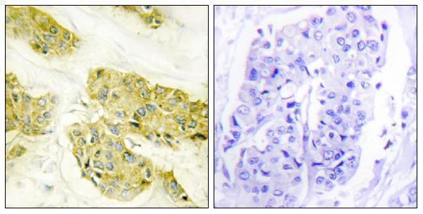
| Sample | Paraffin-embedded human breast carcinoma tissue; fixative not specified (datasheet A10525) |
| Fixation | Image fixative and duration unreported (datasheet A10525); verify before use. |
| Sectioning | 4–5 µm sections on charged slides (standard) |
| Deparaffinisation | Xylene, graded ethanol series to water (standard) |
| Antigen retrieval | Heat-induced epitope retrieval in citrate buffer, pH 6.0, 20 min at 95–98 °C (standard rule: cytoplasmic / membrane antigen) |
| Peroxidase block | 3% H2O2, 10 min, room temperature (standard) |
| Blocking | 10% normal serum of the secondary host, 30 min, room temperature (standard) |
| Primary antibody | Rabbit anti-COL5A3, 1:100-1:300 (datasheet A10525) |
| Primary incubation | Overnight at 4 °C (standard) |
| Detection | HRP-polymer secondary, DAB chromogen 5–10 min (standard) |
| Counterstain | Hematoxylin, blue, dehydrate and mount (standard) |
| Expected result | COL5A3-positive staining in squamous epithelial cells of cervix (HPA tissue IHC: High). HPA tissue profile: General cytoplasmic expression. No signal in the no-primary control. |
COL5A3 is secreted and assigned to the extracellular matrix, with no transmembrane segment (UniProt P25940 localization and topology). In tissue IHC, HPA reports general cytoplasmic expression, including high staining in cervical and tonsillar squamous epithelial cells, intestinal and parathyroid glandular cells, and testicular Leydig cells (HPA: tissue IHC). Its IHC reliability is Approved, with low agreement between staining and RNA data and external verification pending (HPA: reliability).
| Strong cytoplasmic staining in cervical squamous epithelial cells, with a clear counterstain (HPA: High in cervical squamous epithelial cells; general cytoplasmic expression). | This matches a reported positive tissue and cell type (HPA: tissue IHC). Record the fraction and intensity of stained cells, then compare the pattern with a negative reagent control (standard IHC practice). It does not establish exclusively intracellular localization: COL5A3 is secreted (UniProt P25940 localization). |
| A predominantly nuclear pattern, or staining confined to an unexpected intracellular structure, replaces the reported cytoplasmic tissue pattern (HPA: general cytoplasmic expression). | Treat the compartment mismatch as a possible artefact and review the control slide, antibody concentration, and detection conditions (standard IHC practice). HPA reports plasma membrane localization in ICC-IF, so a membrane outline alone cannot be dismissed across applications (HPA: subcellular ICC-IF). |
| Strong staining appears in adipocytes or lung alveolar cells, both reported as not detected by HPA tissue IHC (HPA: adipose tissue and lung). | Check for cross-reactivity or endogenous detection activity before interpreting those cells as COL5A3 positive (standard IHC practice). HPA's entries refer to those specific cell types; they do not establish that every cell in either tissue is negative (HPA: tissue IHC). |
| Brown signal spreads across the section, including areas without a discernible cell or matrix pattern (standard IHC practice). | Consider nonspecific reagent binding, endogenous detection activity, or inadequate washing (standard IHC practice). Compare negative reagent and known-positive controls, and score localized staining separately from diffuse background (standard IHC practice). |
| No signal is seen in cervical squamous epithelial cells or testicular Leydig cells, both reported High by HPA (HPA: cervix and testis). | First check that the positive control and detection system worked, then review antibody dilution and the validated IHC-P workflow if available (standard IHC practice). A failed stain alone cannot establish absent COL5A3; HPA reports variable tissue staining and pending external verification (HPA: reliability; tissue IHC). |
| Tissue pattern versus protein location | HPA describes general cytoplasmic tissue staining, while UniProt assigns the secreted protein to extracellular space and matrix (HPA: tissue IHC; UniProt P25940 localization). Interpret the observed cell pattern without requiring both sources to describe the same compartment; HPA notes that RNA and protein tissue locations can differ (HPA: reliability). |
| Strength of IHC evidence | The tissue IHC profile is Approved, but antibody staining has low agreement with RNA data and awaits external verification (HPA: reliability). HPA048256 is IHC Approved; the supplied record does not label it Enhanced (HPA: antibodies). Use controls when applying the reported pattern to a new section (standard IHC practice). |
| Processing and epitope | UniProt lists a signal peptide at residues 1–29 and a chain at 30–1745 (UniProt P25940 processing). The supplied sources give no antibody epitope, so they cannot predict whether processing changes staining with the chosen antibody (UniProt P25940 processing; HPA: antibodies). |
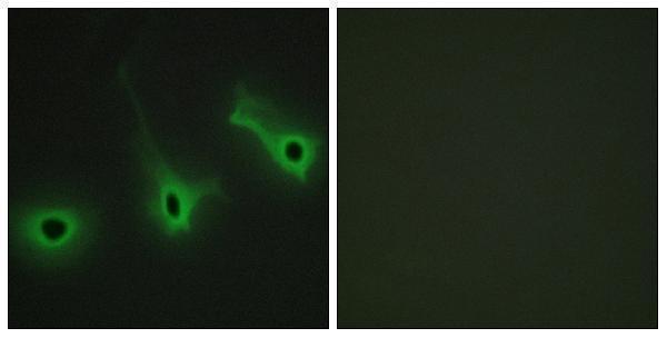
| What should IF/ICC show? | HPA reports approved plasma membrane localization in ICC-IF images and lists HPA041695 as ICC Approved (HPA: subcellular ICC-IF; antibodies). The tissue IHC summary instead describes general cytoplasmic expression, with HPA048256 IHC Approved (HPA: tissue IHC; antibodies). Interpret each application against its own evidence. |
| Situation | Likely cause | Next action |
|---|---|---|
| The known-positive section has no staining. | A run-level detection failure or unsuitable working conditions are possible (standard IHC practice). | Check control performance, detection reagents, antibody dilution, and the antibody's validated IHC-P instructions before calling the specimen negative (standard IHC practice; HPA: High in cervical squamous epithelial cells). |
| All tissue compartments show a similar brown haze. | Nonspecific binding, endogenous detection activity, or incomplete washing may obscure a cell-specific pattern (standard IHC practice). | Inspect a negative reagent control, verify blocking and washing, and evaluate the intended cells only after the background is controlled (standard IHC practice). |
| Nuclei stain strongly while cytoplasm is faint. | The distribution conflicts with HPA's general cytoplasmic tissue profile (HPA: tissue IHC). | Compare the positive and negative controls and reassess detection conditions before scoring nuclear signal as COL5A3 (standard IHC practice; HPA: tissue IHC). |
| Unexpected cells stain strongly, including adipocytes. | The result differs from HPA's not-detected adipocyte entry; cross-reactivity or endogenous detection activity is possible (HPA: adipose tissue; standard IHC practice). | Confirm cell identity and compare controls; do not extend HPA's adipocyte result to every cell in adipose tissue (standard IHC practice; HPA: tissue IHC). |
| The IHC and ICC-IF compartment calls differ. | HPA reports cytoplasmic tissue IHC and plasma membrane ICC-IF, with different antibodies approved for the two applications (HPA: tissue IHC; subcellular ICC-IF; antibodies). | Report the application and antibody used, and assess each image against its application-specific HPA pattern (HPA: tissue IHC; subcellular ICC-IF; antibodies). |
| Staining varies between tissue sections. | HPA reports High, Medium, Low, and not-detected observations across specified tissues and cells; its IHC reliability note flags low agreement with RNA data (HPA: tissue IHC; reliability). | Compare the same cell type across sections and include an HPA-reported positive control in the staining run (HPA: tissue IHC; standard IHC practice). |


Comprehensive Human Protein Atlas IHC scoring per tissue (reliability: Approved — Low consistency between antibody staining and RNA expression data. Secreted protein, tissue location of RNA and protein is expected to differ. Pending external verification.). Rows are taken directly from the HPA tissue chart — click any row's HPA link to view the source.
| Tissue | Cell type | Level | Evidence | Source |
|---|---|---|---|---|
| Cervix | Squamous epithelial cells | High | Protein (IHC) | HPA → |
| Parathyroid gland | Glandular cells | High | Protein (IHC) | HPA → |
| Small intestine | Glandular cells | High | Protein (IHC) | HPA → |
| Testis | Leydig cells | High | Protein (IHC) | HPA → |
| Tonsil | Squamous epithelial cells | High | Protein (IHC) | HPA → |
| Tissue | Cell type | Level | Evidence | Source |
|---|---|---|---|---|
| Adipose tissue | Adipocytes | Not detected | Protein (IHC) | HPA → |
| Endometrium | Cells in endometrial stroma | Not detected | Protein (IHC) | HPA → |
| Epididymis | Glandular cells | Not detected | Protein (IHC) | HPA → |
| Hippocampus | Glial cells | Not detected | Protein (IHC) | HPA → |
| Lung | Alveolar cells | Not detected | Protein (IHC) | HPA → |
Troubleshoot COL5A3 staining in paraffin section IHC by checking retrieval, compartment, controls, and scoring before interpreting chromogenic signal.
A10525 has IHC data from paraffin-embedded human breast carcinoma tissue and IF data from HeLa cells (catalog image captions); its listed reactivity is Human (catalog).
A10525 will render with an IHC figure from paraffin-embedded human breast carcinoma tissue, including a peptide-blocked comparison (catalog IHC image caption). Its listed applications include IHC and IF (catalog), and its IF image shows HeLa cells with a peptide-blocked comparison (catalog IF image caption).
Which to pick: For tissue IHC, choose A10525: its IHC image documents paraffin-embedded human breast carcinoma tissue (catalog IHC image caption), with a listed IHC dilution of 1:100–1:300 (catalog); the fixative is unreported (catalog IHC image caption). For IF/ICC, A10525 is the available choice because IF is listed and shown in HeLa cells (catalog applications and IF image caption); it is polyclonal (catalog). There is no cross-species choice in this payload: A10525 lists Human reactivity only (catalog).