This website uses cookies to ensure you get the best experience on our website.


- Table of Contents
Plan COL6A3 paraffin IHC around extracellular matrix positivity and cytoplasmic staining in a few tissues (HPA tissue IHC). Use ovarian stroma as a positive tissue control (HPA tissue IHC), start the catalog antibody at 1:100–1:300 (datasheet), and score matrix and cellular staining separately.

Expected localisation, validated protocols, controls and antibodies — the at-a-glance facts below, then the full design guide.
| Expected localisation | Extracellular matrix; some cytoplasmic tissue staining (HPA tissue IHC) | |
| Staining pattern | Cytoplasmic in a few tissues; extracellular matrix positive (HPA tissue IHC) | |
| Antigen retrieval | Citrate pH 6.0 HIER, 95–98 °C, 20 min (rule: cytoplasmic / membrane antigen) | |
| Positive control | Bone marrow+1 more · see all | |
| Negative control | Adipose tissue+4 more · see all |
| Fixation | Keep formalin fixation consistent across sections. (standard IHC practice; not target-specific) | |
| Caveat | Secreted variants can uncouple tissue RNA and protein staining (HPA tissue IHC) | |
| Regulation | Tissue-enhanced RNA in smooth muscle (HPA tissue RNA) | |
| Isoform / epitope | Five isoforms; mature chain starts at residue 26 (UniProt) |
The catalog antibody’s IHC-P protocol is accompanied by four published COL6A3 IHC protocols (PMC3163121; PMC7061042; PMC13424786; PMC4745773).
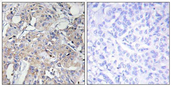
| Sample | Paraffin-embedded human breast carcinoma tissue; fixative not specified (datasheet A02844) |
| Fixation | Image fixative and duration unreported (datasheet A02844); verify before use. |
| Sectioning | 4–5 µm sections on charged slides (standard) |
| Deparaffinisation | Xylene, graded ethanol series to water (standard) |
| Antigen retrieval | Heat-induced epitope retrieval in citrate buffer, pH 6.0, 20 min at 95–98 °C (standard rule: cytoplasmic / membrane antigen) |
| Peroxidase block | 3% H2O2, 10 min, room temperature (standard) |
| Blocking | 10% normal serum of the secondary host, 30 min, room temperature (standard) |
| Primary antibody | Rabbit anti-COL6A3, 1:100 - 1:300 (datasheet A02844) |
| Primary incubation | Overnight at 4 °C (standard) |
| Detection | HRP-polymer secondary, DAB chromogen 5–10 min (standard) |
| Counterstain | Hematoxylin, blue, dehydrate and mount (standard) |
| Expected result | COL6A3-positive staining in hematopoietic cells of bone marrow (HPA tissue IHC: Medium). HPA tissue profile: Cytoplasmic expression in a few tissues with positivity in extracellular matrix. No signal in the no-primary control. |
COL6A3 is a secreted extracellular matrix protein with no transmembrane segment (UniProt P12111 topology). In paraffin IHC, expect extracellular matrix positivity and cytoplasmic staining in a few tissues, including medium staining in ovarian stromal cells and bone marrow hematopoietic cells (HPA tissue IHC). HPA rates the tissue profile Supported but reports low consistency between staining and RNA expression; a secreted variant can separate the protein signal from its site of production (HPA tissue IHC).
| Extracellular matrix staining accompanies cytoplasmic staining in some cells. | This fits the reported IHC profile (HPA tissue IHC) and the protein’s secreted, extracellular matrix location (UniProt P12111). Assess the cell staining and surrounding matrix separately: secreted protein can appear beyond the cells that produced it (HPA tissue IHC). |
| Ovarian stromal cells or bone marrow hematopoietic cells show moderate signal. | These are the supplied medium-positive tissue and cell examples (HPA tissue IHC). Use the named cell population when judging a positive section; positivity in another compartment of the same tissue does not establish the expected pattern (HPA tissue IHC). |
| Staining is predominantly nuclear, with little cytoplasmic or matrix signal. | A nuclear-dominant pattern does not match the reported tissue IHC profile (HPA tissue IHC) or secreted extracellular matrix location (UniProt P12111). Treat it as a suspect signal and check controls and detection conditions before assigning it to COL6A3. |
| Strong staining appears in an HPA-listed negative cell population, such as adipocytes. | HPA reports adipocytes in adipose tissue as not detected (HPA tissue IHC). Check for nonspecific binding or endogenous detection activity using appropriate controls (general IHC practice). This observation alone cannot identify which source caused the signal. |
| A known-positive cell population lacks signal, or brown color is diffuse across the section. | Absent signal in ovarian stromal or bone marrow hematopoietic cells conflicts with their reported medium staining (HPA tissue IHC); diffuse color without a defined cell or matrix pattern is hard to interpret. Review the positive and negative controls, retrieval, antibody concentration, and detection background (general IHC practice). |
| Secreted protein and tissue location | COL6A3 is secreted and associated with extracellular matrix (UniProt P12111). HPA cautions that at least one variant is secreted, so the stained location and the cells expressing its RNA can differ (HPA tissue IHC). Score deposited matrix without assuming the adjacent cell synthesized it. |
| Strength of the tissue reference | The HPA tissue IHC profile is Supported, while HPA also reports low consistency between antibody staining and RNA expression (HPA tissue IHC). Use the named positive cell populations as practical references; avoid treating RNA abundance alone as an IHC pass or fail criterion. |
| Cell population used as a negative reference | HPA reports no detected staining in adipocytes of adipose tissue and several other specified cell populations (HPA tissue IHC). A negative call applies to the listed cells, not automatically to every structure in that organ (HPA tissue IHC). |
| Protein variants and maturation | UniProt lists five isoforms and a signal peptide at residues 1–25 (UniProt P12111). Those facts alone do not establish which isoforms a particular antibody detects or whether processing changes its IHC signal. Confirm the catalog antibody’s epitope information before making an isoform-specific interpretation. |
| Antibody validation scope | The listed HPA IHC antibody, HPA010080, is Supported for IHC; HPA010007 is Approved for ICC and has no listed IHC status (HPA antibodies). An ICC result does not independently validate a paraffin IHC pattern. Assess the IHC-validated antibody against the tissue references and controls. |
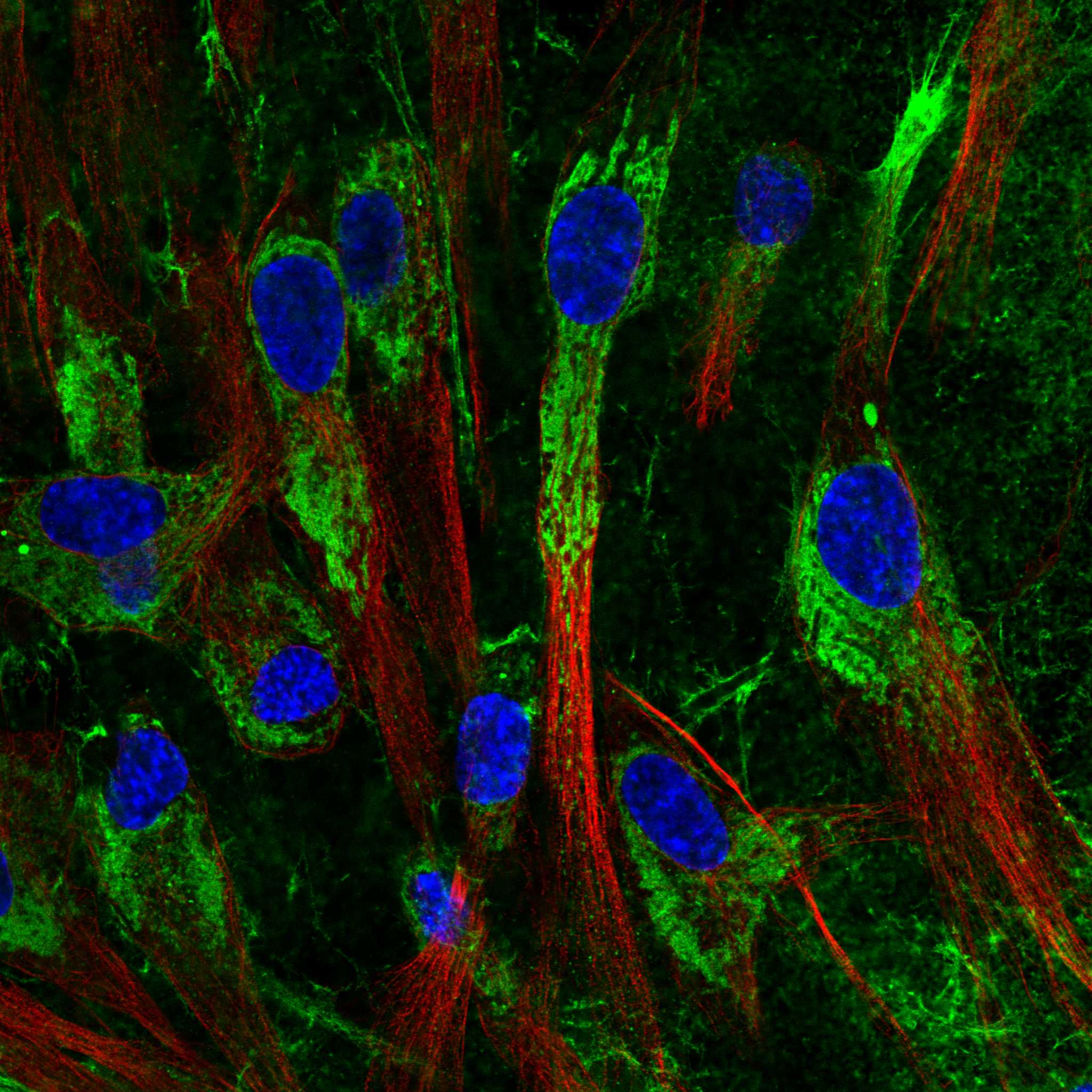
| IF/ICC Q: Why might cells show an intracellular pattern? | A: HPA reports mainly endoplasmic reticulum and additional vesicle localization in ICC-IF (HPA subcellular). This can describe intracellular protein before secretion (UniProt P12111); it does not replace the extracellular matrix and cytoplasmic tissue IHC reference (HPA tissue IHC). |
| Situation | Likely cause | Next action |
|---|---|---|
| Ovarian stromal cells or bone marrow hematopoietic cells are unstained. | These populations have medium staining in the HPA reference (HPA tissue IHC); the failed signal has no established target-specific cause from the supplied evidence. | Check that the intended cells are present and review a positive control, retrieval conditions, antibody dilution, and detection reagents (general IHC practice). Interpret a negative test section only after the control works. |
| The whole section has diffuse brown background. | Diffuse color lacks the defined cell or matrix distribution in the reported profile (HPA tissue IHC). Excess reagent, incomplete blocking, or endogenous detection activity are possible general IHC causes. | Compare the test slide with a no-primary control; review blocking, washes, detection chemistry, and antibody concentration (general IHC practice). Do not score diffuse background as extracellular matrix positivity. |
| Nuclei are the main stained structures. | Nuclear-dominant staining is inconsistent with the tissue profile (HPA tissue IHC) and the secreted extracellular matrix annotation (UniProt P12111); its mechanism is undetermined. | Check the no-primary control and the expected positive tissue pattern, then review antibody specificity and detection conditions (general IHC practice). Avoid a COL6A3-positive call from nuclear staining alone. |
| Adipocytes in adipose tissue stain strongly. | HPA reports adipocytes in adipose tissue as not detected (HPA tissue IHC). Nonspecific binding or endogenous detection activity may explain an unexpected signal (general IHC practice). | Confirm the stained cells are adipocytes, inspect the no-primary control, and review blocking and detection chemistry (general IHC practice). Do not extend the adipocyte reference to every other structure in adipose tissue. |
| Matrix stains, but nearby cells have little cytoplasmic signal. | Secreted COL6A3 can reside in extracellular matrix (UniProt P12111), and HPA cautions that protein and RNA locations can differ for a secreted variant (HPA tissue IHC). | Document matrix and cellular staining separately. Compare morphology and a reference positive population before calling the section negative; absence from adjacent cells alone does not invalidate matrix staining (HPA tissue IHC). |
| An ICC-IF image appears vesicular while the IHC slide highlights matrix. | HPA reports endoplasmic reticulum and vesicle localization in ICC-IF (HPA subcellular), while tissue IHC includes extracellular matrix positivity (HPA tissue IHC). | Judge the paraffin IHC slide against the tissue IHC pattern and its controls. Record the ICC-IF observation in its own context; do not use intracellular ICC localization as the sole IHC scoring rule. |

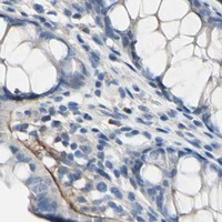


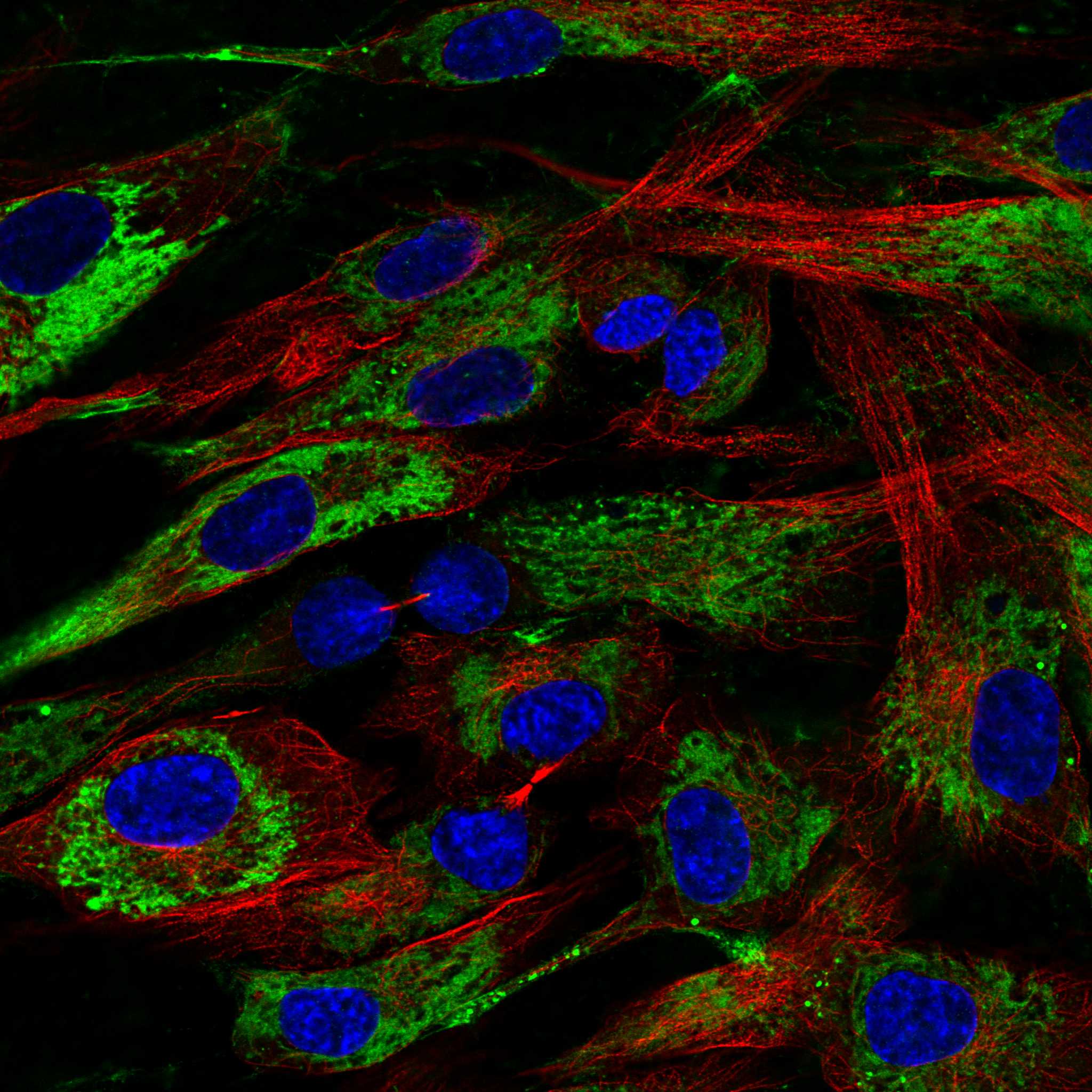
Comprehensive Human Protein Atlas IHC scoring per tissue (reliability: Supported — Low consistency between antibody staining and RNA expression data. At least one protein variant secreted, tissue location of RNA and protein might differ and correlation is complex. Antibody staining in cells/structures not annotated, view images.). Rows are taken directly from the HPA tissue chart — click any row's HPA link to view the source.
| Tissue | Cell type | Level | Evidence | Source |
|---|---|---|---|---|
| Bone marrow | Hematopoietic cells | Medium | Protein (IHC) | HPA → |
| Ovary | Ovarian stroma cells | Medium | Protein (IHC) | HPA → |
| Tissue | Cell type | Level | Evidence | Source |
|---|---|---|---|---|
| Adipose tissue | Adipocytes | Not detected | Protein (IHC) | HPA → |
| Adrenal gland | Glandular cells | Not detected | Protein (IHC) | HPA → |
| Appendix | Glandular cells | Not detected | Protein (IHC) | HPA → |
| Breast | Adipocytes | Not detected | Protein (IHC) | HPA → |
| Bronchus | Respiratory epithelial cells | Not detected | Protein (IHC) | HPA → |
Troubleshoot COL6A3 staining in paraffin-section chromogenic IHC by assessing retrieval, tissue handling, extracellular localisation and compartment-specific scoring (UniProt P12111; HPA tissue IHC).
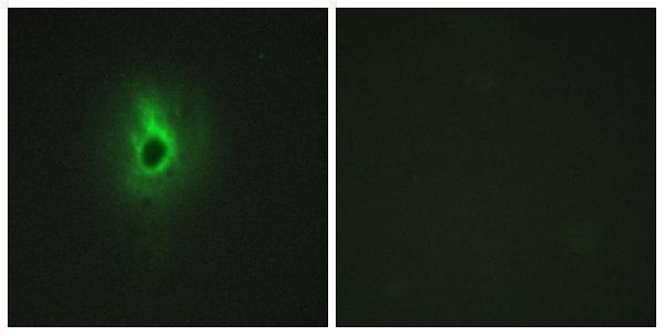
A02844 has IHC data from paraffin-embedded human breast carcinoma tissue and IF data from HeLa cells (catalog: image captions); listed reactivity is human and mouse (catalog: reactivity).
A02844 was shown in IHC on paraffin-embedded human breast carcinoma tissue, with peptide-blocked comparison (catalog: IHC image caption). The same SKU was shown in IF on HeLa cells, with peptide-blocked comparison (catalog: IF image caption).
Which to pick: Choose A02844 for tissue IHC on paraffin sections, the preparation shown in its IHC image; the fixative is unreported (catalog: IHC image caption). Choose it for IF/ICC because both applications are listed and its IF image shows HeLa cells (catalog: applications and IF image caption). For human or mouse samples, it is the listed rabbit polyclonal option, although the supplied images show human samples only (catalog: host, polyclonal designation, reactivity and image captions).