This website uses cookies to ensure you get the best experience on our website.


- Table of Contents
Plan chromogenic COPS5 IHC in paraffin sections around nuclear and cytoplasmic staining (HPA tissue IHC). Score the compartments separately because IFIT3 can diminish nuclear localisation (UniProt).

Expected localisation, validated protocols, controls and antibodies — the at-a-glance facts below, then the full design guide.
| Expected localisation | Nuclear and cytoplasmic staining across tissues (HPA tissue IHC) | |
| Staining pattern | Cells across tissues show nuclear and cytoplasmic signal (HPA tissue IHC) | |
| Antigen retrieval | EDTA pH 8.0 HIER, heat-mediated (datasheet M01849-2) | |
| Positive control | Adipose tissue+4 more · see all | |
| Negative control | None in HPA (detected in all 45 tissues); use no-primary + isotype controls |
| Fixation | Keep fixation consistent across sections (standard IHC practice; not target-specific) | |
| Caveat | IFIT3 can diminish nuclear localisation (UniProt) | |
| Regulation | Expression regulation not specified (UniProt) | |
| Isoform / epitope | 0 annotated isoforms; no extracellular domain to map (UniProt) |
Compare the catalog antibody's IHC-P protocol (datasheet M01849-2) with four published COPS5 IHC methods for paraffin-embedded tissues (PMC4932188; PMC5945527; PMC5722579; PMC5777718).
| Sample | Paraffin-embedded human esophageal squamous carcinoma tissue; fixative not specified (datasheet M01849-2) |
| Fixation | Image fixative and duration unreported (datasheet M01849-2); verify before use. |
| Sectioning | 4–5 µm sections on charged slides (standard) |
| Deparaffinisation | Xylene, graded ethanol series to water (standard) |
| Antigen retrieval | Heat retrieval: EDTA pH 8.0 (datasheet M01849-2); 20 min, 95–100 °C (standard) |
| Peroxidase block | 3% H2O2, 10 min, room temperature (standard) |
| Blocking | 10% goat serum (datasheet M01849-2) |
| Primary antibody | Mouse monoclonal (clone 4G9) anti-COPS5, 2μg/ml (datasheet M01849-2) |
| Primary incubation | Overnight at 4 °C (datasheet M01849-2) |
| Detection | Streptavidin-biotin complex (SABC), DAB chromogen (datasheet M01849-2) |
| Counterstain | Hematoxylin, blue, dehydrate and mount (standard) |
| Expected result | COPS5-positive staining in adipocytes of adipose tissue (HPA tissue IHC: High). HPA tissue profile: Cytoplasmic and nuclear expression in all tissues. No signal in the no-primary control. |
COPS5 is expected in the nucleus and cytoplasm, including the perinuclear region (UniProt Q92905). HPA reports cytoplasmic and nuclear IHC staining across tissues, with high staining in several listed cell populations; its tissue IHC assessment is Supported, with medium consistency between staining and RNA expression (HPA tissue IHC). COPS5 has no transmembrane segment (UniProt Q92905 topology).
| Nuclear and cytoplasmic staining appears in the expected cells of a reference section. | This fits the reported tissue IHC pattern (HPA tissue IHC) and COPS5 localisation (UniProt Q92905). Compare cells and compartments with the reference; a single positive area alone does not establish antibody specificity. |
| Staining is confined to cell membranes or extracellular material, with little nuclear or cytoplasmic signal. | Treat a predominantly membranous or extracellular pattern as suspect: COPS5 has no transmembrane segment, and its reported locations are intracellular (UniProt Q92905 topology and subcellular location). Inspect morphology and detection controls before interpreting it as COPS5. |
| Strong staining appears chiefly in an unexpected cell population within a reference tissue. | Check cell identity against the HPA cell-level pattern; examples of high staining include adipocytes in adipose tissue and hematopoietic cells in bone marrow (HPA tissue IHC). Unexpected staining can reflect cross-reactivity or endogenous detection activity (general IHC practice). |
| Diffuse color covers tissue spaces, obscures cell boundaries, or is similar in many structures. | This prevents a reliable compartment call. Consider nonspecific antibody binding, endogenous detection activity, or excessive chromogen development (general IHC practice). The expected signal should be attributable to nuclei and/or cytoplasm (HPA tissue IHC; UniProt Q92905). |
| A reference section shows no convincing signal in cells reported as strongly positive. | A negative result in, for example, adrenal glandular cells or bronchial respiratory epithelial cells conflicts with their high HPA staining (HPA tissue IHC). Check section integrity, detection controls, and the validated IHC workflow before concluding that COPS5 is absent. |
| Which tissue and cells provide a reference? | HPA lists high staining in adipocytes, adrenal and breast glandular cells, bronchial respiratory epithelial cells, bone marrow hematopoietic cells, caudate glia, and cerebellar granular-layer cells (HPA tissue IHC). Use the corresponding cell population when judging a section. |
| How broadly should staining be expected? | HPA describes cytoplasmic and nuclear expression across tissues and low RNA tissue specificity (HPA tissue IHC). Its supplied record lists no negative tissue reference; lack of staining in an unlisted population is therefore not a validated negative control. |
| How much weight should the IHC pattern carry? | The tissue IHC assessment is Supported and reports medium agreement with RNA expression (HPA tissue IHC). HPA004845 and CAB004242 have Supported IHC status, while no IHC status is supplied for HPA051531 (HPA antibodies). Interpret borderline staining with controls and morphology. |
| Does processing or topology predict surface staining? | UniProt reports no signal peptide, propeptide, transmembrane segment, or glycosylation sites for COPS5 (UniProt Q92905). These annotations support an intracellular interpretation; they do not establish tissue-specific staining strength or a fixation effect. |
| What should IF/ICC show? | HPA reports enhanced nucleoplasmic localisation in ICC-IF images from A-431, K-562, and U2OS (HPA subcellular ICC-IF). UniProt also lists cytosolic and perinuclear locations (UniProt Q92905). Compare IF localisation with its own controls; this ICC-IF observation is not an IHC staining intensity standard. |
| Situation | Likely cause | Next action |
|---|---|---|
| A listed high-staining reference population is blank. | The assay may have failed; absence conflicts with the reported high reference staining (HPA tissue IHC). | Confirm the intended cells are present, then check the IHC-validated antibody, antigen retrieval, detection reagents, and a working positive control (general IHC practice). |
| Signal appears only along cell borders. | A border-only pattern does not match reported nuclear and cytoplasmic COPS5 localisation (HPA tissue IHC; UniProt Q92905). | Inspect an adjacent section and detection controls for nonspecific edge or membrane staining; score intracellular signal only when cell boundaries and compartments are clear (general IHC practice). |
| Chromogen is widespread in cell-free areas. | Diffuse deposition may arise from nonspecific binding or detection background (general IHC practice). | Review the no-primary control, blocking, washes, antibody concentration, and chromogen development (general IHC practice); reassess localisation after background is reduced. |
| A cell population looks positive despite a discordant reference pattern. | Cross-reactivity or endogenous detection activity is possible (general IHC practice); HPA's tissue assessment has medium RNA agreement (HPA tissue IHC). | Verify morphology and compare an appropriate no-primary or detection control; avoid assigning COPS5 from color alone (general IHC practice). |
| Nuclear staining is present but cytoplasmic staining is faint. | Nuclear enrichment can be compatible with the HPA ICC-IF nucleoplasm result; tissue IHC reports both compartments (HPA subcellular ICC-IF; HPA tissue IHC). | Assess the sampled cell type and control section before calling a compartment absent; record nuclear and cytoplasmic staining separately (general IHC practice). |
| Nuclei and cytoplasm are too dark to distinguish. | Overdeveloped chromogen or concentrated antibody can obscure localisation (general IHC practice). | Use the IHC-validated antibody's documented conditions as a starting point, then adjust concentration or development within the assay workflow and retain a positive control (general IHC practice). |


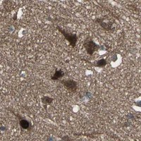


Comprehensive Human Protein Atlas IHC scoring per tissue (reliability: Supported — Medium consistency between antibody staining and RNA expression data.). Rows are taken directly from the HPA tissue chart — click any row's HPA link to view the source.
| Tissue | Cell type | Level | Evidence | Source |
|---|---|---|---|---|
| Adipose tissue | Adipocytes | High | Protein (IHC) | HPA → |
| Adrenal gland | Glandular cells | High | Protein (IHC) | HPA → |
| Appendix | Glandular cells | High | Protein (IHC) | HPA → |
| Bone marrow | Hematopoietic cells | High | Protein (IHC) | HPA → |
| Breast | Glandular cells | High | Protein (IHC) | HPA → |
| Tissue | Cell type | Level | Evidence | Source |
|---|---|---|---|---|
| None in HPA: COPS5 is detected in all 45 scored tissues. Use a no-primary (secondary-only) and an isotype control instead. | ||||
Troubleshoot COPS5 staining in paraffin sections by checking retrieval, compartment patterns, controls and scoring before interpreting differences between specimens.
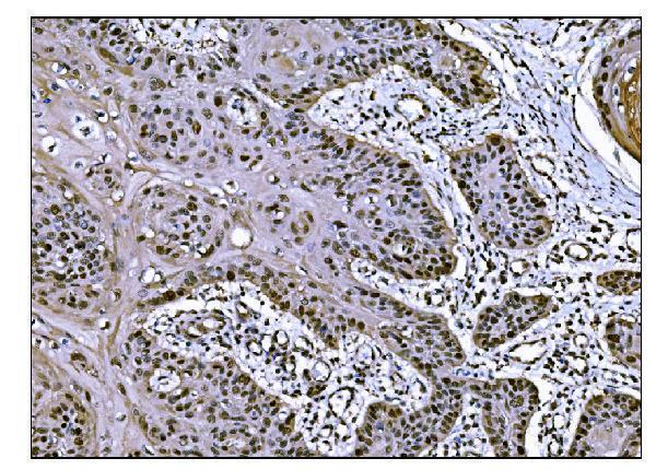
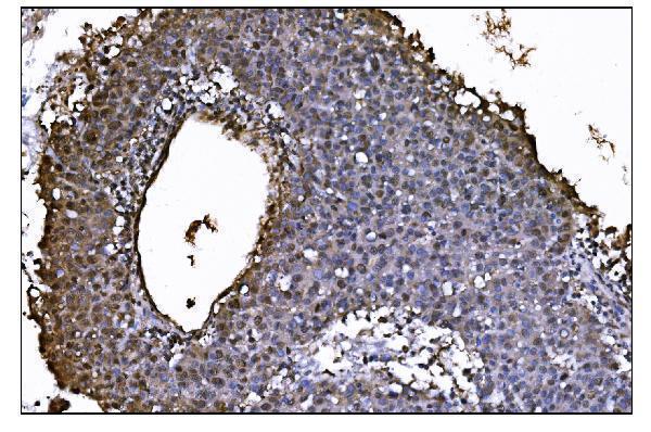
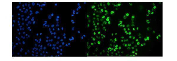
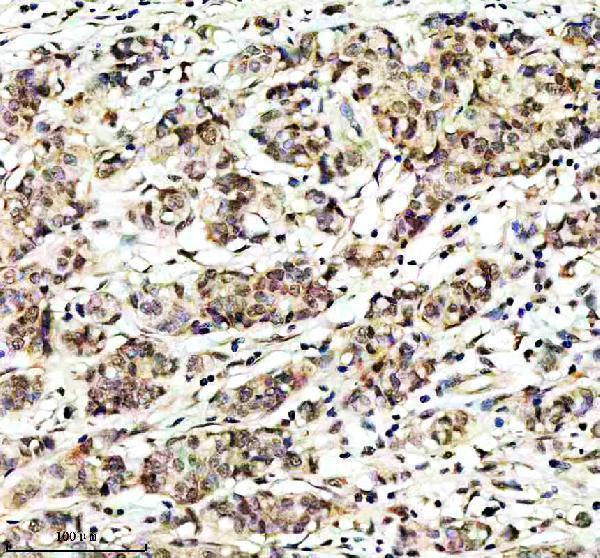
The catalog shows COPS5 staining in human paraffin sections and human cells, with additional mouse and rat tissue IHC images (catalog image captions).
M01849-2 has a human esophageal squamous carcinoma paraffin IHC image and a T-47D IF image (catalog image captions); M01849 has a human breast cancer paraffin IHC image (catalog image caption). PB9490 has paraffin IHC images from human mammary cancer, mouse testis and rat testis, plus a chromogenic A431 ICC image (catalog image captions).
Which to pick: For tissue IHC, M01849-2 is a mouse monoclonal used at 2 μg/ml in its paraffin image, while M01849 is a rabbit monoclonal used at 1:50 in its paraffin image (catalog metadata and image captions). For IF/ICC, M01849-2 has a T-47D fluorescence image; M01849 lists IF/ICC applications, while PB9490 lists IF/ICC but its A431 image uses chromogenic detection (catalog applications and image captions). For cross-species tissue work, PB9490 has human, mouse and rat paraffin IHC images (catalog image captions); the fixative is unreported in these captions (catalog image captions).