This website uses cookies to ensure you get the best experience on our website.


- Table of Contents
Plan chromogenic CPE IHC in paraffin sections using the cytoplasmic tissue pattern reported for islets, enteroendocrine cells and brain (HPA tissue IHC). Use that pattern to select positive controls, and check whether the antibody epitope lies in a removed precursor segment (UniProt).

Expected localisation, validated protocols, controls and antibodies — the at-a-glance facts below, then the full design guide.
| Expected localisation | Cytoplasmic staining in islets, gut endocrine cells and brain (HPA tissue IHC) | |
| Staining pattern | Cytoplasmic signal in islets, enteroendocrine cells and brain (HPA tissue IHC) | |
| Antigen retrieval | Citrate pH 6.0 HIER, 95–98 °C, 20 min (rule: cytoplasmic / membrane antigen) | |
| Positive control | Appendix+4 more · see all | |
| Negative control | Adipose tissue+4 more · see all |
| Fixation | Keep fixation consistent across paraffin sections. (standard IHC practice; not target-specific) | |
| Caveat | Secreted CPE may extend staining beyond secretory granules (UniProt) | |
| Regulation | Expression regulation is not annotated (UniProt) | |
| Isoform / epitope | 2 isoforms; signal peptide and propeptide are removed—check epitope (UniProt) |
The catalog antibody’s IHC-P protocol is accompanied by published CPE IHC protocols for human liver and gastric tissue (PMC4542421; PMC11058891).
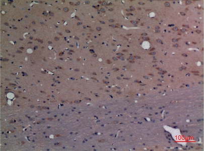
| Sample | Paraffin-embedded Rat Brain tissue; fixative not specified (datasheet A01407) |
| Fixation | Image fixative and duration unreported (datasheet A01407); verify before use. |
| Sectioning | 4–5 µm sections on charged slides (standard) |
| Deparaffinisation | Xylene, graded ethanol series to water (standard) |
| Antigen retrieval | Heat-induced epitope retrieval in citrate buffer, pH 6.0, 20 min at 95–98 °C (standard rule: cytoplasmic / membrane antigen) |
| Peroxidase block | 3% H2O2, 10 min, room temperature (standard) |
| Blocking | 10% normal serum of the secondary host, 30 min, room temperature (standard) |
| Primary antibody | Rabbit anti-CPE, 1:100 (datasheet A01407) |
| Primary incubation | Overnight at 4 °C (standard) |
| Detection | HRP-polymer secondary, DAB chromogen 5–10 min (standard) |
| Counterstain | Hematoxylin, blue, dehydrate and mount (standard) |
| Expected result | CPE-positive staining in endocrine cells of appendix (HPA tissue IHC: High). HPA tissue profile: Distinct cytoplasmic expression mainly in islets of Langerhans, enteroendocrine cells and brain. No signal in the no-primary control. |
CPE should stain mainly in the cytoplasm of pancreatic islet cells, gut endocrine cells and selected brain cells (HPA: Supported tissue IHC; High in pancreatic endocrine cells, intestinal endocrine cells and hippocampal neurons). Its secretory-vesicle and secreted locations support a cytoplasmic granular pattern; CPE has no transmembrane segment, although it can associate with granule membranes (UniProt P16870 localisation and topology).
| Strong, distinct cytoplasmic staining in pancreatic islets, with much less staining in surrounding tissue. | This fits the strongest documented control: HPA reports High staining in pancreatic endocrine cells and says external characterization supports islet staining (HPA: Supported tissue IHC). Judge the cell distribution as well as intensity; a brown deposit across the whole section is less convincing (general IHC interpretation). |
| Cytoplasmic staining in scattered intestinal endocrine cells or hippocampal neurons. | These are additional expected positives: HPA reports High staining in endocrine cells of the colon, duodenum, rectum and small intestine, and in hippocampal neuronal cells (HPA: tissue IHC). A punctate cytoplasmic appearance is compatible with secretory-vesicle localisation, but granules need not be individually resolved by chromogenic IHC (UniProt P16870 localisation; general IHC interpretation). |
| Predominantly nuclear staining, or a continuous rim on most cell surfaces. | Neither is the expected tissue-IHC pattern: HPA describes distinct cytoplasmic expression, while UniProt lists secretory vesicles, vesicle membranes and secretion, with no transmembrane segment (HPA: tissue IHC profile; UniProt P16870 localisation and topology). Reassess morphology and controls before scoring this as CPE (general IHC practice). |
| Strong staining in adipocytes or esophageal squamous epithelium. | HPA reports CPE as Not detected in those specified cells (HPA: adipose tissue adipocytes; esophagus squamous epithelial cells). Unexpected signal can reflect nonspecific antibody binding or endogenous detection activity; compare a negative-control section before attributing it to CPE (general IHC practice). |
| Diffuse brown haze across positive and negative areas. | A widespread haze obscures the cell-selective pattern reported by HPA (HPA: distinct cytoplasmic tissue-IHC profile). It can arise from nonspecific binding, inadequate washing or endogenous detection activity (general IHC practice). Treat a faint haze separately from clear cytoplasmic staining in the documented positive cells. |
| Tissue choice and validation | Pancreatic islets provide the most directly characterized positive comparison: HPA rates tissue IHC Supported and cites external characterization of islet staining (HPA: reliability description). The listed HPA antibodies are IHC Supported; this does not establish identical performance for an untested catalog antibody (HPA: antibody validation). |
| Secretory pathway and processing | CPE has a signal peptide at residues 1–25, a propeptide at 26–42 and a mature chain at 43–476 (UniProt P16870 processing). It is found in secretory vesicles, on granule membranes and as secreted protein (UniProt P16870 localisation). Without an epitope map, these facts cannot predict which processed form the antibody detects. |
| Isoforms and glycosylation | UniProt lists two isoforms and glycosylation sites at residues 139 and 390 (UniProt P16870 isoforms and glycosylation). Their effects on this antibody’s IHC staining cannot be assigned without epitope and validation data; do not explain an unexpected tissue pattern by invoking an isoform alone. |
| Antigen retrieval | Use the catalog antibody’s validated IHC-P retrieval conditions if available, then assess the pancreatic-islet positive control (general IHC practice; HPA: Supported islet staining). The supplied HPA and UniProt records do not report a CPE-specific retrieval requirement or fixation sensitivity. |
| IF/ICC Q&A: should the same pattern be expected? | HPA reports vesicles and centrosome as approved ICC-IF locations, with images from A-431, U-251MG and U2OS (HPA: subcellular ICC-IF). These cell-image findings provide context for IF interpretation; they do not make centrosomal dots a required feature of paraffin-section IHC. |
| Chromogenic detection background | Endogenous enzyme activity or nonspecific reagent binding can create color unrelated to the primary antibody (general chromogenic IHC practice). A no-primary control helps distinguish that background from the HPA-supported, cell-selective cytoplasmic pattern (HPA: tissue IHC profile; general IHC practice). |
| Situation | Likely cause | Next action |
|---|---|---|
| Pancreatic islets show no convincing signal. | The positive-control pattern has failed; antibody performance, retrieval or detection may be inadequate (HPA: Supported islet staining; general IHC practice). | Confirm that islets are present, then check the catalog antibody’s IHC-P instructions, retrieval, dilution and detection controls (general IHC practice). Do not label another tissue negative until the positive control works. |
| Signal is mainly nuclear or outlines nearly every cell. | The compartment conflicts with HPA’s cytoplasmic tissue pattern and UniProt’s vesicle and secreted localisation (HPA: tissue IHC profile; UniProt P16870 localisation). | Review a no-primary control and staining distribution, and repeat with the antibody’s validated IHC-P conditions if needed (general IHC practice). Score only convincing signal in expected cells. |
| Adipocytes or esophageal squamous cells stain strongly. | Those cell types are listed as Not detected by HPA; cross-reactivity or detection background is possible (HPA: adipose tissue and esophagus IHC; general IHC practice). | Compare with a no-primary control and a documented positive area such as pancreatic islets; reassess antibody specificity if the unexpected cell staining persists (general IHC practice; HPA: Supported islet staining). |
| Brown haze covers both positive and negative areas. | Diffuse background can reflect nonspecific binding, washing or endogenous detection activity (general IHC practice). | Inspect the no-primary control, blocking and wash steps, and detection reagents; accept a result only when the expected cells remain distinguishable (general IHC practice; HPA: distinct cytoplasmic tissue-IHC profile). |
| Intestinal staining appears only in scattered cells. | A scattered pattern can be appropriate because HPA assigns High staining specifically to intestinal endocrine cells, rather than to every epithelial cell (HPA: colon, duodenum, rectum and small-intestine IHC). | Check whether the stained cells have endocrine-cell morphology and cytoplasmic signal; compare them with the pancreatic-islet control before calling the section weak (HPA: tissue IHC; general IHC interpretation). |
| IF shows centrosomal dots that are absent from chromogenic IHC. | Centrosome is an approved HPA ICC-IF location, while the tissue-IHC profile emphasizes cytoplasmic staining (HPA: subcellular ICC-IF; tissue IHC profile). | Interpret each application against its own HPA evidence. Do not require visible centrosomal dots to accept cell-selective cytoplasmic staining in paraffin-section IHC (HPA: ICC-IF and tissue IHC; general IHC interpretation). |


Comprehensive Human Protein Atlas IHC scoring per tissue (reliability: Supported — External characterization data supports antibody staining in islets of Langerhans.). Rows are taken directly from the HPA tissue chart — click any row's HPA link to view the source.
| Tissue | Cell type | Level | Evidence | Source |
|---|---|---|---|---|
| Appendix | Endocrine cells | High | Protein (IHC) | HPA → |
| Cerebellum | Bergmann glia - cytoplasm/membrane | High | Protein (IHC) | HPA → |
| Colon | Endocrine cells | High | Protein (IHC) | HPA → |
| Duodenum | Endocrine cells | High | Protein (IHC) | HPA → |
| Hippocampus | Neuronal cells | High | Protein (IHC) | HPA → |
| Tissue | Cell type | Level | Evidence | Source |
|---|---|---|---|---|
| Adipose tissue | Adipocytes | Not detected | Protein (IHC) | HPA → |
| Adrenal gland | Glandular cells | Not detected | Protein (IHC) | HPA → |
| Bone marrow | Hematopoietic cells | Not detected | Protein (IHC) | HPA → |
| Breast | Adipocytes | Not detected | Protein (IHC) | HPA → |
| Caudate | Glial cells | Not detected | Protein (IHC) | HPA → |
Troubleshoot CPE staining in paraffin sections by checking retrieval, cell type and compartment before comparing chromogenic signal across samples.
The IHC-validated anti-CPE antibody has a paraffin-section rat-brain image (A01407 catalog image caption); IF is listed without an image (A01407 catalog applications and image records).
A01407 will render with its rat-brain paraffin-section IHC image at 1:100 (A01407 catalog image caption); its listed reactivity is human, mouse and rat (A01407 catalog reactivity). A01407-2 is listed for IHC, with paraffin-section use specified for human and rat, but has no IHC image and will not render as a card (A01407-2 catalog application and image records).
Which to pick: Choose A01407 for tissue IHC when an imaged paraffin-section example matters; its caption reports rat brain at 1:100 and does not report the fixative (A01407 catalog image caption). Choose A01407 for IF because IF is listed at 1:50, while no IF image or separate ICC validation is reported (A01407 catalog applications, dilution and image records). Both antibodies are rabbit polyclonals with human, mouse and rat reactivity listed, but the IHC image documents only A01407 in rat brain (A01407 and A01407-2 catalog host, clonality and reactivity; A01407 catalog image caption).