This website uses cookies to ensure you get the best experience on our website.
- Table of Contents


1 Citations
Facts about Alpha-crystallin B chain.

.
| Human | |
|---|---|
| Gene Name: | CRYAB |
| Uniprot: | P02511 |
| Entrez: | 1410 |

| Belongs to: |
|---|
| small heat shock protein (HSP20) family |

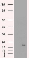
alpha B crystallin; alpha(B)-crystallin; AlphaB Crystallin; alpha-crystallin B chain; CRYA2; CRYA2alpha crystallin B chain; CRYAB; crystallin, alpha B; CTPP2; Heat shock protein beta-5; heat-shock 20 kD like-protein; HSPB5; Renal carcinoma antigen NY-REN-27; Rosenthal fiber component
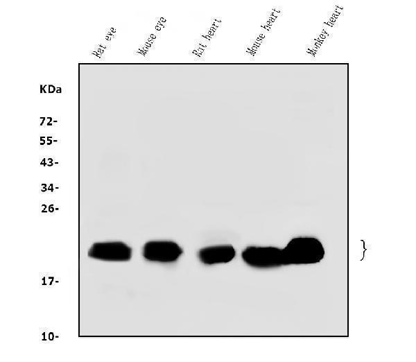
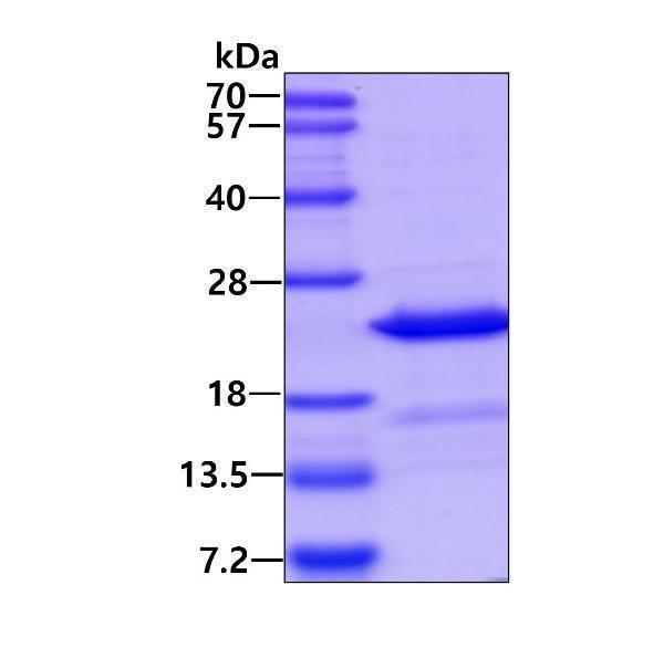
Mass (kDA):
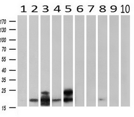
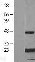
20.159 kDA

| Human | |
|---|---|
| Location: | 11q23.1 |
| Sequence: | 11; NC_000011.10 (111908564..111923740, complement) |
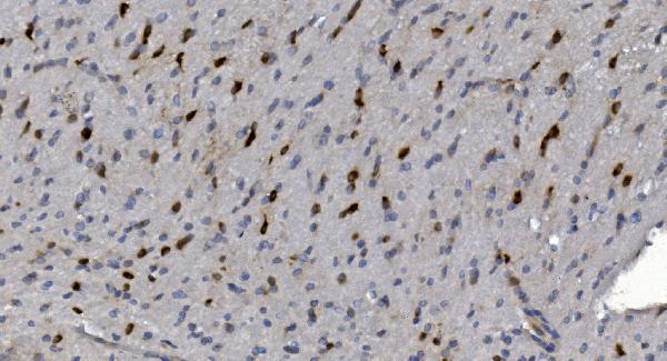
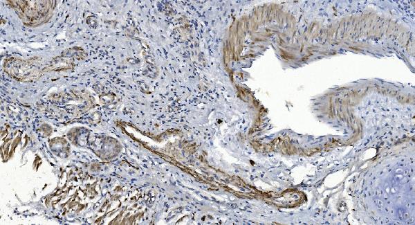
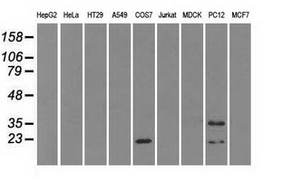
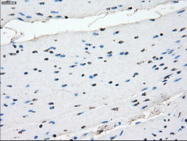
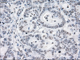
Lens as well as other tissues (PubMed:838078, PubMed:2387586). Expressed in myocardial tissue (PubMed:28493373).
Cytoplasm. Nucleus. Translocates to the nucleus during heat shock and resides in sub-nuclear structures known as SC35 speckles or nuclear splicing speckles (PubMed:19464326). Localizes at the Z-bands and the intercalated disk in cardiomyocytes (PubMed:28493373).


PMID: 838078 by Kramps J.A., et al. The primary structure of the B2 chain of human alpha-crystallin.
PMID: 2387586 by Dubin R.A., et al. Human alpha B-crystallin gene and preferential promoter function in lens.