This website uses cookies to ensure you get the best experience on our website.
- Table of Contents
4 Citations 9 Q&As
1 Citations 5 Q&As
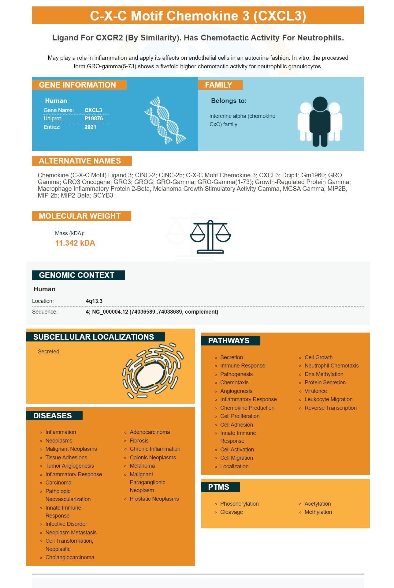
Facts about C-X-C motif chemokine 3.
May play a role in inflammation and apply its effects on endothelial cells in an autocrine fashion. In vitro, the processed form GRO-gamma(5-73) shows a fivefold higher chemotactic activity for neutrophilic granulocytes.
Human | |
---|---|
Gene Name: | CXCL3 |
Uniprot: | P19876 |
Entrez: | 2921 |
Belongs to: |
---|
intercrine alpha (chemokine CxC) family |
chemokine (C-X-C motif) ligand 3; CINC-2; CINC-2b; C-X-C motif chemokine 3; CXCL3; Dcip1; Gm1960; GRO gamma; GRO3 oncogene; GRO3; GROG; GRO-gamma; GRO-gamma(1-73); Growth-regulated protein gamma; Macrophage inflammatory protein 2-beta; melanoma growth stimulatory activity gamma; MGSA gamma; MIP2B; MIP-2b; MIP2-beta; SCYB3
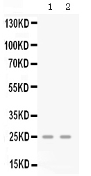
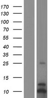
Mass (kDA):
11.342 kDA
Human | |
---|---|
Location: | 4q13.3 |
Sequence: | 4; NC_000004.12 (74036589..74038689, complement) |
Secreted.
If you're looking for a new anti-CXCL3 antibody, you've come to the right place. In this article, we'll discuss what this marker is and its uses in the field. We'll also touch on CXCL9, a related chemokine, as well as Boster Bio's CXCL3 antibody. Ultimately, we'll see if Boster Bio's CXCL3 antibody is a good choice.
One of the best uses for the CXCL3 marker is as a surrogate for early treatment response in TB patients. A recent study has shown that CXCL9 was a highly sensitive marker for predicting TB treatment response, and the other CXCR3 ligands showed similar sensitivity. This finding is important for the development of new treatments against TB. In addition, this biomarker has a range of clinical applications, including in clinical trials.
This study was limited by several limitations. In order for it to be effective in predicting treatment response, it needs to affect two or more markers. IFN-g was excluded from this study because of its poor predictive value. The study's criteria for significant changes in the CXCL3 marker were subjective and arbitrary. Furthermore, it failed to investigate changes in the serum CXCL3 ligand level during other inflammatory responses and infections. Thus, the findings from this study may be biased in practical application.
To investigate the effectiveness of this biomarker, a number of studies were performed. The CXCR3-CXCL3 axis is proposed as a target for behavioral changes during virus infection or type I IFN treatment. Moreover, CXCL3 is a ligand for other chemokines, which may influence the severity of the disease. These results suggest that the CXCR3 ligand has a therapeutic role in TB.
As a renowned provider of antibodies and ELISA kits, Boster Bio is committed to improving the quality of research and innovation by developing biomarkers and research tools for cancer, neurosciences, and inflammation. Their antibodies are manufactured in-house and meet stringent quality standards, making them an excellent choice for researchers and scientists alike. In addition, Boster antibodies have high level sensitivity and maintain biological activity for extended testing.
The canonical CXCR3 protein is 368 amino acids long and 40.7 kDa in mass. The protein is membrane-subcellularly located and belongs to the G-protein coupled receptor (GPCR) family. It plays a role in GPCR signaling and mediates C-C chemokine binding and activity. The chemokine CXCR3 is expressed in various tissues, including the liver, kidney, heart, skeletal muscle, and the placenta.
Its role in non-melanoma skin cancers is not fully understood. This may be related to the relatively benign nature of cutaneous skin cancers. These cancers are typically removed through surgical procedure, though a subset can spread and metastasize. Approximately 0.025 percent of premalignant actinic keratosis (PAK) lesions develop into invasive SCC. This risk increases in immunocompromised patients by 65-250-fold, suggesting that the immune system plays a role in controlling SCC.
The CXCR3 molecule plays a dual role in skin cancer. It is responsible for attracting effector T cells to the skin of mice with hyperplastic skin. In humans, HPV16E7 protein binding results in dysregulation of the cell cycle of keratinocytes and precancerous hyperplastic epithelium. Furthermore, actinic keratosis may progress to squamous cell carcinoma.
When you're researching the various uses of the CXCL3 marker, you can use a quality antibody from Boster Bio. This company specializes in producing picogram-sensitivity ELISA kits and IHC-optimized polyclonal antibodies. Boster Bio offers more than 12,000 antibodies, many of which have been validated for IHC, WB, or Flow. The antibodies have been tested quantitatively against panels of 250 tissues and un-transfected cell lines.
The chemokine CXCL10 was identified in blood plasma of patients with ueRA and healthy controls. The CXCL10 marker was also associated with clinical disease activity, including SJC in 66 joints, CDAI, and DAS28-CRP. These results support the potential for clinical applications of the CXCL3 marker. Its clinical utility is now being explored in multiple indications, including the treatment of osteoporosis, rheumatoid arthritis, and inflammatory diseases.
Several studies have evaluated the diagnostic utility of the CXCL3 marker in various types of patients with arthritis, and the IP-10 marker exhibited the best diagnostic utilities. However, the optimal cutoff value for this marker was based on sensitivity, specificity, positive and negative predictive values, and area under the ROC curve. In the trial, the IP-10-based assay missed three cases, while detecting three grew Stenotrophomonas spp.
Moreover, the fAb increased CXCL10 secretion in microglia. It was equally effective as LPS and TNF-a stimulation at the cellular level. The findings are encouraging and suggest the possibility of CXCL3 ligands as clinically useful surrogate markers for active pulmonary tuberculosis. These findings are a step toward better understanding the mechanism and therapeutic benefits of the CXCR3 marker in AD.
The vitiligo molecule plays an important role in the progression of depigmentation in a mouse model. However, there is no consensus on the correlation of CXCL10 expression with disease severity. Further, CXCR3 and its ligands play important roles in T-lymphocytosis and recruitment of T-lymphocytes in vitiligo. In the present study, eighty patients with vitiligo were evaluated for CXCR3 expression by immunohistology and flow cytometry.
The expression of CXCL3 in the serum of HCC patients was found to be significantly correlated with tumor outcomes. In this study, high levels of CXCL3 were associated with tumors with vascular invasion, and low levels of CXCL3 were found in HCC patients with a capsule surrounding the tumor tissue. These findings suggest that CXCL3 may serve as a promising biomarker for HCC.
Specifically, CXCL3 expression is required for growth of human breast cancer CSCs. CXCL3 knockdown was shown to decrease tumor-sphere size in Huh7 and PLC/PRF/5 cells. These cells are characterized by high self-renewal and tumorigenesis. However, knockdown of CXCL3 prevented tumor formation in HCC cell lines with high or low expression of CD133.
In a separate study, we investigated the association between CXCL3 and immune infiltration. This resulted in the identification of four hub genes, one of which is the CXCL3 marker. In addition, we found that CXCL3 and has-miR-1-3p were significantly correlated, which suggested that these hub genes may serve as significant biomarkers in rectal cancer. The two genes were found to be highly expressed at both the gene and RNA levels, and CXCL3 was associated with both neutrophils and macrophages. In addition, our study also revealed that CXCL3 was differentially expressed between different tumor stages.
Despite this association, CXCL3 has the potential to be a biomarker for colorectal cancer. Its high levels are associated with more aggressive disease states. In addition to breast cancer, CXCL3 has been associated with breast and prostate cancer. It has also been implicated in liver injury and inflammation in human melanoma. These findings have important implications for diagnosis and molecular targeted therapy.
The human CXCL3 ELISA kit is a sandwich ELISA designed for the quantitative detection of CXCL3 protein in serum. The kit uses SimpleStep ELISA(r) technology and has a sensitivity of 1.1 pg/mL. It is available as a 384-well version. You can also use a 96-well microplate.
A recent study used a GEO dataset to assess its prognostic value. Gene set enrichment analysis was also used to investigate potential molecular mechanisms. CXCL3 expression was found to be higher in tumor tissues compared to the adjacent normal tissues. Although CXCL3 expression was associated with increased risk of CC, a multivariate Cox analysis revealed no association between the presence of the marker and overall survival (OS).
The cytometric and immunohistochemical staining techniques used to determine the CXCR3 protein level were performed on 15 human cases. The use of different anti-CXCR3 antibodies enabled researchers to examine the presence of CXCR3 expression in a diverse population of cells. They were able to detect CXCR3 in a variety of samples including peripheral blood T cells and human B cells.
PMID: 2201751 by Tekamp-Olson P., et al. Cloning and characterization of cDNAs for murine macrophage inflammatory protein 2 and its human homologues.
PMID: 2217207 by Haskill S., et al. Identification of three related human GRO genes encoding cytokine functions.
*More publications can be found for each product on its corresponding product page