This website uses cookies to ensure you get the best experience on our website.


- Table of Contents
Plan paraffin-section CYGB IHC around the predominantly cytoplasmic tissue pattern (HPA tissue IHC). Use skeletal-muscle myocytes as a high-staining reference and interpret intensity with the reported medium consistency between staining and RNA (HPA tissue IHC).

Expected localisation, validated protocols, controls and antibodies — the at-a-glance facts below, then the full design guide.
| Expected localisation | Predominantly cytoplasmic in tissue IHC (HPA tissue IHC) | |
| Staining pattern | Cytoplasmic staining, prominent in some immune cells (HPA tissue IHC) | |
| Antigen retrieval | Citrate pH 6 HIER, heat-mediated (datasheet A01197) | |
| Positive control | Skeletal muscle+4 more · see all | |
| Negative control | Bone marrow+4 more · see all |
| Fixation | Keep fixation consistent across sections (standard IHC practice; not target-specific) | |
| Caveat | Staining and RNA show only medium consistency (HPA tissue IHC) | |
| Regulation | Expression regulation is unreported (UniProt) | |
| Isoform / epitope | No listed isoforms; one 1–190 chain (UniProt) |
The catalog antibody protocol is paired with published CYGB IHC methods for human liver and glioma sections (PMC5849339; PMC3663650).
| Sample | Paraffin-embedded mouse intestine tissues; fixative not specified (datasheet A01197) |
| Fixation | Image fixative and duration unreported (datasheet A01197); verify before use. |
| Sectioning | 4–5 µm sections on charged slides (standard) |
| Deparaffinisation | Xylene, graded ethanol series to water (standard) |
| Antigen retrieval | Heat retrieval: Citrate pH 6, 20 min (datasheet A01197) |
| Peroxidase block | 3% H2O2, 10 min, room temperature (standard) |
| Blocking | 10% goat serum (datasheet A01197) |
| Primary antibody | Rabbit anti-CYGB, 0.5-1μg/ml (datasheet A01197) |
| Primary incubation | Overnight at 4 °C (datasheet A01197) |
| Detection | Streptavidin-biotin complex (SABC), DAB chromogen (datasheet A01197) |
| Counterstain | Hematoxylin, blue, dehydrate and mount (standard) |
| Expected result | CYGB-positive staining in myocytes of skeletal muscle (HPA tissue IHC: High). HPA tissue profile: General cytoplasmic expression, most abundant in a subset of immune cells. No signal in the no-primary control. |
CYGB can localize to the cytoplasm and nucleus and has no transmembrane segment (UniProt Q8WWM9). In tissue IHC, expect predominantly cytoplasmic staining, especially in a subset of immune cells (HPA tissue IHC). Skeletal muscle myocytes are reported as high, while several glandular and neuronal cell populations are medium (HPA tissue IHC). The tissue profile is Approved, with medium consistency between antibody staining and RNA expression (HPA tissue IHC).
| Strong cytoplasmic staining in skeletal muscle myocytes, with weaker or variable staining in other cells. | This fits the reported high myocyte signal and general cytoplasmic tissue pattern (HPA tissue IHC). Compare intensity within identified cell types; a uniformly dark section does not reproduce this cell-specific profile (HPA tissue IHC). |
| Signal lies mainly on cell borders, in extracellular material, or as a surface rim. | Treat this as a localization mismatch requiring investigation: CYGB has no transmembrane segment, and the reported tissue pattern is cytoplasmic (UniProt Q8WWM9; HPA tissue IHC). Check morphology and detection controls before assigning the signal to CYGB (general IHC practice). |
| Strong staining appears in a cell population reported as undetected, such as skin fibroblasts. | This conflicts with the reference cell-level observation (HPA tissue IHC: skin fibroblasts not detected). Cross-reactivity or endogenous detection activity are possibilities, not diagnoses; assess matched controls and whether the stained cells were identified correctly (general IHC practice). |
| Color spreads across cells, stroma, and blank areas without clear cellular boundaries. | A diffuse deposit cannot establish CYGB-positive cells; the reference describes cellular, predominantly cytoplasmic staining (HPA tissue IHC). Review reagent background, blocking, washing, and the no-primary control (general IHC practice). |
| No staining is visible in skeletal muscle myocytes on an otherwise interpretable section. | The expected high signal is absent (HPA tissue IHC: skeletal muscle myocytes High). First check tissue identity and assay controls, then review retrieval, primary incubation, and detection as general IHC troubleshooting steps; the sources do not establish a CYGB-specific retrieval requirement. |
| Cell-level reference | HPA reports high skeletal muscle myocytes and medium bronchial respiratory epithelial, colon glandular, and cerebral cortical neuronal cells (HPA tissue IHC). Select a comparator by its named cell population, since a tissue contains multiple cell types (general IHC practice). |
| Negative comparator | HPA reports no detection in bone marrow hematopoietic cells, skin fibroblasts, and prostate glandular cells (HPA tissue IHC). These are cell-specific observations, not a promise that every cell or structure in those sections will be unstained. |
| Evidence strength | The tissue profile is Approved with medium staining–RNA consistency (HPA tissue IHC). HPA017757 has IHC Approved status; HPA061642 has no listed IHC status (HPA antibodies). Treat an unexpected pattern as needing controls rather than as definitive biology. |
| Protein features | CYGB is a single 1–190 chain with no signal peptide, propeptide, or transmembrane segment (UniProt Q8WWM9). Those features support an intracellular expectation; they do not identify an antibody epitope or establish antigen retrieval conditions. |
| IF/ICC Q: Is a nuclear-speckle signal compatible with CYGB? | A: Yes in the reported ICC-IF images: nuclear speckles are the supported main location, with additional cytosol staining (HPA subcellular). UniProt also lists nucleus and cytoplasm (UniProt Q8WWM9). Interpret that observation in its IF/ICC context; it does not replace the predominantly cytoplasmic tissue-IHC reference (HPA tissue IHC). |
| Detection chemistry | Endogenous activity can contribute chromogenic staining, depending on the detection system (general IHC practice). Use controls appropriate to the chosen detection chemistry; neither HPA nor UniProt supplies a CYGB-specific interference claim. |
| Situation | Likely cause | Next action |
|---|---|---|
| The expected skeletal muscle myocyte signal is absent. | The section may lack evaluable myocytes, or an assay step may have failed; HPA reports high myocyte staining (HPA tissue IHC). | Confirm cell identity and a working positive control, then review retrieval, primary incubation, and detection records (general IHC practice). Do not infer a CYGB-specific fixation effect from this result. |
| Most of the section has diffuse chromogen. | Background from the detection workflow may obscure the cellular pattern (general IHC practice). HPA describes general cytoplasmic expression with enrichment in a subset of immune cells (HPA tissue IHC). | Inspect a no-primary control and review blocking, washes, and chromogen development (general IHC practice). Score CYGB only where cells and their cytoplasm can be resolved. |
| Skin fibroblasts or bone marrow hematopoietic cells stain strongly. | The result differs from HPA's not-detected observations for those cells (HPA tissue IHC). Cell misidentification, cross-reactivity, or endogenous detection activity may explain it (general IHC practice). | Recheck morphology and matched controls; if signal persists, seek independent antibody or orthogonal evidence before calling those cells CYGB-positive (general IHC practice). |
| Signal forms a membrane rim or coats extracellular structures. | That pattern is difficult to reconcile with CYGB's lack of a transmembrane segment and the cytoplasmic tissue profile (UniProt Q8WWM9; HPA tissue IHC). | Check the no-primary control and inspect the deposit against cell boundaries (general IHC practice). Do not score a rim alone as the expected CYGB pattern. |
| A nuclear signal accompanies cytoplasmic staining. | Nuclear localization is listed by UniProt, and nuclear speckles are supported in HPA ICC-IF (UniProt Q8WWM9; HPA subcellular). The tissue-IHC profile emphasizes cytoplasm (HPA tissue IHC). | Record nuclear and cytoplasmic compartments separately. Compare the tissue signal with cell identity and controls before deciding whether the nuclear component is specific (general IHC practice). |
| Heart muscle RNA enrichment seems inconsistent with a slide's staining intensity. | HPA lists tissue-enhanced RNA in heart muscle but only medium consistency between antibody staining and RNA expression (HPA tissue IHC). RNA abundance does not specify the staining intensity of each cell (general IHC interpretation). | Score the observed cell-level IHC pattern and document the discrepancy. Avoid converting RNA enrichment into an expected chromogenic intensity for an individual cell (HPA tissue IHC). |


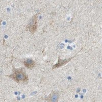


Comprehensive Human Protein Atlas IHC scoring per tissue (reliability: Approved — Medium consistency between antibody staining and RNA expression data.). Rows are taken directly from the HPA tissue chart — click any row's HPA link to view the source.
| Tissue | Cell type | Level | Evidence | Source |
|---|---|---|---|---|
| Skeletal muscle | Myocytes | High | Protein (IHC) | HPA → |
| Adrenal gland | Glandular cells | Medium | Protein (IHC) | HPA → |
| Appendix | Glandular cells | Medium | Protein (IHC) | HPA → |
| Bronchus | Respiratory epithelial cells | Medium | Protein (IHC) | HPA → |
| Cerebellum | Cells in molecular layer | Medium | Protein (IHC) | HPA → |
| Tissue | Cell type | Level | Evidence | Source |
|---|---|---|---|---|
| Bone marrow | Hematopoietic cells | Not detected | Protein (IHC) | HPA → |
| Caudate | Glial cells | Not detected | Protein (IHC) | HPA → |
| Cervix | Glandular cells | Not detected | Protein (IHC) | HPA → |
| Liver | Cholangiocytes | Not detected | Protein (IHC) | HPA → |
| Lymph node | Germinal center cells | Not detected | Protein (IHC) | HPA → |
Troubleshoot CYGB staining in paraffin sections by checking retrieval, cell identity, subcellular pattern and matched controls before scoring.
A01197 has IHC images from paraffin sections of mouse intestine and cardiac muscle, rat intestine, and human intestinal cancer tissue (catalog A01197 IHC captions). No IF/ICC data are supplied (catalog A01197 applications).
A01197 has IHC images from paraffin sections of mouse intestine and cardiac muscle, rat intestine, and human intestinal cancer tissue (catalog A01197 IHC captions). It is listed for IHC in human, mouse, and rat samples (catalog A01197 applications and reactivity).
Which to pick: Choose A01197 for paraffin-section IHC; its captions document citrate pH 6 retrieval and 1 μg/ml primary antibody, but do not report the fixative (catalog A01197 IHC captions). A01197 is also the cross-species choice listed for human, mouse, and rat; it is a rabbit antibody with no clone specified (catalog A01197 reactivity, host, and clone). No IF/ICC choice is supported by the supplied application list or images (catalog A01197 applications and IF images).