This website uses cookies to ensure you get the best experience on our website.


- Table of Contents
Plan CYP27A1 staining in paraffin sections using the catalog antibody’s IHC protocol. Compare the observed cytoplasmic pattern with tissue IHC evidence, including high staining in lung macrophages (HPA tissue IHC).

Expected localisation, validated protocols, controls and antibodies — the at-a-glance facts below, then the full design guide.
| Expected localisation | Cytoplasmic tissue staining (HPA tissue IHC) | |
| Staining pattern | Cytoplasmic in most tissues; high in lung macrophages (HPA tissue IHC) | |
| Antigen retrieval | EDTA pH 8.0 HIER, heat-mediated (datasheet A02121-2) | |
| Positive control | Adrenal gland+4 more · see all | |
| Negative control | Adipose tissue+2 more · see all |
| Fixation | Keep fixation consistent across sections. (standard IHC practice; not target-specific) | |
| Caveat | Staining and RNA show medium consistency (HPA tissue IHC) | |
| Regulation | Expression regulation not specified (UniProt) | |
| Isoform / epitope | No annotated isoforms; mature chain spans residues 34–531 (UniProt) |
The catalog antibody’s IHC-P protocol is paired with three published CYP27A1 chromogenic IHC protocols (datasheet A02121-2; PMC4618091; PMC8399082; PMC10494299).
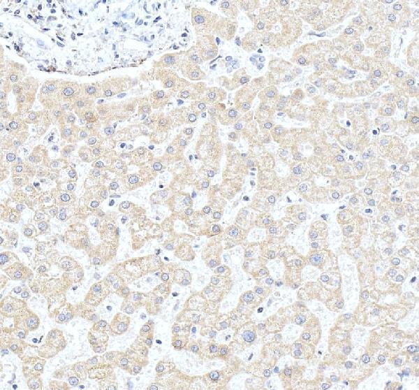
| Sample | Paraffin-embedded human liver tissue; fixative not specified (datasheet A02121-2) |
| Fixation | Image fixative and duration unreported (datasheet A02121-2); verify before use. |
| Sectioning | 4–5 µm sections on charged slides (standard) |
| Deparaffinisation | Xylene, graded ethanol series to water (standard) |
| Antigen retrieval | Heat retrieval: EDTA pH 8.0 (datasheet A02121-2); 20 min, 95–100 °C (standard) |
| Peroxidase block | 3% H2O2, 10 min, room temperature (standard) |
| Blocking | 10% goat serum (datasheet A02121-2) |
| Primary antibody | Rabbit anti-CYP27A1, 2-5μg/ml (datasheet A02121-2) |
| Primary incubation | Overnight at 4 °C (datasheet A02121-2) |
| Detection | HRP-conjugated secondary, DAB chromogen (datasheet A02121-2) |
| Counterstain | Hematoxylin, blue, dehydrate and mount (standard) |
| Expected result | CYP27A1-positive staining in glandular cells of adrenal gland (HPA tissue IHC: High). HPA tissue profile: Cytoplasmic expression in most tissues. No signal in the no-primary control. |
CYP27A1 staining in paraffin sections should be predominantly cytoplasmic, with strong signal in cell populations such as lung macrophages and kidney tubule cells (HPA: High in both; cytoplasmic expression in most tissues). The protein associates with the mitochondrial inner membrane after import and has no transmembrane segment (UniProt Q02318 topology). HPA rates tissue IHC reliability Enhanced, while reporting medium consistency between staining and RNA data (HPA tissue IHC).
| Strong cytoplasmic staining in lung macrophages or kidney tubule cells, with tissue structures still clear (HPA: High in both). | This fits the reported cell distribution and is a useful positive pattern for IHC interpretation (HPA tissue IHC). Chromogenic IHC can show cytoplasmic staining without resolving individual mitochondria (standard IHC practice). |
| Staining is mainly nuclear, or restricted to cell surfaces rather than cytoplasm. | That pattern conflicts with the reported cytoplasmic tissue profile and mitochondrial location (HPA tissue IHC; HPA subcellular; UniProt Q02318). Check staining controls and localization before assigning it to CYP27A1 (standard IHC practice). |
| Adipocytes, marrow hematopoietic cells, or ovarian stroma stain strongly (HPA: Not detected in these cells). | The result conflicts with the listed HPA tissue IHC observations; nonspecific antibody binding or endogenous chromogen activity are possibilities, not diagnoses (HPA tissue IHC; standard IHC practice). Review controls and cell identity. |
| Color spreads across tissue structures, including areas outside the expected positive cells. | Diffuse background prevents a reliable cell-specific call (standard IHC practice). Evaluate the negative detection control, blocking, washing, and development conditions before interpreting the distribution (standard IHC practice). |
| No signal appears in lung macrophages or kidney tubule cells (HPA: High in both). | A failed positive control is possible, but an HPA High designation does not guarantee every section will stain (HPA tissue IHC; standard IHC practice). Verify tissue identity and assay performance before calling the sample negative. |
| Compartment and optical resolution | CYP27A1 associates with the mitochondrial inner membrane as a membrane-extrinsic protein and has no transmembrane segment (UniProt Q02318); assess chromogenic sections for cytoplasmic distribution rather than requiring visible mitochondrial puncta (standard IHC practice). |
| Choice of positive and low-signal comparison cells | Lung macrophages and kidney tubule cells are reported High, while urothelial cells and skeletal myocytes are Low (HPA tissue IHC). Compare the named cell populations; a tissue label alone does not specify which cells should stain. |
| Interpretive confidence | The listed antibody HPA059155 has Enhanced IHC validation, but HPA describes only medium consistency between antibody staining and RNA expression (HPA antibodies; HPA tissue IHC). Treat a discordant pattern as requiring confirmation rather than assuming its identity. |
| Processing and epitope assumptions | UniProt lists a mature chain at residues 34–531 and three acetylated lysines, but no antibody epitope is supplied (UniProt Q02318). These annotations cannot predict which retrieval condition will work; target-specific fixation sensitivity is unreported in the supplied sources. |
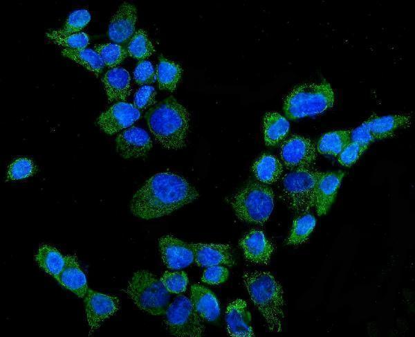
| IF/ICC Q&A: what location should an image show? | Mainly mitochondrial signal, with additional cytosolic signal, is approved in HPA ICC-IF images (HPA subcellular). This localization helps interpret an image; it does not specify an IF/ICC preparation or detection protocol. |
| Situation | Likely cause | Next action |
|---|---|---|
| No chromogenic signal in an HPA High cell population (HPA tissue IHC). | The positive control or detection run may have failed; the supplied sources give no CYP27A1-specific fixation or retrieval effect (standard IHC practice; HPA tissue IHC). | Confirm the sampled cells, run an established positive section, and review the antibody's IHC-P instructions and detection controls (standard IHC practice). |
| Weak signal appears in cells listed as HPA High (HPA tissue IHC). | Underdevelopment, an unsuitable working dilution, or incomplete assay optimization can reduce visible chromogen (standard IHC practice); no dilution is supplied here. | Check control performance and optimize detection or dilution within the catalog antibody's IHC-P instructions (standard IHC practice); do not infer a CYP27A1-specific retrieval defect. |
| Nuclei dominate the staining while cytoplasm is faint. | The distribution disagrees with HPA's cytoplasmic tissue profile and approved mitochondrial ICC-IF location (HPA tissue IHC; HPA subcellular). | Inspect the negative detection control and compare a known positive cell population; repeat with validated IHC conditions if the mismatch persists (standard IHC practice). |
| Strong color appears in adipocytes or marrow hematopoietic cells (HPA: Not detected). | Nonspecific binding or endogenous detection activity may account for unexpected color (standard IHC practice); the observation alone cannot distinguish them. | Check the no-primary detection control and antibody blocking or dilution, then compare the named cells with a positive control section (standard IHC practice). |
| Background obscures cell boundaries across the section. | Insufficient washing, blocking, or excessive development can produce widespread background in chromogenic IHC (standard IHC practice). | Review the negative control and adjust washing, blocking, and chromogen development using routine IHC optimization (standard IHC practice); score only interpretable cells. |
| A low-signal cell population is scored negative after a faint stain. | HPA lists urothelial cells and skeletal myocytes as Low, which is distinct from Not detected (HPA tissue IHC). | Keep Low and Not detected categories separate; score the identified cell type against controls under the same detection conditions (HPA tissue IHC; standard IHC practice). |


Comprehensive Human Protein Atlas IHC scoring per tissue (reliability: Enhanced — Medium consistency between antibody staining and RNA expression data.). Rows are taken directly from the HPA tissue chart — click any row's HPA link to view the source.
| Tissue | Cell type | Level | Evidence | Source |
|---|---|---|---|---|
| Adrenal gland | Glandular cells | High | Protein (IHC) | HPA → |
| Bronchus | Respiratory epithelial cells | High | Protein (IHC) | HPA → |
| Duodenum | Glandular cells | High | Protein (IHC) | HPA → |
| Kidney | Cells in tubules | High | Protein (IHC) | HPA → |
| Lung | Macrophages | High | Protein (IHC) | HPA → |
Troubleshoot CYP27A1 staining in paraffin section chromogenic IHC using the catalog antibody’s tissue image, protein annotation, and tissue expression data.
A02121-2 has IHC data from human liver paraffin sections and IF data from A431 cells; listed reactivity includes human, mouse, rat and monkey (A02121-2 image captions; catalog reactivity).
The sole rendered card is A02121-2, whose IHC image shows human liver paraffin sections (cards payload; A02121-2 IHC caption). Its IF image shows A431 cells; M02121-1 has no supplied IHC or IF image (A02121-2 IF caption; M02121-1 image fields).
Which to pick: Choose A02121-2 for tissue IHC using the documented human liver paraffin-section example; the fixative is unreported (A02121-2 IHC caption). Choose A02121-2 for IF/ICC when an image-backed example is useful, based on its A431-cell IF image (A02121-2 IF caption). For cross-species work, A02121-2 lists human, mouse, rat and monkey reactivity, while M02121-1 is a rabbit monoclonal listing human, mouse and rat; the supplied IHC image documents human tissue only (catalog reactivity; M02121-1 catalog entry; A02121-2 IHC caption).