This website uses cookies to ensure you get the best experience on our website.


- Table of Contents
Use liver hepatocytes as the positive reference for selective cytoplasmic CYP2E1 staining (HPA tissue IHC). This paraffin-section IHC guide covers fixation, the IHC-validated antibody at 0.5–1 μg/mL (datasheet: A00672), chromogenic detection, and endogenous peroxidase blocking (standard IHC practice).

Expected localisation, validated protocols, controls and antibodies — the at-a-glance facts below, then the full design guide.
| Expected localisation | Hepatocyte cytoplasm (HPA tissue IHC); ER and mitochondrial inner membrane (UniProt) | |
| Staining pattern | Selective cytoplasmic staining in hepatocytes (HPA tissue IHC) | |
| Antigen retrieval | Citrate pH 6 HIER, heat-mediated (datasheet A00672) | |
| Positive control | Liver | |
| Negative control | Adipose tissue+4 more · see all |
| Fixation | Keep paraffin-section fixation consistent (standard IHC practice; not target-specific); Matched tissue-IHC evidence does not establish the fixation claim. Validate the specimen-specific method before use. (selected-SKU IHC image A00672) | |
| Caveat | Endogenous peroxidase can confound liver DAB staining (standard IHC practice) | |
| Regulation | Expression regulation is not annotated (UniProt) | |
| Isoform / epitope | No annotated isoforms; one 1–493 chain (UniProt) |
The catalog antibody’s IHC-P protocol (datasheet: A00672) is followed by 4 published CYP2E1 IHC protocols (PMC3447587; PMC6682552; PMC5177661; PMC4660393).
| Sample | Paraffin-embedded human liver cancer tissues; fixative not specified (datasheet A00672) |
| Fixation | Image fixative and duration unreported (datasheet A00672); verify before use. |
| Sectioning | 4–5 µm sections on charged slides (standard) |
| Deparaffinisation | Xylene, graded ethanol series to water (standard) |
| Antigen retrieval | Heat retrieval: Citrate pH 6, 20 min (datasheet A00672) |
| Peroxidase block | 3% H2O2, 10 min, room temperature (standard) |
| Blocking | 10% goat serum (datasheet A00672) |
| Primary antibody | Rabbit anti-CYP2E1, 0.5-1μg/ml (datasheet A00672) |
| Primary incubation | Overnight at 4 °C (datasheet A00672) |
| Detection | Streptavidin-biotin complex (SABC), DAB chromogen (datasheet A00672) |
| Counterstain | Hematoxylin, blue, dehydrate and mount (standard) |
| Expected result | CYP2E1-positive staining in hepatocytes of liver (HPA tissue IHC: High). HPA tissue profile: Selective cytoplasmic expression in hepatocytes. No signal in the no-primary control. |
CYP2E1 should show selective cytoplasmic staining in hepatocytes, with high signal in liver (HPA tissue IHC: Enhanced; HPA: High in hepatocytes). Its reported locations are the endoplasmic reticulum and mitochondria (UniProt P05181; HPA subcellular: approved). UniProt lists no transmembrane segment despite its membrane association (UniProt P05181 topology).
| Strong cytoplasmic chromogen in hepatocytes, with surrounding structures readily distinguishable. | This matches the selective, High hepatocyte pattern reported for liver (HPA tissue IHC: Enhanced). Score the stained cells and their compartment together; intensity alone cannot establish specificity (general IHC practice). |
| Predominantly nuclear staining, or a sharp cell-surface outline without convincing hepatocyte cytoplasm. | That compartment conflicts with the reported cytoplasmic tissue pattern and ER/mitochondrial locations (HPA tissue IHC; HPA subcellular). Treat it as suspect and check the detection controls before assigning CYP2E1 positivity (general IHC practice). |
| Strong signal in cells other than hepatocytes, including a listed HPA-negative cell type. | HPA describes selective hepatocyte expression and reports several specified cell types as Not detected (HPA tissue IHC). Cross-reactivity or endogenous detection activity is possible; a negative result in one listed cell type does not establish that every other cell type must be negative (general IHC practice). |
| A uniform haze covers tissue, blank regions, or many cell types without a recognizable cytoplasmic pattern. | This is difficult to reconcile with selective hepatocyte staining (HPA tissue IHC). Background from detection or incomplete blocking is plausible; compare a no-primary control and assess whether signal follows tissue structures (general IHC practice). |
| No hepatocyte signal in an otherwise intact liver section. | Liver is the supplied positive reference, with High hepatocyte staining (HPA tissue IHC). First verify that hepatocytes and the detection system are evaluable; absence of signal alone cannot distinguish a technical failure from a specimen-specific result (general IHC practice). |
| Tissue reference | Liver hepatocytes provide the strongest supplied positive reference (HPA tissue IHC: High; Enhanced). The listed Not detected observations apply to the named cells in those tissues, not to every cell in each organ (HPA tissue IHC). |
| Subcellular interpretation | ER and mitochondrial localization supports a cytoplasmic readout; UniProt also describes post-translational mitochondrial targeting and inner-membrane association (HPA subcellular: approved; UniProt P05181). Routine chromogenic IHC cannot reliably resolve those organelles individually (general IHC practice). |
| Antibody evidence | Both listed antibodies have Enhanced IHC validation; only HPA029564 also has Approved ICC status (HPA antibodies). These ratings support their reported applications, but do not supply a dilution, retrieval condition, or proof that every observed signal is specific. |
| IF/ICC: where should signal appear? | Expect ER- and mitochondria-associated signal (HPA subcellular: approved). HPA lists ICC-IF images from HEK293, HaCaT, and serum-starved hTERT-RPE1 cells; the supplied record gives no cell-specific intensity comparison or IF protocol (HPA subcellular). |
| Retrieval and fixation | The supplied HPA and UniProt records give no CYP2E1-specific retrieval condition or fixation-sensitivity result. If optimization is needed, evaluate retrieval against liver-positive and no-primary controls as general IHC practice; do not infer a target-specific fixation effect. |
| Situation | Likely cause | Next action |
|---|---|---|
| Liver hepatocytes are blank. | The expected positive reference is missing (HPA tissue IHC: High in hepatocytes); the cause cannot be identified from staining absence alone. | Check section integrity, antibody application, retrieval, and chromogenic detection with appropriate controls (general IHC practice). Reassess a liver reference before calling the specimen negative. |
| Nuclei dominate the signal. | A nuclear pattern conflicts with HPA's cytoplasmic liver staining and approved ER/mitochondrial locations (HPA tissue IHC; HPA subcellular). | Compare a no-primary control and inspect the counterstain and chromogen separately; repeat with an IHC-validated antibody if the pattern persists (general IHC practice; HPA antibodies: Enhanced IHC). |
| Non-hepatocyte cells stain as strongly as hepatocytes. | The pattern departs from selective hepatocyte expression (HPA tissue IHC); cross-reactivity or endogenous chromogenic activity may contribute (general IHC practice). | Identify the stained cell type, compare it with HPA's named cell-level observations, and run a no-primary control. If using peroxidase detection, check endogenous peroxidase blocking (general IHC practice). |
| Brown haze obscures cell boundaries. | Diffuse background prevents assessment of the selective cytoplasmic pattern (HPA tissue IHC); detection background is one possible cause (general IHC practice). | Compare tissue with a no-primary control, then review blocking, wash steps, and detection exposure one variable at a time (general IHC practice). Preserve a clear hepatocyte reference while adjusting conditions. |
| A proposed negative control shows staining. | HPA's Not detected calls concern specific tissue–cell pairs; an unmatched cell population may still be interpretable differently (HPA tissue IHC). | Confirm that the observed cells match a supplied Not detected pair. Then compare no-primary staining and hepatocyte positivity before attributing the signal to CYP2E1 (HPA tissue IHC; general IHC practice). |
| IF/ICC shows only diffuse fluorescence. | That appearance does not establish the ER and mitochondrial locations approved by HPA (HPA subcellular); background or limited spatial resolution may contribute (general IF practice). | Check the no-primary image and image acquisition settings, then assess whether signal follows ER and mitochondrial reference markers (general IF practice; HPA subcellular: approved). |


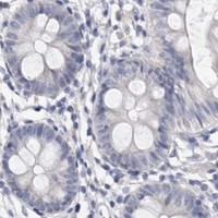


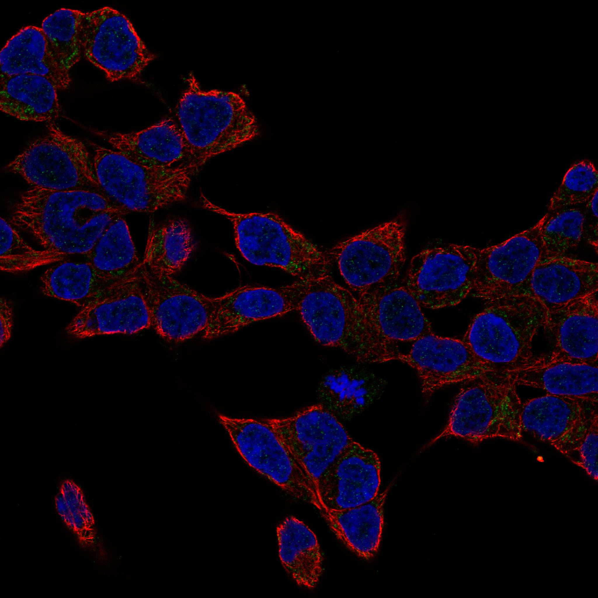
Comprehensive Human Protein Atlas IHC scoring per tissue (reliability: Enhanced — High consistency between antibody staining and RNA expression data.). Rows are taken directly from the HPA tissue chart — click any row's HPA link to view the source.
| Tissue | Cell type | Level | Evidence | Source |
|---|---|---|---|---|
| Liver | Hepatocytes | High | Protein (IHC) | HPA → |
| Tissue | Cell type | Level | Evidence | Source |
|---|---|---|---|---|
| Adipose tissue | Adipocytes | Not detected | Protein (IHC) | HPA → |
| Adrenal gland | Glandular cells | Not detected | Protein (IHC) | HPA → |
| Appendix | Glandular cells | Not detected | Protein (IHC) | HPA → |
| Bone marrow | Hematopoietic cells | Not detected | Protein (IHC) | HPA → |
| Breast | Adipocytes | Not detected | Protein (IHC) | HPA → |
Use hepatocyte cytoplasmic staining as the reference pattern when troubleshooting CYP2E1 in paraffin sections (HPA tissue IHC).
A00672 has IHC images from human liver cancer and mouse liver paraffin sections (catalog image captions); both antibodies list human, mouse, and rat reactivity (catalog reactivity). No IF/ICC data are supplied (catalog applications/images).
A00672 lists IHC for human, mouse, and rat, with images from human liver cancer and mouse liver paraffin sections (catalog applications/reactivity; image captions). M00672 lists IHC for human, mouse, and rat, but supplies no IHC image or tissue validation (catalog applications/reactivity; image data).
Which to pick: For tissue IHC, choose A00672 when a documented paraffin-section example is useful; its captions report citrate retrieval and 1 μg/ml primary antibody, but do not report the fixative (A00672 image captions). Neither SKU lists IF/ICC validation, so there is no evidence-based IF/ICC pick here (catalog applications/images). For cross-species IHC, both list human, mouse, and rat reactivity; M00672 is rabbit monoclonal clone AADC-3, while A00672 has the documented human and mouse paraffin-section images (catalog reactivity/clone; A00672 image captions).