This website uses cookies to ensure you get the best experience on our website.


- Table of Contents
Plan DAPK1 paraffin IHC around cytoplasmic tissue staining, including high staining in squamous epithelial cells (HPA tissue IHC). The catalog antibody has a starting range of 2–5 μg/ml for IHC (datasheet A01161-1).

Expected localisation, validated protocols, controls and antibodies — the at-a-glance facts below, then the full design guide.
| Expected localisation | Cytoplasmic staining in most tissues (HPA tissue IHC) | |
| Staining pattern | High cytoplasmic staining in squamous epithelial cells (HPA tissue IHC) | |
| Antigen retrieval | EDTA pH 8.0 HIER, heat-mediated (datasheet A01161-1) | |
| Positive control | Esophagus+4 more · see all | |
| Negative control | Adipose tissue+3 more · see all |
| Fixation | Keep fixation consistent across sections. (standard IHC practice; not target-specific) | |
| Caveat | Staining has medium consistency with RNA expression (HPA tissue IHC) | |
| Regulation | Isoform 2 occurs in colorectal carcinomas (UniProt) | |
| Isoform / epitope | Four isoforms; epitope coverage is unspecified (UniProt) |
The catalog antibody’s IHC-P protocol (datasheet: A01161-1) is accompanied by four published DAPK1 IHC protocols (PMC6825784; PMC5428482; PMC5520959; PMC5429897).
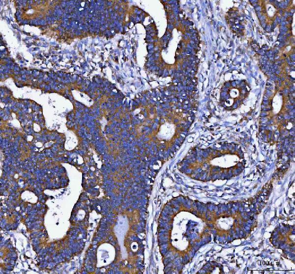
| Sample | Paraffin-embedded human colorectal adenocarcinoma tissue; fixative not specified (datasheet A01161-1) |
| Fixation | Image fixative and duration unreported (datasheet A01161-1); verify before use. |
| Sectioning | 4–5 µm sections on charged slides (standard) |
| Deparaffinisation | Xylene, graded ethanol series to water (standard) |
| Antigen retrieval | Heat retrieval: EDTA pH 8.0 (datasheet A01161-1); 20 min, 95–100 °C (standard) |
| Peroxidase block | 3% H2O2, 10 min, room temperature (standard) |
| Blocking | 10% goat serum (datasheet A01161-1) |
| Primary antibody | Rabbit anti-DAPK1, 2-5 μg/ml (datasheet A01161-1) |
| Primary incubation | Overnight at 4 °C (datasheet A01161-1) |
| Detection | HRP-conjugated secondary, DAB chromogen (datasheet A01161-1) |
| Counterstain | Hematoxylin, blue, dehydrate and mount (standard) |
| Expected result | DAPK1-positive staining in squamous epithelial cells of esophagus (HPA tissue IHC: High). HPA tissue profile: Cytoplasmic expression in most tissues. No signal in the no-primary control. |
DAPK1 is a cytoplasmic, cytoskeleton-associated protein with no transmembrane segment (UniProt P53355). In paraffin-section IHC, expect cytoplasmic staining in many tissues, including squamous epithelial cells of the esophagus and keratinocytes of the skin, both reported as High (HPA tissue IHC). HPA rates the tissue profile Approved, with medium consistency between staining and RNA expression (HPA tissue IHC).
| Cytoplasmic staining in esophageal squamous epithelial cells or skin keratinocytes. | This fits the expected compartment and a reported High-staining cell population (UniProt P53355; HPA tissue IHC). Compare cells within the section; the HPA rating is Approved with medium RNA–staining consistency, so appearance alone does not establish specificity (HPA tissue IHC). |
| Predominantly nuclear or sharply membranous staining, with little cytoplasmic signal. | An IHC pattern confined to those compartments conflicts with UniProt's cytoplasmic and cytoskeletal annotation and HPA's mainly cytoplasmic tissue profile (UniProt P53355; HPA tissue IHC). Review morphology and detection controls before interpreting it as DAPK1 (general IHC practice). |
| Strong staining in adipocytes while an expected-positive cell population is weak. | HPA reports DAPK1 as Not detected in adipocytes, so the reversal warrants a specificity check; it does not prove cross-reactivity (HPA tissue IHC). Compare a primary-antibody-omission control to assess endogenous detection activity, then assess antibody-dependent staining (general IHC practice). |
| Uniform haze across cells, extracellular space and section edges. | A haze that obscures cell boundaries cannot support a cell-specific DAPK1 call (general IHC practice). Check background in a primary-antibody-omission control and review blocking, washing and detection conditions; HPA's tissue profile does not diagnose the cause of haze (HPA tissue IHC; general IHC practice). |
| No cytoplasmic signal in esophageal squamous epithelium on an otherwise evaluable section. | That differs from HPA's High result for this cell population (HPA tissue IHC). First confirm the relevant cells are present and a positive-control section stained in the same run; absent signal could reflect the assay or the specimen, and the supplied sources do not identify a DAPK1-specific failure mechanism (general IHC practice). |
| Cell-specific tissue profile | HPA describes cytoplasmic expression in most tissues and low tissue specificity, yet scores individual populations differently: esophageal squamous epithelial cells High, adrenal glandular cells Medium, and adipocytes Not detected (HPA tissue IHC). Score the named cell population rather than treating an entire tissue section as uniformly positive or negative (general IHC practice). |
| Antibody validation and interpretation | HPA rates its tissue IHC profile Approved with medium consistency against RNA data; listed IHC antibodies HPA040472, HPA048436 and CAB037302 are Approved (HPA tissue IHC; HPA antibodies). These labels support use of the reported pattern as a reference but do not establish that every unexpected stain is DAPK1 (HPA tissue IHC; general IHC practice). |
| Isoforms and target structure | UniProt lists 4 isoforms and reports isoform 2 in normal intestinal tissue and colorectal carcinomas (UniProt P53355). The supplied record gives no antibody epitope, so it cannot establish which isoforms an IHC antibody detects or explain a staining difference between specimens (UniProt P53355; general IHC practice). |
| Processing and topology | The annotated DAPK1 chain spans residues 1–1430, with no signal peptide, propeptide or transmembrane segment in the supplied record (UniProt P53355). Its topology supports a cellular cytoplasmic expectation; it provides no basis here to predict membrane staining or a released extracellular fragment (UniProt P53355). |
| IF/ICC Q: Why might a punctum appear? | A: HPA's ICC-IF summary places DAPK1 mainly at the centrosome, whereas its tissue IHC profile is cytoplasmic and UniProt also notes cytoskeletal association (HPA subcellular ICC-IF; HPA tissue IHC; UniProt P53355). Interpret a centrosomal punctum in the separate IF/ICC context; it is not a required chromogenic tissue-IHC pattern (HPA subcellular ICC-IF; HPA tissue IHC). |
| Situation | Likely cause | Next action |
|---|---|---|
| Expected-positive epithelium is unstained. | The run may have failed, or the relevant cells may be absent from the assessed area; HPA reports High staining in esophageal squamous epithelial cells (HPA tissue IHC; general IHC practice). | Confirm cell identity and section quality, then compare a known-positive section processed in the same run. Review the antibody's IHC-P instructions and run controls before changing retrieval or dilution (general IHC practice). |
| Signal is mainly nuclear or outlines cell membranes. | This distribution does not match the cytoplasmic tissue profile or UniProt localization (HPA tissue IHC; UniProt P53355). The supplied sources do not identify which assay step caused it. | Recheck compartment assignment against morphology and the expected-positive control; assess primary-antibody-dependent signal with an omission control (general IHC practice). Do not score an unsupported compartment as DAPK1 on appearance alone (UniProt P53355; HPA tissue IHC). |
| Adipocytes or smooth muscle cells stain strongly. | HPA lists both populations as Not detected, so strong staining needs investigation; endogenous detection activity or antibody cross-reactivity are possibilities, not established diagnoses (HPA tissue IHC; general IHC practice). | Compare adjacent negative and expected-positive cell populations, then run a primary-antibody-omission control. Persistent staining without primary antibody points to the detection system; staining only with primary antibody needs further specificity assessment (general IHC practice). |
| The entire section has diffuse brown background. | Widespread background prevents a reliable cell-specific score; its source cannot be assigned from HPA or UniProt (general IHC practice). | Examine the omission control, review blocking and washing, and check whether the detection reagents themselves color the section. Reassess the cytoplasmic pattern only after cell boundaries are readable (general IHC practice; HPA tissue IHC). |
| A low or absent signal is called a failed stain in every cell. | HPA reports genuine differences by cell population, including Not detected adipocytes and parathyroid glandular cells, and Low rectal glandular cells (HPA tissue IHC). | Name the cell population being scored and compare it with HPA's matching population. Use a reported High population in the same run to judge whether the assay worked (HPA tissue IHC; general IHC practice). |
| A centrosomal IF/ICC punctum is expected in chromogenic tissue IHC. | The centrosome summary comes from HPA ICC-IF; HPA tissue IHC instead describes a cytoplasmic profile (HPA subcellular ICC-IF; HPA tissue IHC). | Judge this IHC section by cytoplasmic staining in the appropriate cell types. Consult the separate IF/ICC guide when interpreting punctate fluorescence (HPA tissue IHC; HPA subcellular ICC-IF). |


Comprehensive Human Protein Atlas IHC scoring per tissue (reliability: Approved — Medium consistency between antibody staining and RNA expression data.). Rows are taken directly from the HPA tissue chart — click any row's HPA link to view the source.
| Tissue | Cell type | Level | Evidence | Source |
|---|---|---|---|---|
| Esophagus | Squamous epithelial cells | High | Protein (IHC) | HPA → |
| Nasopharynx | Respiratory epithelial cells | High | Protein (IHC) | HPA → |
| Placenta | Trophoblastic cells | High | Protein (IHC) | HPA → |
| Skeletal muscle | Myocytes | High | Protein (IHC) | HPA → |
| Skin | Keratinocytes | High | Protein (IHC) | HPA → |
| Tissue | Cell type | Level | Evidence | Source |
|---|---|---|---|---|
| Adipose tissue | Adipocytes | Not detected | Protein (IHC) | HPA → |
| Hippocampus | Glial cells | Not detected | Protein (IHC) | HPA → |
| Parathyroid gland | Glandular cells | Not detected | Protein (IHC) | HPA → |
| Smooth muscle | Smooth muscle cells | Not detected | Protein (IHC) | HPA → |
Troubleshoot DAPK1 staining in paraffin sections by checking retrieval, cell type, subcellular pattern, and controls before scoring (datasheet A01161-1; HPA tissue IHC).
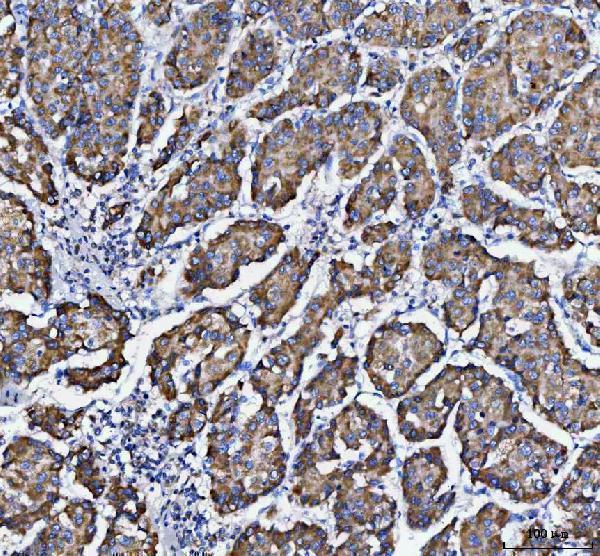
Catalog figures show DAPK1 IHC in human paraffin sections of colorectal, liver, lung, and breast cancers (A01161-1 and M01161 image captions), plus IF in HeLa cells (M01161 IF image caption).
A01161-1 has IHC images from human paraffin sections of colorectal adenocarcinoma, liver cancer, and lung cancer (A01161-1 image captions). M01161 has an IHC image from a human breast cancer paraffin section and an IF image from HeLa cells (M01161 image captions).
Which to pick: For tissue IHC, choose A01161-1 for its documented EDTA retrieval at pH 8.0 and 2 μg/ml primary antibody in a human colorectal adenocarcinoma paraffin section (A01161-1 image caption); M01161 offers a rabbit monoclonal option with a human breast cancer paraffin-section image (catalog: M01161 clone COA-4; M01161 image caption). For IF/ICC, choose M01161 because both applications are listed and its IF image uses HeLa cells (M01161 applications; M01161 IF image caption). For cross-species studies, both list Human, Mouse, and Rat reactivity (catalog: A01161-1 and M01161 reactivity), although their supplied IHC images show human tissue only and neither caption reports the fixative (A01161-1 and M01161 image captions).