DAPK3 / Death-associated protein kinase 3 · IHC design guide

Design Immunohistochemistry for DAPK3

Plan chromogenic DAPK3 IHC in paraffin sections using the IHC-validated antibody at 1:50–1:200 (datasheet: A03300-2). Compare distinctly stained smooth muscle cells with liver cholangiocytes reported as not detected, and score cytoplasmic and membranous signal by cell type (HPA tissue IHC).

Evidence assembled Oct 2026 · For research use; verify linked source records and product datasheet before use
Immunohistochemistry protocol sheet for DAPK3 (IHC for DAPK3): expected localisation Cytoplasmic and membranous tissue signal (HPA tissue IHC), antibody A03300-2, validated IHC image, and IHC protocol steps
Printable DAPK3 IHC protocol sheet — expected localisation Cytoplasmic and membranous tissue signal (HPA tissue IHC), antibody A03300-2, controls and protocol steps. Open the full DAPK3 IHC guide →

DAPK3 Immunohistochemistry Experimental Design Guide

Expected localisation, validated protocols, controls and antibodies — the at-a-glance facts below, then the full design guide.

Must know before staining
Expected localisation Cytoplasmic and membranous tissue signal (HPA tissue IHC)
Staining pattern Smooth muscle cells stand out amid widespread cytoplasmic and membranous staining (HPA tissue IHC)
Antigen retrieval Tris-EDTA pH 9.0 HIER, 95–98 °C, 20 min (rule: nuclear antigen)
Positive control ⓘ Adipose tissue+4 more · see all
Negative control ⓘ Liver+2 more · see all
Important caveats
Reasons your staining may differ from the expected pattern.
Fixation Keep fixation consistent across sections. (standard IHC practice; not target-specific)
Caveat Smooth muscle content can affect mixed-tissue scores (HPA tissue IHC)
Regulation No expression regulator specified (UniProt)
Isoform / epitope 2 isoforms; antibody epitope coverage is unspecified (UniProt; datasheet)
Section 1

Recommended DAPK3 IHC & IF Protocols

The catalog antibody’s IHC-P protocol is accompanied by two published chromogenic DAPK3 protocols: prostate sections (PMC4884664) and testis sections (PMC12061289).

Recommended immunohistochemistry (IHC-P) protocol parameters
SampleParaffin-embedded human breast carcinoma tissue; fixative not specified (datasheet A03300-2)
FixationImage fixative and duration unreported (datasheet A03300-2); verify before use.
Sectioning4–5 µm sections on charged slides (standard)
DeparaffinisationXylene, graded ethanol series to water (standard)
Antigen retrievalHeat-induced epitope retrieval in Tris-EDTA buffer, pH 9.0, 20 min at 95–98 °C (standard rule: nuclear antigen)
Peroxidase block3% H2O2, 10 min, room temperature (standard)
Blocking10% normal serum of the secondary host, 30 min, room temperature (standard)
Primary antibodyRabbit anti-DAPK3, 1:50-1:200 (datasheet A03300-2)
Primary incubationOvernight at 4 °C (standard)
DetectionHRP-polymer secondary, DAB chromogen 5–10 min (standard)
CounterstainHematoxylin, blue, dehydrate and mount (standard)
Expected resultDAPK3-positive staining in adipocytes of adipose tissue (HPA tissue IHC: High). HPA tissue profile: Cytoplasmic and membranous expression in most tissues, distinct expression in smooth muscle cells. No signal in the no-primary control.
💡Decision noteStart with Tris-EDTA pH 9.0 HIER at 95–98 °C for 20 min (page antigen retrieval); neither emitted article specifies retrieval conditions (PMC4884664; PMC12061289).
Section 2

What Is the Expected DAPK3 Staining Pattern?

DAPK3 staining should be most apparent in the cytoplasm of smooth muscle cells, although HPA reports cytoplasmic and membranous staining across most tissues (HPA tissue IHC). Nuclear staining can also be plausible because DAPK3 shuttles between cytoplasm and nucleus (UniProt O43293). Interpret these patterns with care: the HPA tissue IHC profile is Approved, with medium consistency between antibody staining and RNA expression (HPA tissue IHC). DAPK3 has no transmembrane segment (UniProt O43293 topology).

What am I looking at on my slide?
Distinct cytoplasmic staining in smooth muscle cells, with some membranous appearance.This fits the reported strong smooth muscle signal and the broader cytoplasmic and membranous tissue pattern (HPA tissue IHC). Score the smooth muscle cells themselves; staining elsewhere in the section does not establish their intensity.
Predominantly nuclear staining, or signal concentrated near mitotic structures.Nuclear signal can be plausible because DAPK3 shuttles between nucleus and cytoplasm; UniProt also lists centrosome, spindle, centromere and midbody locations (UniProt O43293). Check cellular context before calling this an artefact. A uniform nuclear-only pattern across unrelated cells needs further validation against the expected tissue IHC pattern (HPA tissue IHC).
Strong signal in cholangiocytes or parathyroid glandular cells.HPA reports DAPK3 as not detected in those cell types (HPA tissue IHC). Recheck cell identification and compare controls; unexpected staining can reflect cross-reactivity or endogenous detection activity (general IHC practice). One unexpected section cannot establish which cause applies.
Diffuse colour over cells and extracellular spaces, obscuring cell boundaries.This does not allow confident assignment of DAPK3 to the cytoplasmic and membranous pattern reported by HPA (HPA tissue IHC). Background from nonspecific reagent binding or the detection system is possible (general IHC practice); assess controls before scoring.
No staining in a smooth muscle positive control.Smooth muscle cells are reported High by HPA (HPA tissue IHC). A blank result therefore warrants checking the IHC workflow and control section (general IHC practice). It does not, by itself, show that the experimental tissue lacks DAPK3.
💡Expected DAPK3 appearanceCall a convincing positive when identifiable smooth muscle cells show strong, chiefly cytoplasmic staining, with possible membranous appearance (HPA tissue IHC); widespread diffuse colour without cell definition or strong staining confined to HPA not-detected cells warrants investigation (HPA tissue IHC; general IHC practice).
How each factor affects the staining
Tissue and cell selectionDAPK3 RNA has low tissue specificity, while HPA tissue IHC levels vary by cell type (HPA tissue IHC). Use a named positive population, such as smooth muscle cells reported High, and score it separately from neighbours.
Compartment interpretationHPA tissue IHC describes cytoplasmic and membranous expression; UniProt describes predominantly cytoplasmic DAPK3 with nuclear shuttling (HPA tissue IHC; UniProt O43293). Membranous appearance does not establish membrane insertion: DAPK3 has no transmembrane segment (UniProt O43293 topology).
Isoforms and antibody coverageUniProt lists two isoforms and reports both in bladder smooth muscle (UniProt O43293). The supplied record gives no antibody epitope or isoform coverage, so an IHC result cannot be assigned to either isoform without separate validation.
Strength of tissue evidenceThe HPA tissue IHC profile is Approved with medium staining–RNA consistency; the listed HPA028569 antibody is IHC Approved (HPA tissue IHC; HPA antibody record). Treat reported levels as reference observations, not a guaranteed result for every specimen or detection workflow.
IF/ICC Q&A: should its pattern match IHC?For IF/ICC, HPA reports supported nucleoplasm and cytosol locations, with approved vesicles and uncertain primary cilium localization (HPA subcellular). Those cell-image observations can inform compartment checks, but do not replace the tissue IHC pattern (HPA tissue IHC).
Why is my staining missing, weak or wrong?
SituationLikely causeNext action
Smooth muscle is blank or unexpectedly weak.The positive population may be missed, or the IHC detection workflow may have failed (HPA: High in smooth muscle cells; general IHC practice).Confirm smooth muscle is present on the section, then inspect the positive control, primary-antibody step, detection reagents and counterstain (general IHC practice). The supplied sources give no DAPK3-specific retrieval setting.
Colour covers the slide, including empty areas.Nonspecific binding or detection background can obscure the reported cellular pattern (general IHC practice; HPA tissue IHC).Review a no-primary control, blocking, wash steps and detection chemistry (general IHC practice). Judge DAPK3 only where cell boundaries and compartments remain interpretable.
A reported not-detected cell type stains strongly.Cell misidentification, cross-reactivity or endogenous detection activity are possibilities (HPA tissue IHC; general IHC practice).Recheck the cell type and compare the no-primary control with a separate positive control (general IHC practice). HPA reports cholangiocytes, parathyroid glandular cells and splenic red-pulp cells as not detected (HPA tissue IHC).
Staining is exclusively nuclear throughout the section.DAPK3 can enter the nucleus, but a uniform nuclear-only tissue pattern differs from the HPA cytoplasmic and membranous IHC profile (UniProt O43293; HPA tissue IHC).Compare identifiable smooth muscle cells with the positive control and check background controls (HPA tissue IHC; general IHC practice). Do not reject nuclear signal solely because of its compartment (UniProt O43293).
Membranous staining is interpreted as a surface-only protein.HPA reports a membranous staining appearance, while UniProt lists no transmembrane segment (HPA tissue IHC; UniProt O43293 topology).Describe the observed pattern as membranous appearance and record any concurrent cytoplasmic signal (HPA tissue IHC). Avoid inferring membrane insertion or extracellular DAPK3 from chromogenic staining alone (UniProt O43293 topology).
IF/ICC and tissue IHC appear different.HPA reports nucleoplasm and cytosol in ICC-IF images, while its tissue IHC summary emphasizes cytoplasmic and membranous staining (HPA subcellular; HPA tissue IHC).Interpret each result against its own HPA reference and the identifiable cells being examined (HPA subcellular; HPA tissue IHC). Do not use the IF/ICC images as an IHC-P protocol or as proof of a tissue result.

Sample controls for DAPK3 IHC & IF

🧪Run lung first: macrophages should stain (HPA: High in lung macrophages); use liver cholangiocytes as the negative tissue compartment (HPA: Not detected in liver cholangiocytes). On the lung slide, cells without specific staining should show background signal only, but identify them by morphology rather than assuming all nonmacrophages are negative (UniProt O43293: widely expressed).
Positive control tissue: Adipose tissue (Adipocytes, HPA High)
Negative control tissue: Liver (HPA Not detected)
ICC-IF cell lines (HPA subcellular resource): HPA ICC-IF images show DAPK3 in A-431, U-251MG, U2OS, ASC52telo, RPTEC/TERT1, hTERT-RPE1 (serum starved), KOLF2.1J, with annotated localisation: Nucleoplasm (supported), Cytosol (supported) (HPA subcellular).
Technical controls: Include a no-primary, secondary-only control; host-matched nonimmune IgG for the polyclonal catalog antibody; and DAPK3 knockout material or a validated immunizing-peptide block (selected IHC caption: pAb; standard IHC specificity controls). For lung macrophages, quench endogenous peroxidase and inspect pigment-associated background in chromogenic IHC (HPA: High in lung macrophages; standard IHC practice).
⚠️Feasibility: A target-specific fixation window or fixation effect is unreported, and the selected A03300-2 paraffin-section caption does not state a fixative (selected IHC caption: fixative unreported). Antigen retrieval requirements are unreported, so optimize retrieval empirically; check lung macrophage pigment when interpreting chromogenic signal (standard IHC practice). The supplied evidence does not establish whether frozen sections or IF/ICC are easier, although HPA ICC-IF images support nucleoplasm and cytosol localization (HPA subcellular).

HPA tissue IHC evidence for DAPK3

Comprehensive Human Protein Atlas IHC scoring per tissue (reliability: Approved — Medium consistency between antibody staining and RNA expression data.). Rows are taken directly from the HPA tissue chart — click any row's HPA link to view the source.

Positive expression · recommended positive controls

TissueCell typeLevelEvidenceSource
Adipose tissue Adipocytes High Protein (IHC) HPA →
Bone marrow Hematopoietic cells High Protein (IHC) HPA →
Breast Adipocytes High Protein (IHC) HPA →
Gallbladder Glandular cells High Protein (IHC) HPA →
Lung Macrophages High Protein (IHC) HPA →

Undetected expression · recommended negative controls

TissueCell typeLevelEvidenceSource
Liver Cholangiocytes Not detected Protein (IHC) HPA →
Parathyroid gland Glandular cells Not detected Protein (IHC) HPA →
Spleen Cells in red pulp Not detected Protein (IHC) HPA →
Section 3

Advanced DAPK3 IHC Tips

Troubleshoot DAPK3 staining in paraffin sections by checking retrieval, compartment patterns, cell type, controls and scoring before interpreting chromogenic signal.

What retrieval should I try first when DAPK3 staining is weak?
Start with Tris-EDTA pH 9.0 heat-induced epitope retrieval at 95–98 °C for 20 min (page retrieval setting). Keep section thickness, heating vessel and cooling procedure consistent across the test and control sections, then compare signal in the same cell types (standard IHC practice). If staining remains weak, test a shorter or longer heating time on adjacent sections while checking whether tissue morphology and background deteriorate (standard IHC practice). Nuclear and cytoplasmic signal are both biologically plausible for DAPK3, so judge retrieval by interpretable cell-level staining rather than by nuclear intensity alone (UniProt O43293 subcellular; HPA subcellular).
Could fixation explain weak or uneven DAPK3 staining?
The selected antibody caption documents staining in paraffin-embedded human breast carcinoma at 1:100, but does not report the fixative (catalog antibody A03300-2 caption). Target-specific DAPK3 sensitivity to fixation is therefore unknown from the supplied evidence; record the actual fixative, fixation duration and processing history for each specimen before comparing staining (catalog antibody A03300-2 caption; standard IHC practice). When uneven staining tracks thick tissue, folds or poorly preserved areas, compare intact regions and a separately processed control section (standard IHC practice). Optimise retrieval and antibody concentration on adjacent sections, and retain morphology as a criterion for accepting either adjustment (standard IHC practice).
Should I expect DAPK3 staining in the nucleus or cytoplasm?
Score nuclear and cytoplasmic staining separately: DAPK3 is predominantly cytoplasmic but can shuttle between nucleus and cytoplasm (UniProt O43293 subcellular). Supported immunofluorescence locations include nucleoplasm and cytosol, while tissue IHC describes cytoplasmic and membranous expression in most tissues (HPA subcellular; HPA tissue IHC). Its cytoplasmic localisation is promoted by phosphorylation at Thr-299 and involves Rho/Rock signalling, so a compartment shift should be assessed in matched cell populations (UniProt O43293 subcellular). Check nuclear counterstain alignment and distinguish discrete intracellular signal from staining along section edges or damaged tissue before calling a compartment change (standard IHC practice).
Can this stain distinguish DAPK3 isoforms or phosphorylation states?
Do not assign a positive chromogenic signal to one isoform without an antibody epitope map or isoform-specific validation (standard IHC practice). DAPK3 has 2 annotated isoforms, and both are expressed in bladder smooth muscle; the supplied tissue-IHC caption identifies the antibody and dilution but gives no epitope sequence (UniProt O43293 isoforms and tissue specificity; catalog antibody A03300-2 caption). Phosphorylation sites include Thr-265 and Thr-299, but a pan-DAPK3 stain cannot establish site occupancy without phosphosite-specific validation (UniProt O43293 modified residues; standard IHC practice). Compare staining with an independently validated assay if the study requires an isoform or phosphorylation claim (standard IHC practice).
How can I check a DAPK3 IHC pattern with multiplex immunofluorescence?
Use immunofluorescence as a separate confirmation experiment and multiplex DAPK3 with a validated marker for the cell type being scored, such as smooth muscle cells where tissue IHC reports distinct expression (HPA tissue IHC; standard IF practice). Choose spectrally separated fluorophores after examining unstained tissue autofluorescence, and assign the dimmer detection channel according to measured background rather than colour preference (standard IF practice). DAPK3 has no transmembrane segment, so optimise permeabilisation for intracellular epitope access while preserving nuclear and cytoplasmic boundaries (UniProt O43293 topology and subcellular; standard IF practice). Include single-stain controls to identify bleed-through before comparing fluorescence with chromogenic cell-level patterns (standard IF practice).
How do I reduce diffuse brown background without losing DAPK3 signal?
Compare a no-primary control with the stained section to locate detection-system or tissue-derived colour before changing the primary antibody (standard IHC practice). Apply a peroxidase block before chromogenic detection, use appropriate protein blocking and washing, and check whether precipitate appears outside cells after development (standard IHC practice). The selected paraffin-section caption reports 1:100 for the catalog antibody; use that as a documented starting concentration and titrate around it if diffuse staining persists (catalog antibody A03300-2 caption; standard IHC practice). Preserve interpretable cytoplasmic and nuclear cell detail while reducing background, since both compartments are supported DAPK3 locations (UniProt O43293 subcellular; HPA subcellular).
What should I measure when DAPK3 staining varies across a section? ⚠ ANSWER MARKED FOR VERIFICATION
Define the cell population and compartment before scoring, then record the percentage of positive cells and an H-score from staining intensity categories, or count positive cells per mm² when density is the endpoint (standard IHC practice). Normalise positive-cell counts to the number of evaluable cells of the same type, or density to evaluable tissue area, excluding folds and necrosis by a prespecified rule (standard IHC practice). Report nuclear and cytoplasmic scores separately because DAPK3 can occupy both compartments (UniProt O43293 subcellular). Use identical detection and imaging settings across comparison sections, and document the scoring threshold and control performance (standard IHC practice).
Which DAPK3 staining patterns support a true positive call?
Prioritise reproducible intracellular staining in intact, identifiable cells over isolated colour at edges, folds or necrotic regions (standard IHC practice). Cytoplasmic and nuclear localisation are plausible, and tissue IHC reports distinct smooth muscle expression; macrophages in lung are also reported at high staining levels (UniProt O43293 subcellular; HPA tissue IHC). Treat an unexpected cell or compartment pattern as a finding to verify with controls, rather than as proof of altered kinase activity (standard IHC practice; UniProt O43293 function). A no-primary control helps identify endogenous enzyme or detection background, while matched morphology and cell-type context help distinguish true staining from artefact (standard IHC practice).
Boster reagents

Best DAPK3 / Death-associated protein kinase 3 IHC Antibodies

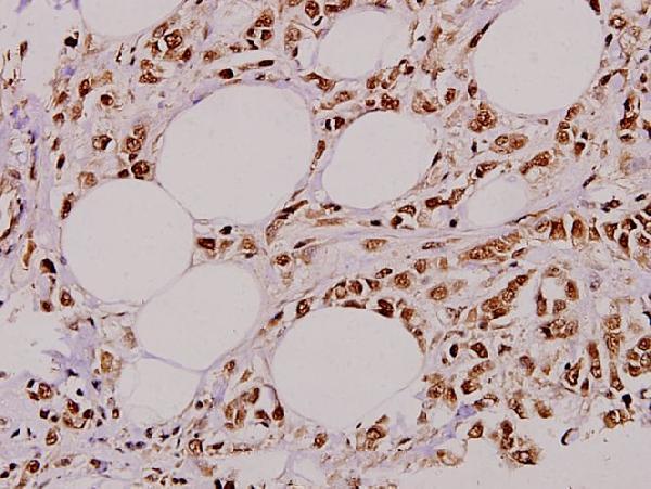
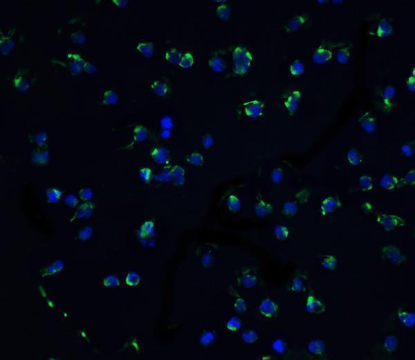
Anti-DAPK3 antibodies have IHC data in paraffin-embedded human breast carcinoma and IF data in Jurkat cells (catalog image captions); both list Human, Mouse, and Rat reactivity (catalog reactivity).

Real IHC data Immunohistochemistry (IHC) analyzes of ZIP-kinase (K261) pAb in paraffin-embedded human breast carcinoma tissue at 1:100.
Anti-ZIP-kinase (K261) DAPK3 Antibody
Cat # A03300-2
Real IF data Immunofluorescence of ZIPK in Jurkat cells with ZIPK antibody at 10 μg/mL.
Anti-ZIPK DAPK3 Antibody
Cat # A03300-1

A03300-2 is listed for IHC and pictured staining paraffin-embedded human breast carcinoma at 1:100 (catalog applications; A03300-2 image caption). A03300-1 is listed for IF/ICC and pictured staining Jurkat cells by fluorescence at 10 μg/mL (catalog applications; A03300-1 image caption).

Which to pick: Choose A03300-2 for paraffin-section IHC because it lists IHC and has a matching tissue image; the caption does not report the fixative (catalog applications; A03300-2 image caption). Choose A03300-1 for IF/ICC because it lists both applications and has Jurkat-cell IF data (catalog applications; A03300-1 image caption). Both list Human, Mouse, and Rat reactivity and a rabbit host; neither reports a clone, so choose by application for those species (catalog reactivity; catalog host/clone).

Each figure is that product's own IHC / IF validation image from its datasheet.

References

  1. UniProt Consortium. UniProt entry O43293 (DAPK3_HUMAN, Death-associated protein kinase 3).
  2. Human Protein Atlas. DAPK3 tissue IHC expression (reliability: Approved).
  3. Human Protein Atlas. DAPK3 subcellular location (ICC-IF): Mainly localized to the nucleoplasm and cytosol. In addition localized to vesicles and primary cilium..
  4. Human Protein Atlas. DAPK3 antibody validation summary (1 antibodies).
  5. Activation of AKT negatively regulates the pro-apoptotic function of death-associated protein kinase 3 (DAPK3) in prostate cancer. Cancer letters 2016 — PMC4884664.
  6. An Inhibitor of Death-Associated Protein Kinase 3 (DAPK3) Disrupts Hippo Signaling and Intestinal Epithelial Regeneration in Murine DSS-Induced Colitis. Digestive diseases and sciences 2026 — PMC12969504.
  7. DAPK3 is Essential for DBP-Induced Autophagy of Mouse Leydig Cells. Advanced science (Weinheim, Baden-Wurttemberg, Germany) 2025 — PMC12061289.
  8. PubMed PMID:9488481 — UniProt-cited evidence.
  9. PubMed PMID:10356987 — UniProt-cited evidence.
  10. PubMed PMID:14702039 — UniProt-cited evidence.