This website uses cookies to ensure you get the best experience on our website.


- Table of Contents
Plan chromogenic DCLK3 IHC on paraffin sections around the reported caudate axonal pattern (HPA tissue IHC). Interpret staining alongside the annotated cytoplasmic and nuclear localisation (UniProt), and keep fixation consistent across sections (standard IHC practice).

Expected localisation, validated protocols, controls and antibodies — the at-a-glance facts below, then the full design guide.
| Expected localisation | Caudate axons (HPA tissue IHC); cytoplasm and nucleus (UniProt) | |
| Staining pattern | Mainly caudate axons; Bergmann glia cytoplasm/membrane (HPA tissue IHC) | |
| Antigen retrieval | Tris-EDTA pH 9.0 HIER, 95–98 °C, 20 min (rule: nuclear antigen) | |
| Positive control | Breast+4 more · see all | |
| Negative control | Adipose tissue+4 more · see all |
| Fixation | Keep fixation consistent across paraffin sections. (standard IHC practice; not target-specific) | |
| Caveat | Caudate glia stain, but hippocampal glia were undetected (HPA tissue IHC) | |
| Regulation | Brain/testis tissue-enhanced RNA (HPA RNA specificity) | |
| Isoform / epitope | No annotated isoforms, signal peptide or propeptide (UniProt) |
The catalog antibody’s IHC-P protocol is paired with published DCLK3 staining protocols for macaque brain (PMC5917821) and colorectal cancer tumor sections (PMC13602744).
| Sample | Paraffin-embedded human brain tissue; fixative not specified (datasheet A15882) |
| Fixation | Image fixative and duration unreported (datasheet A15882); verify before use. |
| Sectioning | 4–5 µm sections on charged slides (standard) |
| Deparaffinisation | Xylene, graded ethanol series to water (standard) |
| Antigen retrieval | Heat-induced epitope retrieval in Tris-EDTA buffer, pH 9.0, 20 min at 95–98 °C (standard rule: nuclear antigen) |
| Peroxidase block | 3% H2O2, 10 min, room temperature (standard) |
| Blocking | 10% normal serum of the secondary host, 30 min, room temperature (standard) |
| Primary antibody | Rabbit anti-DCLK3, 1:100-1:300 (datasheet A15882) |
| Primary incubation | Overnight at 4 °C (standard) |
| Detection | HRP-polymer secondary, DAB chromogen 5–10 min (standard) |
| Counterstain | Hematoxylin, blue, dehydrate and mount (standard) |
| Expected result | DCLK3-positive staining in myoepithelial cells of breast (HPA tissue IHC: Medium). HPA tissue profile: Expressed in several tissues, mainly expressed in axons in caudate. No signal in the no-primary control. |
In paraffin-section IHC, expect DCLK3 staining in several tissues, with axonal staining prominent in caudate and medium staining reported in caudate glial cells, cerebral cortex neuropil, and selected non-neural cells (HPA tissue IHC: Approved; pending external verification). DCLK3 is annotated in the cytoplasm and nucleus and has no transmembrane segment (UniProt Q9C098: subcellular location; topology). Assess compartment and cell identity together.
| Caudate shows axonal staining, with medium staining in glial cells; cerebral cortex neuropil also stains (HPA tissue IHC). | This fits the reported brain pattern (HPA tissue IHC). Score the structures and cells actually stained rather than treating every cell in the section as positive. An Approved IHC profile remains pending external verification (HPA tissue IHC: reliability); agreement with the reference pattern supports interpretation but does not establish specificity on its own. |
| Signal appears solely along cell surfaces or in extracellular spaces, without a plausible intracellular pattern. | Review this as a possible artifact: DCLK3 is annotated in the cytoplasm and nucleus and lacks a transmembrane segment (UniProt Q9C098: location; topology). A nuclear component alone cannot be rejected solely on compartment grounds because UniProt also lists the nucleus (UniProt Q9C098: location). Compare morphology and controls before assigning a compartment. |
| Strong staining is widespread in a cell population reported as undetected, such as adipocytes or cardiomyocytes (HPA tissue IHC). | Check for cross-reactivity or endogenous detection activity (general IHC practice). HPA reports DCLK3 as undetected in those specific cell populations, not necessarily throughout their tissues (HPA tissue IHC). A discrepant result warrants comparison with a no-primary control and an expected-positive section; it does not, by itself, identify the source of signal. |
| Brown chromogen broadly covers tissue and obscures cell boundaries or the expected axonal pattern. | Diffuse background makes localization unreliable (general chromogenic IHC practice). Evaluate the no-primary control, tissue pigment, and whether staining remains associated with recognizable cells or processes. The HPA caudate profile emphasizes axons and also reports medium glial staining (HPA tissue IHC); uniform haze cannot be scored as that pattern. |
| An expected-positive section has no interpretable signal, including in caudate axons or reported medium-positive cells (HPA tissue IHC). | First check assay performance with appropriate controls, reagent handling, and detection steps (general IHC practice). HPA reports expression across several tissues, but its IHC reliability is Approved and pending external verification (HPA tissue IHC). A blank section alone cannot distinguish assay failure from variation in the sampled tissue. |
| Tissue and cell selection | Brain and testis are tissue-enhanced at the RNA level (HPA tissue RNA). For protein IHC, HPA specifically reports caudate axons and medium staining in caudate glia, cerebral cortex neuropil, breast myoepithelial cells, kidney tubule cells, and other listed populations (HPA tissue IHC). RNA enrichment alone does not specify which cells will stain on a slide. |
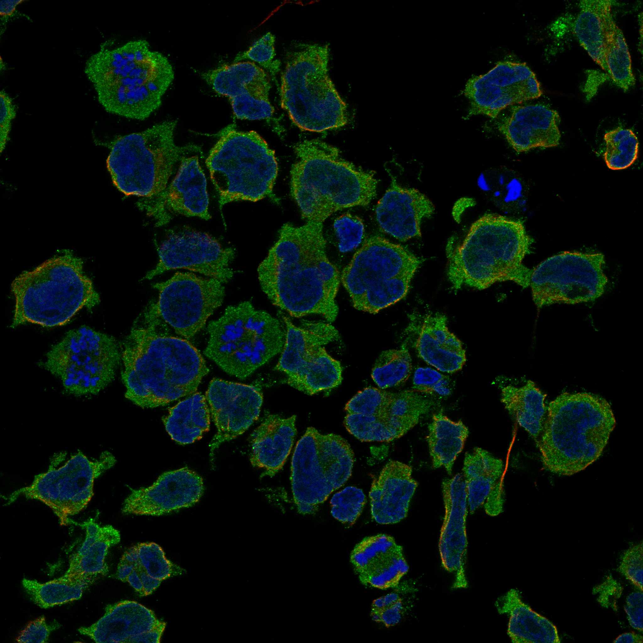
| Compartment evidence | UniProt annotates cytoplasm and nucleus (UniProt Q9C098: location). HPA ICC-IF instead reports mainly cytosol plus vesicles (HPA subcellular: approved). Those ICC-IF observations can inform a compartment check, but they do not establish how vesicles or nuclei must appear in chromogenic paraffin-section IHC. |
| Protein topology and processing | DCLK3 has no transmembrane segment, signal peptide, or annotated propeptide; UniProt lists one chain spanning residues 1–648 (UniProt Q9C098: topology; processing). This supports caution with a purely surface or secreted-looking pattern. The record does not provide an antibody epitope or target-specific retrieval behavior. |
| Antibody validation scope | HPA040685 is listed as IHC Approved, while HPA053234 is listed as ICC Approved without an IHC status in this payload (HPA antibodies). The tissue IHC profile is Approved but pending external verification (HPA tissue IHC: reliability). Do not transfer the ICC antibody's status to paraffin-section IHC. |
| Reported absence and low staining | HPA lists adipocytes, cardiomyocytes, and several other specified populations as not detected, while lung macrophages and several glandular or epithelial populations are low (HPA tissue IHC). Use the named cell population when comparing sections. A low or undetected reference result is less useful for diagnosing a failed assay than a reported medium-positive population. |
| Situation | Likely cause | Next action |
|---|---|---|
| Caudate lacks the reported axonal staining (HPA tissue IHC). | Possible assay failure, sampling difference, or weak signal; the image pattern alone cannot separate these causes (general IHC practice). | Check a reported medium-positive population in the same run and review primary-antibody, detection, and counterstain records (general IHC practice). If changing retrieval conditions, treat that as routine assay optimization; no DCLK3-specific fixation or retrieval effect is supplied. |
| Only sharp nuclear staining is visible. | Nuclear location is annotated, but the supplied HPA tissue pattern does not establish nuclear-only IHC as the expected result (UniProt Q9C098: location; HPA tissue IHC). | Compare with caudate axons and other reported positive cells, then check control sections (HPA tissue IHC; general IHC practice). Avoid calling nuclei inherently wrong: UniProt lists the nucleus (UniProt Q9C098: location). |
| Adipocytes or cardiomyocytes stain strongly and widely (HPA tissue IHC: not detected). | Cross-reactivity or endogenous detection activity is possible (general IHC practice); disagreement with HPA does not identify which occurred. | Compare an adjacent no-primary control and the expected-positive section; score the named cell population separately from surrounding tissue (general IHC practice; HPA tissue IHC). |
| Diffuse chromogen makes cell types and processes hard to distinguish. | Background deposition, tissue pigment, or endogenous activity can obscure specific staining (general chromogenic IHC practice). | Inspect the no-primary control and unstained tissue appearance, then review blocking and detection conditions (general IHC practice). Score DCLK3 only where the pattern remains anatomically interpretable against the HPA tissue reference. |
| Punctate intracellular staining is treated as proof of a positive paraffin section. | HPA reports vesicles in ICC-IF, while its tissue IHC summary emphasizes caudate axons (HPA subcellular; HPA tissue IHC). | Evaluate puncta alongside cell identity, tissue morphology, and IHC controls (general IHC practice). ICC-IF localization is supporting context, not a required chromogenic IHC pattern. |
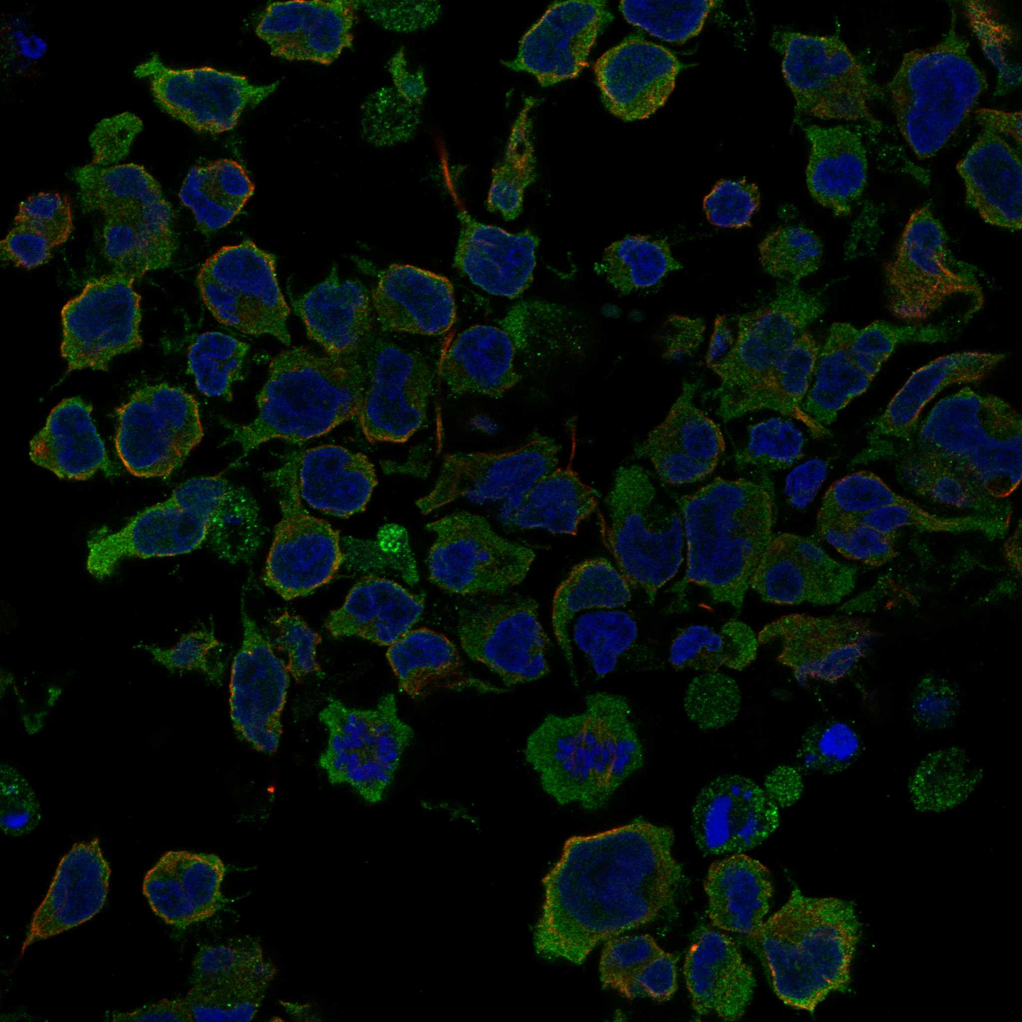
| Can the ICC-IF images define the expected IHC result? | HPA reports cytosol and vesicles in ICC-IF, and lists ICC approval for HPA053234; its listed IHC approval belongs to HPA040685 (HPA subcellular; HPA antibodies). | Use the HPA tissue IHC profile to judge paraffin-section staining. Consult the separate IF/ICC guide for fluorescence interpretation; do not infer an IHC protocol or interchangeable antibody validation from the ICC-IF images. |
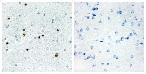
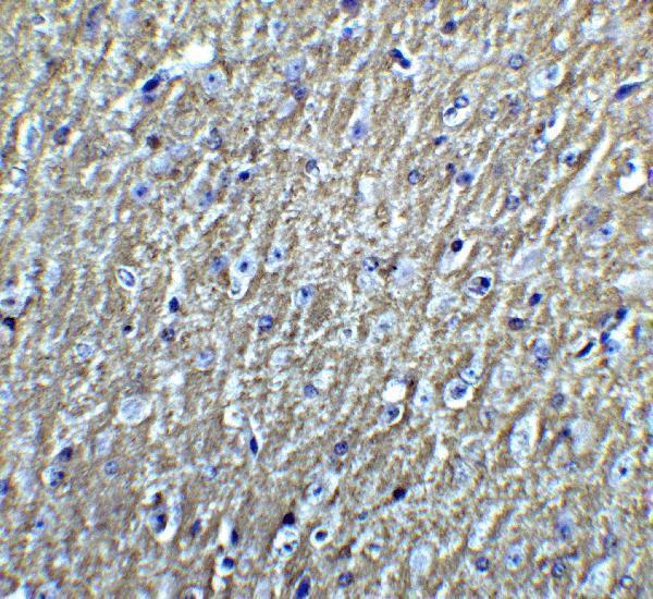


Comprehensive Human Protein Atlas IHC scoring per tissue (reliability: Approved — Pending external verification.). Rows are taken directly from the HPA tissue chart — click any row's HPA link to view the source.
| Tissue | Cell type | Level | Evidence | Source |
|---|---|---|---|---|
| Breast | Myoepithelial cells | Medium | Protein (IHC) | HPA → |
| Caudate | Glial cells | Medium | Protein (IHC) | HPA → |
| Cerebellum | Bergmann glia - cytoplasm/membrane | Medium | Protein (IHC) | HPA → |
| Cerebral cortex | Neuropil | Medium | Protein (IHC) | HPA → |
| Kidney | Cells in tubules | Medium | Protein (IHC) | HPA → |
| Tissue | Cell type | Level | Evidence | Source |
|---|---|---|---|---|
| Adipose tissue | Adipocytes | Not detected | Protein (IHC) | HPA → |
| Bone marrow | Hematopoietic cells | Not detected | Protein (IHC) | HPA → |
| Colon | Endothelial cells | Not detected | Protein (IHC) | HPA → |
| Endometrium | Cells in endometrial stroma | Not detected | Protein (IHC) | HPA → |
| Esophagus | Squamous epithelial cells | Not detected | Protein (IHC) | HPA → |
Troubleshoot DCLK3 staining in paraffin sections by checking retrieval, controls, cellular location, and scoring before interpreting chromogenic signal.
Two anti-DCLK3 antibodies have IHC images from human or mouse brain; one also has IF images from both species (catalog: image captions).
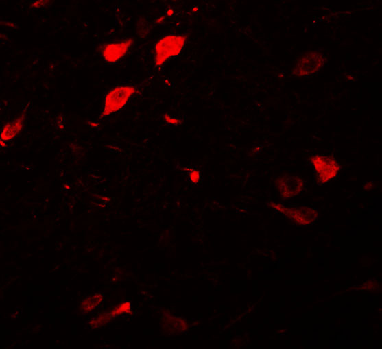
A15882 has IHC data from paraffin-embedded human brain and lists human and mouse reactivity (catalog: IHC image caption and reactivity). A15882-1 has IHC data from mouse brain and IF data from human and mouse brain (catalog: IHC and IF image captions).
Which to pick: For paraffin-section IHC, choose A15882 when a human-brain example is most relevant; its caption does not report the fixative (catalog: A15882 IHC image caption). For tissue IF, choose A15882-1 because it has images from both species; ICC is listed under neither product's applications (catalog: A15882-1 IF image captions; both application lists). Both list human and mouse reactivity, so use the specimen-matched images to guide a cross-species choice; both are rabbit antibodies, and clone status is unreported (catalog: reactivity, host, clone fields).