DDA1 / DET1- and DDB1-associated protein 1 · IHC design guide

Design Immunohistochemistry for DDA1

Plan DDA1 IHC-P around granular cytoplasmic staining in most tissues (HPA tissue IHC). Bone marrow hematopoietic cells show medium staining, while lymph-node germinal-center cells are not detected, providing comparison tissues for scoring (HPA tissue IHC).

Evidence assembled Oct 2026 · For research use; verify linked source records and product datasheet before use
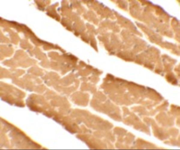
Immunohistochemistry protocol sheet for DDA1 (IHC for DDA1): expected localisation Granular cytoplasm in most tissues (HPA tissue IHC), antibody A10757, validated IHC image, and IHC protocol steps
Printable DDA1 IHC protocol sheet — expected localisation Granular cytoplasm in most tissues (HPA tissue IHC), antibody A10757, controls and protocol steps. Open the full DDA1 IHC guide →

DDA1 Immunohistochemistry Experimental Design Guide

Expected localisation, validated protocols, controls and antibodies — the at-a-glance facts below, then the full design guide.

Must know before staining
Expected localisation Granular cytoplasm in most tissues (HPA tissue IHC)
Staining pattern Granular cytoplasm in glandular and hematopoietic cells (HPA tissue IHC)
Antigen retrieval Citrate pH 6.0 HIER, 95–98 °C, 20 min (rule: cytoplasmic / membrane antigen)
Positive control ⓘ Adrenal gland+4 more · see all
Negative control ⓘ Lymph node+1 more · see all
Important caveats
Reasons your staining may differ from the expected pattern.
Fixation Keep formalin fixation consistent across sections. (standard IHC practice; not target-specific)
Caveat Staining–RNA consistency is medium; verify controls (HPA tissue IHC)
Regulation No expression regulation annotated (UniProt)
Isoform / epitope 0 isoforms annotated; chain spans residues 2–102 (UniProt)
Section 1

Recommended DDA1 IHC & IF Protocols

The catalog antibody’s IHC-P protocol is accompanied by a published DDA1 protocol using breast cancer tissue microarrays (PMC8899920).

Recommended immunohistochemistry (IHC-P) protocol parameters
SampleTissue sections; selected-image fixative not specified (standard IHC workflow)
FixationImage fixative and duration unreported (datasheet A10757); verify before use.
Sectioning4–5 µm sections on charged slides (standard)
DeparaffinisationXylene, graded ethanol series to water (standard)
Antigen retrievalHeat-induced epitope retrieval in citrate buffer, pH 6.0, 20 min at 95–98 °C (standard rule: cytoplasmic / membrane antigen)
Peroxidase block3% H2O2, 10 min, room temperature (standard)
Blocking10% normal serum of the secondary host, 30 min, room temperature (standard)
Primary antibodyRabbit anti-DDA1, 5 μg/mL (datasheet A10757)
Primary incubationOvernight at 4 °C (standard)
DetectionHRP-polymer secondary, DAB chromogen 5–10 min (standard)
CounterstainHematoxylin, blue, dehydrate and mount (standard)
Expected resultDDA1-positive staining in glandular cells of adrenal gland (HPA tissue IHC: Medium). HPA tissue profile: Granular cytoplasmic expression in most tissues. No signal in the no-primary control.
💡Decision noteStart with citrate pH 6.0 retrieval at 95–98 °C for 20 min (page antigen-retrieval specification); the published protocol also uses a low-pH buffer (PMC8899920).
Section 2

What Is the Expected DDA1 Staining Pattern?

In paraffin-section IHC, expect granular cytoplasmic DDA1 staining in most tissues, including medium staining in adrenal glandular cells and bone-marrow hematopoietic cells (HPA tissue IHC). HPA rates the tissue staining Approved, with medium consistency against RNA data and external verification pending (HPA tissue IHC). DDA1 has no annotated transmembrane segment; UniProt does not assign a subcellular location (UniProt Q9BW61).

What am I looking at on my slide?
Granular cytoplasmic staining in glandular or hematopoietic cells.Consistent with the reported IHC pattern; HPA scores adrenal glandular and bone-marrow hematopoietic cells Medium (HPA tissue IHC). Compare staining within the identified cell population, since a positive field can contain cells with different signal levels (general IHC practice).
Predominantly nuclear staining in a paraffin section.The IHC tissue profile reports granular cytoplasm, while separate ICC-IF data place DDA1 mainly in nucleoplasm and nucleoli (HPA tissue IHC; HPA ICC-IF). Treat a nuclear-only IHC result as discordant; review controls and cell identification before calling it specific.
Strong staining in lymph-node germinal-center cells or vaginal squamous epithelium.These particular cell populations are reported Not detected (HPA tissue IHC). Check whether the signal instead belongs to adjacent cells; persistent staining in the named populations raises concern for cross-reactivity or endogenous detection activity (general IHC practice).
Uniform colour over cells and surrounding tissue, without a granular cell pattern.This differs from the reported cytoplasmic granularity (HPA tissue IHC). Diffuse background can arise from nonspecific binding, insufficient washing, or detection chemistry (general IHC practice); it does not establish DDA1 expression.
No staining in a section containing adrenal glandular cells or bone-marrow hematopoietic cells.Both populations have Medium staining in the HPA examples (HPA tissue IHC). Verify that the expected cells are present, then review the run controls, antibody conditions, retrieval and detection steps (general IHC practice). A single negative section cannot establish DDA1 absence.
💡Expected DDA1 appearanceCall an IHC result concordant when identifiable cells show granular cytoplasmic staining, with Medium signal in the cited adrenal glandular or bone-marrow hematopoietic examples (HPA tissue IHC); isolated nuclear-only or diffuse, cell-independent colour warrants review against that pattern (HPA tissue IHC; general IHC practice).
How each factor affects the staining
Tissue and cell selectionHPA describes granular cytoplasmic expression in most tissues, but its examples vary by cell population: Medium in adrenal glandular cells and Not detected in lymph-node germinal-center cells (HPA tissue IHC). Score the named cells rather than a whole organ.
Antibody evidenceThe tissue profile is Approved, with medium RNA–staining consistency and external verification pending (HPA tissue IHC). HPA042134 is IHC Approved; HPA055272 has ICC approval only (HPA antibodies). Keep those application-specific validation labels distinct.
Assay-dependent compartment readoutTissue IHC reports granular cytoplasm, whereas HPA ICC-IF reports mainly nucleoplasm and nucleoli (HPA tissue IHC; HPA ICC-IF). The records do not resolve this difference; interpret an IHC slide against IHC evidence.
Topology and processingUniProt annotates no transmembrane segment, signal peptide or propeptide, and lists the mature chain as residues 2–102 (UniProt Q9BW61). These annotations provide no basis to expect a membrane rim or shed extracellular deposit in IHC.
Fixation evidenceTarget-specific fixation effects are not established by the supplied assay evidence. Verify with a matched IHC source before attributing a result to fixation.
Why is my staining missing, weak or wrong?
SituationLikely causeNext action
The positive-control tissue is blank.Adrenal glandular or bone-marrow hematopoietic cells are Medium in the HPA examples; a blank result conflicts with that observation if the named cells are present (HPA tissue IHC).Confirm cell identity and section quality, then check the run control, antibody application, retrieval and chromogenic detection in sequence (general IHC practice).
The whole section shows a brown haze.A cell-independent haze obscures the granular cytoplasmic pattern reported for tissue IHC (HPA tissue IHC). Nonspecific reagent binding or detection background is possible (general IHC practice).Inspect a control lacking primary antibody, then review blocking, washes and detection conditions before scoring cellular signal (general IHC practice).
Only nuclei are strongly coloured.Nuclear signal differs from the reported tissue-IHC cytoplasmic pattern (HPA tissue IHC), even though HPA ICC-IF identifies nucleoplasm and nucleoli (HPA ICC-IF).Check counterstain and detection controls, confirm the stained cells, and report the IHC–ICC-IF discrepancy rather than assigning the nuclear result to a proven IHC pattern.
A reported negative cell population stains.HPA reports Not detected in lymph-node germinal-center cells and vaginal squamous epithelial cells (HPA tissue IHC); signal there may reflect cell misidentification, cross-reactivity or endogenous detection activity (general IHC practice).Recheck morphology and a control lacking primary antibody; compare the result with a named Medium-staining cell population (HPA tissue IHC; general IHC practice).
The signal is present but too weak to interpret.HPA reports Low staining in several populations, including skin keratinocytes and ovarian follicle cells (HPA tissue IHC). Weak colour alone cannot distinguish a low-expression example from an underperforming run.Use an HPA Medium example as a companion control, verify detection and retrieval performance, and score only cells whose signal is distinguishable from background (HPA tissue IHC; general IHC practice).
What pattern should IF/ICC show?HPA ICC-IF places DDA1 mainly in nucleoplasm and nucleoli; HPA055272 is ICC Approved, while HPA042134 is IHC Approved (HPA ICC-IF; HPA antibodies).Use the separate IF/ICC guide for assay design. Record nuclear IF/ICC localisation on its own terms and avoid using it to override the granular cytoplasmic tissue-IHC profile (HPA ICC-IF; HPA tissue IHC).

Sample controls for DDA1 IHC & IF

🧪Run bone marrow first and look for DDA1 staining in hematopoietic cells (Medium; HPA: bone marrow). Run lymph node as the negative tissue, focusing on germinal center cells (Not detected; HPA: lymph node); on the bone marrow slide, morphologically intact cells without chromogen above background can provide an internal background reference, but HPA does not identify a DDA1-negative cell type there (HPA: bone marrow).
Positive control tissue: Adrenal gland (Glandular cells, HPA Medium)
Negative control tissue: Lymph node (HPA Not detected)
ICC-IF cell lines (HPA subcellular resource): HPA ICC-IF images show DDA1 in PC-3, U-251MG, U2OS, NIH 3T3, with annotated localisation: Nucleoplasm (approved), Nucleoli (approved) (HPA subcellular).
Technical controls: Include a no-primary, secondary-only control, an isotype control matched to the primary antibody’s host species and clonality, and a DDA1 knockout specimen or immunizing-peptide block where available (standard IHC practice). Block endogenous peroxidase and check for residual chromogen in bone marrow, where hematopoietic cells can contribute peroxidase activity (standard IHC practice).
⚠️Feasibility: A target-specific fixation window and antigen-retrieval dependency are unreported in the supplied evidence; optimize retrieval on matched sections (standard IHC practice). The A10757 mouse-heart IHC caption reports 5 μg/mL, but its fixative is unreported (selected tissue-IHC caption). The evidence does not establish that frozen sections or IF are easier; for bone marrow IHC, account for endogenous peroxidase when interpreting hematopoietic-cell staining (standard IHC practice).

HPA tissue IHC evidence for DDA1

Comprehensive Human Protein Atlas IHC scoring per tissue (reliability: Approved — Medium consistency between antibody staining and RNA expression data. Pending external verification.). Rows are taken directly from the HPA tissue chart — click any row's HPA link to view the source.

Positive expression · recommended positive controls

TissueCell typeLevelEvidenceSource
Adrenal gland Glandular cells Medium Protein (IHC) HPA →
Appendix Glandular cells Medium Protein (IHC) HPA →
Bone marrow Hematopoietic cells Medium Protein (IHC) HPA →
Breast Adipocytes Medium Protein (IHC) HPA →
Bronchus Respiratory epithelial cells Medium Protein (IHC) HPA →

Undetected expression · recommended negative controls

TissueCell typeLevelEvidenceSource
Lymph node Germinal center cells Not detected Protein (IHC) HPA →
Vagina Squamous epithelial cells Not detected Protein (IHC) HPA →
Section 3

Advanced DDA1 IHC Tips

Troubleshoot DDA1 chromogenic IHC in paraffin sections using the page retrieval setting, the selected antibody image, and compartment-specific controls.

How should I adjust retrieval if DDA1 staining is weak or uneven?
Start with citrate buffer at pH 6.0, heated to 95–98 °C for 20 min (page antigen-retrieval setting). If staining is weak, check that sections remained submerged and compare a longer citrate incubation or an alternative retrieval buffer on matched sections, while watching for tissue damage (standard IHC practice). Keep antibody concentration and detection conditions constant across that comparison so retrieval is the variable being assessed (standard IHC practice). The selected A10757 mouse-heart image reports 5 µg/mL antibody but does not report retrieval conditions, so its staining cannot establish which retrieval condition produced the image (selected A10757 tissue-IHC caption).
Could fixation explain a weak DDA1 signal in paraffin sections?
Target-specific sensitivity of DDA1 staining to fixation is unknown from the supplied evidence (selected A10757 tissue-IHC caption; HPA tissue IHC). The A10757 mouse-heart caption reports antibody at 5 µg/mL but does not state the fixative, fixation duration, or processing conditions (selected A10757 tissue-IHC caption). For a controlled comparison, use adjacent sections with recorded fixation histories, apply the same citrate pH 6.0 retrieval, and hold antibody and detection conditions constant (page antigen-retrieval setting; standard IHC practice). If signal tracks fixation history, document that observation for the tested specimens; neither the HPA staining pattern nor DDA1 topology or modifications establishes a target-specific fixation effect (HPA tissue IHC; UniProt Q9BW61 record).
How should I judge cytoplasmic staining when DDA1 also appears nuclear?
Assess compartment patterns within identified cells: HPA tissue IHC describes granular cytoplasmic expression in most tissues, whereas HPA ICC/IF places DDA1 mainly in the nucleoplasm and nucleoli (HPA tissue IHC; HPA subcellular). UniProt supplies no subcellular annotation for DDA1, so it does not resolve that difference (UniProt Q9BW61 subcellular record). Compare cytoplasmic granules and nuclear signal separately in intact cells, using the same counterstain and exposure or development conditions across sections (standard IHC practice). Treat diffuse staining that ignores cell boundaries, or a compartment pattern seen only at damaged edges, as a reason to repeat the assay with controls before assigning localisation (standard IHC practice).
What should I check when an antibody gives unexpected DDA1 staining?
Check the antibody's stated immunogen or epitope against the DDA1 sequence before changing retrieval, because the supplied record does not map the catalog antibody's binding site (UniProt Q9BW61 record; selected A10757 tissue-IHC caption). DDA1 is 102 amino acids long, has no annotated transmembrane segment or glycosylation sites, and has no annotated isoforms in this record (UniProt Q9BW61 record). Recorded modifications include N-acetylalanine at position 2 and phosphoserines at positions 33 and 95, but their effect on this antibody's staining is unreported (UniProt Q9BW61 record; selected A10757 tissue-IHC caption). Compare staining with an independently validated antibody or a suitable target-loss control before attributing an unexpected pattern to an epitope or modification (standard IHC validation practice).
How can I compare DDA1 IHC with multiplex IF staining?
Use IF as a separate assay comparison: HPA ICC/IF reports DDA1 mainly in the nucleoplasm and nucleoli, while tissue IHC reports granular cytoplasmic expression in most tissues (HPA subcellular; HPA tissue IHC). Multiplex DDA1 with a validated marker for the cell population under study, and inspect single-channel images before interpreting overlap (standard IF practice). Choose a fluorophore whose emission is separated from the specimen's autofluorescence, then include unstained and single-label controls to evaluate bleed-through and background (standard IF practice). DDA1 has no annotated transmembrane segment, but this antibody's epitope is unmapped; for an intracellular epitope, compare mild permeabilisation conditions while preserving nuclear structure (UniProt Q9BW61 topology; selected A10757 tissue-IHC caption; standard IF practice).
How do I reduce diffuse chromogenic background without losing DDA1 signal?
First inspect a no-primary control alongside the DDA1-stained section to distinguish detection background from antibody-associated staining (standard IHC practice). Block endogenous peroxidase before chromogenic detection, optimise protein blocking and washes, and limit DAB development if the control also becomes brown (standard chromogenic IHC practice). If background follows the primary antibody, titrate around the reported A10757 image concentration of 5 µg/mL on matched sections while keeping retrieval and detection fixed; that concentration is image evidence, not an established optimum for every specimen (selected A10757 tissue-IHC caption; standard IHC practice). Score staining only in intact cells and compare it with the reported granular cytoplasmic tissue pattern, while allowing the reported ICC/IF nuclear pattern to inform review (HPA tissue IHC; HPA subcellular).
How should I quantify heterogeneous DDA1 staining across sections? ⚠ ANSWER MARKED FOR VERIFICATION
Define the cell population and compartment before scoring, because HPA reports granular cytoplasmic tissue staining but mainly nucleoplasmic and nucleolar ICC/IF staining (HPA tissue IHC; HPA subcellular). For chromogenic IHC, record the percentage of positive cells and intensity categories, then calculate an H-score as the sum of percentage at each intensity multiplied by its intensity value (standard IHC scoring practice). If cell abundance varies, also report positive cells per mm² of viable tissue or per total cells in the same compartment (standard IHC quantification practice). Normalise comparisons to the same cell population, viable area, retrieval, development time, and scoring thresholds; exclude folds, edges, and necrosis consistently (standard IHC practice).
What would make an apparent DDA1-positive cell convincing?
A convincing positive has cell-bound signal above the no-primary control, a reproducible compartment pattern, and staining in morphologically intact cells across comparable sections (standard IHC validation practice). Granular cytoplasmic staining is compatible with the reported tissue-IHC profile, while nuclear or nucleolar staining warrants separate review because HPA ICC/IF reports those locations and UniProt does not annotate DDA1 localisation (HPA tissue IHC; HPA subcellular; UniProt Q9BW61 subcellular record). HPA reports medium staining in several specified cell populations and no detection in lymph-node germinal-center cells or vaginal squamous epithelial cells, so evaluate cell identity before comparing specimens (HPA tissue IHC). Discount signal concentrated at section edges, necrotic areas, or sites positive in a no-primary control, and check endogenous peroxidase activity before assigning DAB signal to DDA1 (standard chromogenic IHC practice).
Boster reagents

Best DDA1 / DET1- and DDB1-associated protein 1 IHC Antibodies

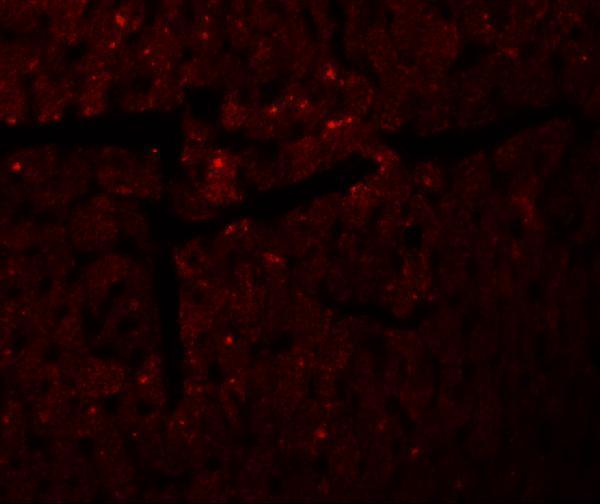
The catalog antibody has IHC and IF images from mouse heart tissue (catalog image captions). Human, mouse and rat are listed as reactive species (catalog: A10757).

Real IHC data Immunohistochemistry of DDA1 in mouse heart tissue with DDA1 antibody at 5 μg/mL.
Anti-DDA DDA1 Antibody
Cat # A10757

A10757 will render with an IHC image of mouse heart tissue at 5 μg/mL (catalog IHC image caption). Its IF image also shows mouse heart tissue at 20 μg/mL; human, mouse and rat are listed as reactive species (catalog IF image caption; catalog reactivity).

Which to pick: Choose A10757 for paraffin-section IHC because IHC-P is listed and its IHC image shows mouse heart tissue; the fixative is unreported (catalog applications; catalog IHC image caption). For fluorescence, A10757 has an IF image from mouse heart tissue, while ICC validation is unreported (catalog IF image caption; catalog applications). A10757 is listed as reactive with human, mouse and rat, but the supplied IHC and IF images demonstrate mouse tissue only; its host is rabbit and clonality is unreported (catalog reactivity; catalog image captions; catalog host and clone fields).

Each figure is that product's own IHC / IF validation image from its datasheet.

References

  1. UniProt Consortium. UniProt entry Q9BW61 (DDA1_HUMAN, DET1- and DDB1-associated protein 1).
  2. Human Protein Atlas. DDA1 tissue IHC expression (reliability: Approved).
  3. Human Protein Atlas. DDA1 subcellular location (ICC-IF): Mainly localized to the nucleoplasm and nucleoli..
  4. Human Protein Atlas. DDA1 antibody validation summary (2 antibodies).
  5. DDA1, a novel oncogene, promotes lung cancer progression through regulation of cell cycle. Journal of cellular and molecular medicine 2017 — PMC5542901.
  6. A clinically compatible drug-screening platform based on organotypic cultures identifies vulnerabilities to prevent and treat brain metastasis. EMBO molecular medicine 2022 — PMC8899920.
  7. PubMed PMID:17452440 — UniProt-cited evidence.
  8. PubMed PMID:15489334 — UniProt-cited evidence.
  9. PubMed PMID:18669648 — UniProt-cited evidence.