This website uses cookies to ensure you get the best experience on our website.


- Table of Contents
Plan paraffin IHC for DDX6 using cytoplasmic staining as the tissue benchmark (HPA tissue IHC). Colon glandular cells offer a high-staining reference (HPA tissue IHC); the IHC-validated antibody has a stated range of 2–5 µg/mL (datasheet A03826-1).

Expected localisation, validated protocols, controls and antibodies — the at-a-glance facts below, then the full design guide.
| Expected localisation | Cytoplasmic staining in tissue sections (HPA tissue IHC) | |
| Staining pattern | General cytoplasmic staining; high in glandular cells (HPA tissue IHC) | |
| Antigen retrieval | EDTA pH 8.0 HIER, heat-mediated (datasheet A03826-1) | |
| Positive control | Appendix+4 more · see all | |
| Negative control | None in HPA (detected in all 45 tissues); use no-primary + isotype controls |
| Fixation | Keep fixation consistent across sections. (standard IHC practice; not target-specific) | |
| Caveat | Cellular stress can shift DDX6 to stress granules (UniProt) | |
| Regulation | Broad expression; tissue intensity varies (HPA tissue IHC) | |
| Isoform / epitope | No annotated isoforms or cleavage products (UniProt) |
The catalog antibody’s IHC-P protocol is paired with a published chromogenic DDX6 protocol for human testicular tissue (PMC6028756).
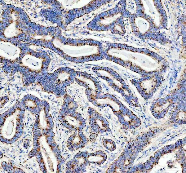
| Sample | Paraffin-embedded human stomach cancer tissue; fixative not specified (datasheet A03826-1) |
| Fixation | Image fixative and duration unreported (datasheet A03826-1); verify before use. |
| Sectioning | 4–5 µm sections on charged slides (standard) |
| Deparaffinisation | Xylene, graded ethanol series to water (standard) |
| Antigen retrieval | Heat retrieval: EDTA pH 8.0 (datasheet A03826-1); 20 min, 95–100 °C (standard) |
| Peroxidase block | 3% H2O2, 10 min, room temperature (standard) |
| Blocking | 10% goat serum (datasheet A03826-1) |
| Primary antibody | Rabbit anti-DDX6, 2-5μg/ml (datasheet A03826-1) |
| Primary incubation | Overnight at 4 °C (datasheet A03826-1) |
| Detection | HRP-conjugated secondary, DAB chromogen (datasheet A03826-1) |
| Counterstain | Hematoxylin, blue, dehydrate and mount (standard) |
| Expected result | DDX6-positive staining in glandular cells of appendix (HPA tissue IHC: High). HPA tissue profile: General cytoplasmic expression. No signal in the no-primary control. |
DDX6 should show general cytoplasmic staining in paraffin sections, with strong staining expected in several glandular and squamous epithelial populations (HPA: tissue IHC). It is a nonmembrane protein that also occupies P-bodies and can enter the nucleus (UniProt P26196: topology and localization). Treat tissue patterns as a guide: HPA rates its IHC reliability Approved, with medium consistency against RNA data and external verification pending (HPA: reliability).
| Cytoplasmic staining is strongest in appendix or colon glandular cells; hematoxylin leaves nuclei distinct. | This fits the reported general cytoplasmic pattern and High staining in those cells (HPA: tissue IHC). Compare staining within the same section before scoring intensity, since chromogenic signal also reflects the IHC workflow (general IHC practice). |
| Staining is predominantly nuclear, with little cytoplasmic signal in an otherwise positive tissue. | Investigate the compartment pattern: cytoplasm is the principal tissue IHC finding (HPA: tissue IHC). Nuclear entry is documented, so nuclear signal alone is not proof of an artefact (UniProt P26196: localization); check controls and staining distribution (general IHC practice). |
| Strong staining appears in glial cells or myocytes while expected epithelial cells are weak. | HPA reports Low staining in hippocampal and caudate glia and skeletal myocytes, but High staining in several glandular epithelia (HPA: tissue IHC). Check for cross-reactivity or endogenous chromogenic activity before assigning the unexpected signal to DDX6 (general IHC practice). |
| Brown deposit covers stroma, lumina, or much of the section without a clear cell pattern. | Diffuse deposit does not match the reported cellular cytoplasmic pattern (HPA: tissue IHC). Review blocking, washes, detection reagents and the negative control for nonspecific staining or endogenous activity (general IHC practice). |
| No signal appears in appendix or colon glandular cells. | Those cells are reported High and are useful positive comparators (HPA: tissue IHC). A blank result calls for review of the positive control, antigen retrieval, primary antibody dilution and detection steps; it cannot establish absent DDX6 on its own (general IHC practice). |
| Choice of tissue comparator | Appendix, colon, duodenum, endometrium, gallbladder and rectum glandular cells are reported High; skeletal myocytes and selected glia are Low (HPA: tissue IHC). Use the named cell population when judging a control, since whole-section staining can hide that distinction (general IHC practice). |
| Granules and compartment resolution | DDX6 occupies cytoplasmic P-bodies and can move to stress granules under cellular stress (UniProt P26196: localization). Tissue IHC is reported as generally cytoplasmic (HPA: tissue IHC); do not require individually resolved puncta to accept a chromogenic section (general IHC practice). |
| Antibody evidence | HPA024201, HPA026644 and CAB004668 have Approved IHC status (HPA: antibody validation). That status supports their reported tissue patterns, while HPA's medium RNA consistency and pending external verification limit how firmly an unexpected pattern can be assigned to DDX6 (HPA: reliability). |
| Topology and processing | DDX6 has no transmembrane segment, signal peptide or propeptide (UniProt P26196: topology and processing). A dominant membrane-outline pattern deserves checking against cytoplasmic positive cells; the uncertain plasma membrane observation in ICC-IF does not establish that pattern for tissue IHC (HPA: subcellular). |
| IF/ICC: what should a positive cell show? | Expect cytoplasmic bodies with additional cytosol signal (HPA: ICC-IF, supported). Plasma membrane and midbody assignments are uncertain (HPA: ICC-IF). This describes cellular localization; it does not define a staining protocol or override the tissue IHC pattern. |
| Situation | Likely cause | Next action |
|---|---|---|
| Known-positive glandular cells are blank. | The expected High signal is missing (HPA: appendix and colon glandular cells); the failed step is undetermined. | Confirm tissue and cell identity, then review retrieval, primary antibody dilution, detection reagents and a positive control in the same run (general IHC practice). |
| Cytoplasm is faint across all tissues. | A weak run may obscure the general cytoplasmic pattern (HPA: tissue IHC); HPA supplies no DDX6-specific fixation-sensitivity finding (HPA: supplied tissue IHC record). | Compare known High cells and run controls; review section handling, retrieval and detection as general IHC variables without attributing the result to a DDX6-specific fixation effect (general IHC practice). |
| Only nuclei stain strongly. | The balance differs from general cytoplasmic tissue staining (HPA: tissue IHC), although nuclear import is possible (UniProt P26196: localization). | Check whether cytoplasmic positive cells stain, inspect the negative control, and repeat with the IHC-validated antibody if the compartment pattern remains discordant (general IHC practice; HPA: IHC Approved). |
| Brown signal is widespread outside cells. | Nonspecific deposit or endogenous chromogenic activity can obscure a cellular pattern (general IHC practice); HPA reports general cytoplasmic expression (HPA: tissue IHC). | Inspect a negative control, blocking and wash steps, then score only interpretable cellular staining (general IHC practice). |
| Low-reference cells appear stronger than expected positive cells. | Cell identity, section variability or an off-target reaction may explain the mismatch (general IHC practice); HPA lists glia and myocytes as Low and several glandular populations as High (HPA: tissue IHC). | Verify the named cell populations on the counterstain and compare controls. Treat a persistent reversal as uncertain, given HPA's medium RNA consistency and pending external verification (HPA: reliability). |
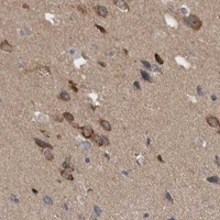

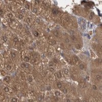
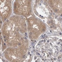
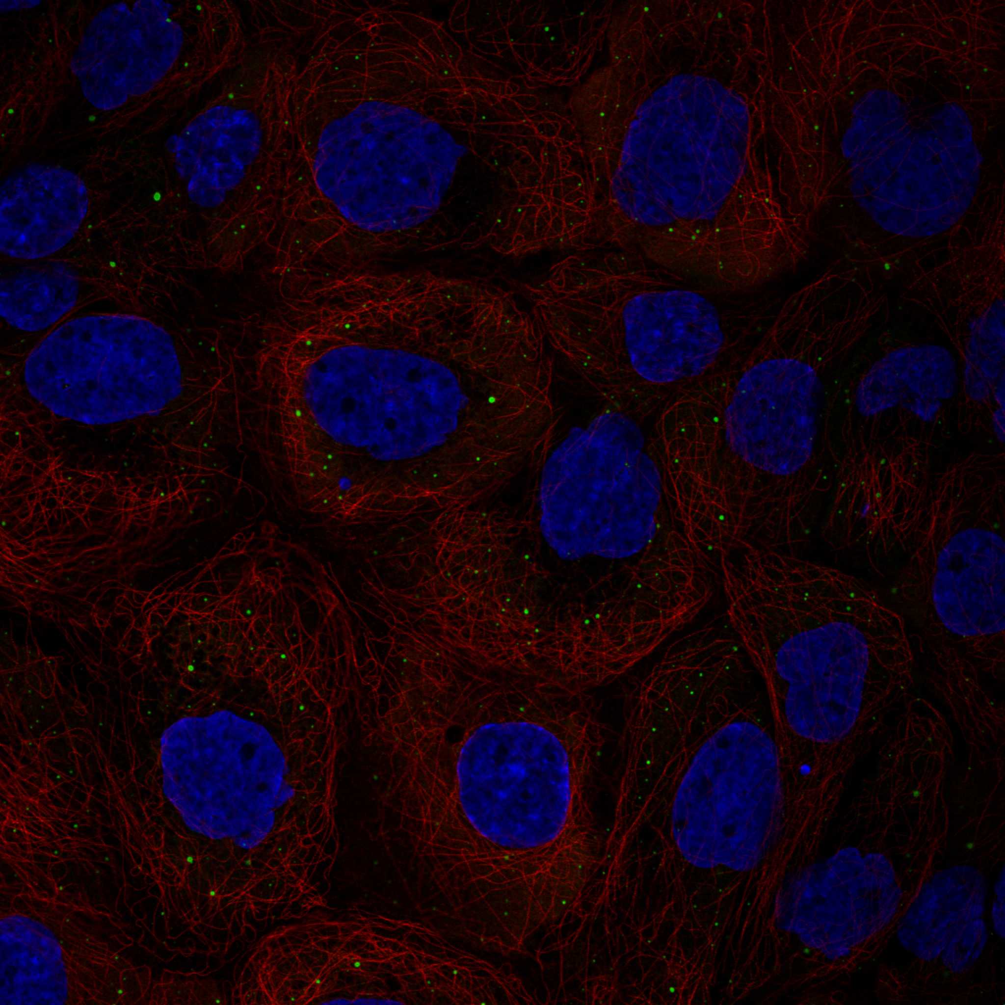
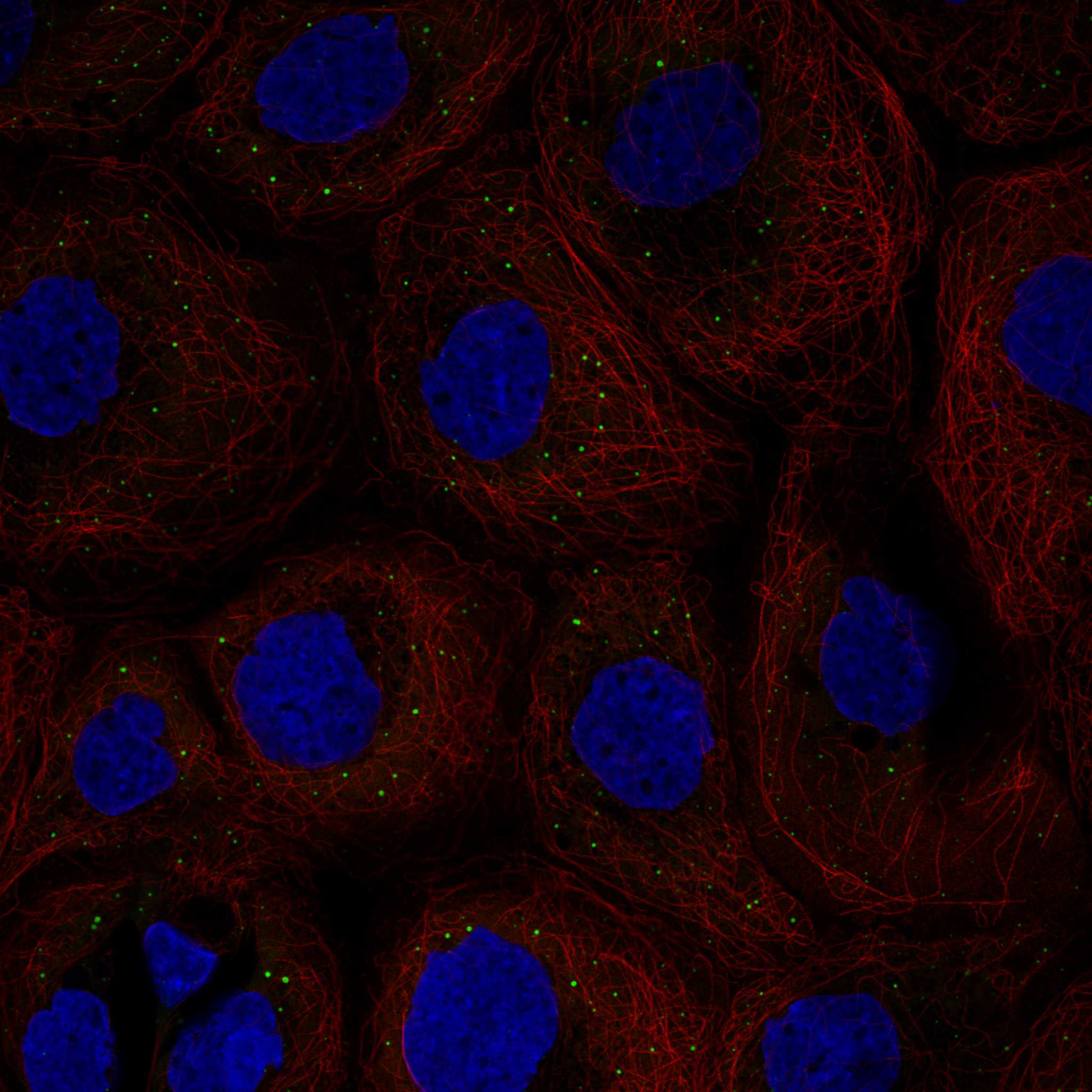
Comprehensive Human Protein Atlas IHC scoring per tissue (reliability: Approved — Medium consistency between antibody staining and RNA expression data. Pending external verification.). Rows are taken directly from the HPA tissue chart — click any row's HPA link to view the source.
| Tissue | Cell type | Level | Evidence | Source |
|---|---|---|---|---|
| Appendix | Glandular cells | High | Protein (IHC) | HPA → |
| Colon | Glandular cells | High | Protein (IHC) | HPA → |
| Duodenum | Glandular cells | High | Protein (IHC) | HPA → |
| Endometrium | Glandular cells | High | Protein (IHC) | HPA → |
| Esophagus | Squamous epithelial cells | High | Protein (IHC) | HPA → |
| Tissue | Cell type | Level | Evidence | Source |
|---|---|---|---|---|
| None in HPA: DDX6 is detected in all 45 scored tissues. Use a no-primary (secondary-only) and an isotype control instead. | ||||
Troubleshoot DDX6 staining in paraffin sections by checking retrieval, cytoplasmic localisation, controls and scoring before interpreting chromogenic signal.
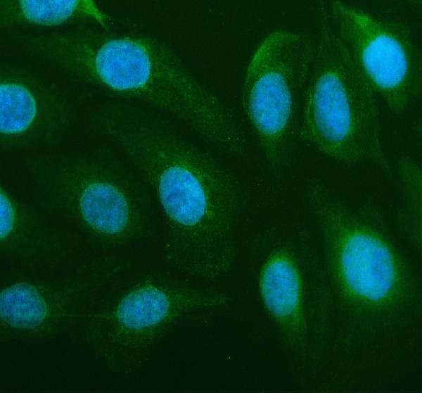
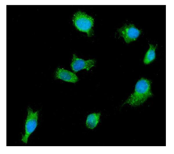
The catalog shows DDX6 IHC in human paraffin stomach cancer tissue and IF/ICC in human U2OS and MCF-7 cells (catalog image captions). Both antibodies list human, mouse and rat reactivity (catalog reactivity).
A03826-1 has IHC data from human paraffin stomach cancer tissue and IF/ICC data from U2OS cells (A03826-1 image captions). M03826-1 has IF/ICC data from MCF-7 cells and no listed IHC application (M03826-1 image caption; catalog applications).
Which to pick: Choose A03826-1 for paraffin-section IHC: its own caption shows staining of human stomach cancer tissue at 2 μg/ml after heat retrieval in EDTA, pH 8.0; the fixative is unreported (A03826-1 IHC caption). For IF/ICC, M03826-1 is a mouse monoclonal with an MCF-7 image, while A03826-1 also has a U2OS IF/ICC image (catalog host and clone; M03826-1 and A03826-1 IF captions). Both list human, mouse and rat reactivity, but the supplied IHC and IF images show human samples only; A03826-1 is the pictured tissue-IHC choice for work involving those species (catalog reactivity; image captions).