DEPDC1 / DEP domain-containing protein 1A · IHC design guide

Design Immunohistochemistry for DEPDC1

Plan DEPDC1 chromogenic IHC-P around nuclear staining in testis germ cells (HPA tissue IHC). This guide covers fixation, antigen retrieval, controls and the catalog antibody’s 1:50–1:100 IHC-P dilution range (datasheet: A08594).

Evidence assembled Oct 2026 · For research use; verify linked source records and product datasheet before use
Immunohistochemistry protocol sheet for DEPDC1 (IHC for DEPDC1): expected localisation Nuclear staining in several tissues (HPA tissue IHC), antibody A08594, validated IHC image, and IHC protocol steps
Printable DEPDC1 IHC protocol sheet — expected localisation Nuclear staining in several tissues (HPA tissue IHC), antibody A08594, controls and protocol steps. Open the full DEPDC1 IHC guide →

DEPDC1 Immunohistochemistry Experimental Design Guide

Expected localisation, validated protocols, controls and antibodies — the at-a-glance facts below, then the full design guide.

Must know before staining
Expected localisation Nuclear staining in several tissues (HPA tissue IHC)
Staining pattern Nuclear signal in testis germ cells; high in pachytene spermatocytes (HPA tissue IHC)
Antigen retrieval Tris-EDTA pH 9.0 HIER, 95–98 °C, 20 min (rule: nuclear antigen)
Positive control ⓘ Testis+4 more · see all
Negative control ⓘ Adipose tissue+4 more · see all
Important caveats
Reasons your staining may differ from the expected pattern.
Fixation Formalin-fixed, paraffin-embedded testis was used (datasheet: A08594 IHC-P) (selected-SKU IHC image A08594)
Caveat Staining has medium consistency with RNA data; verify specificity (HPA tissue IHC)
Regulation Up-regulated in bladder cancer cells at protein level (UniProt)
Isoform / epitope 2 isoforms; N-terminal epitope coverage is unverified (UniProt; datasheet: A08594 N-term)
Section 1

Recommended DEPDC1 IHC & IF Protocols

The catalog antibody’s IHC-P protocol is accompanied by four published DEPDC1 IHC protocols covering lung, breast, nasopharyngeal, and osteosarcoma specimens (PMC11101781; PMC6473048; PMC5609947; PMC10028160).

Recommended immunohistochemistry (IHC-P) protocol parameters
SampleFFPE human testis tissue (datasheet A08594)
FixationImage formalin-fixed; duration unreported (datasheet A08594); verify before use.
Sectioning4–5 µm sections on charged slides (standard)
DeparaffinisationXylene, graded ethanol series to water (standard)
Antigen retrievalHeat-induced epitope retrieval in Tris-EDTA buffer, pH 9.0, 20 min at 95–98 °C (standard rule: nuclear antigen)
Peroxidase block3% H2O2, 10 min, room temperature (standard)
Blocking10% normal serum of the secondary host, 30 min, room temperature (standard)
Primary antibodyRabbit anti-DEPDC1, 1:50-1:100 (datasheet A08594)
Primary incubationOvernight at 4 °C (standard)
DetectionHRP-polymer secondary, DAB chromogen 5–10 min (standard)
CounterstainHematoxylin, blue, dehydrate and mount (standard)
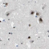
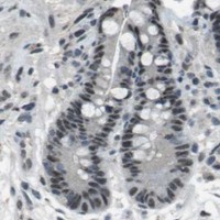
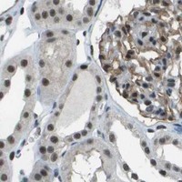
Expected resultDEPDC1-positive staining in pachytene spermatocytes of testis (HPA tissue IHC: High). HPA tissue profile: Nuclear expression in several tissues, mainly expressed in spermatogonia, spermatocytes and early spermatids. No signal in the no-primary control.
💡Decision noteStart with Tris-EDTA pH 9.0 HIER at 95–98 °C for 20 min (page retrieval; UniProt: nuclear localisation).
Section 2

What Is the Expected DEPDC1 Staining Pattern?

DEPDC1 is expected mainly in nuclei (UniProt Q5TB30; HPA tissue IHC), especially spermatogonia, spermatocytes and early spermatids; pachytene spermatocytes show High staining (HPA: Testis). UniProt reports no transmembrane segment (UniProt Q5TB30 topology). HPA rates the tissue IHC pattern Approved, with medium consistency against RNA data and external verification pending (HPA: tissue IHC reliability).

What am I looking at on my slide?
Strong nuclear staining in pachytene spermatocytes, with other germ cells showing nuclear staining (HPA: Testis; HPA: tissue IHC profile).This fits the reported testis pattern: pachytene spermatocytes are High, and the broader nuclear profile includes spermatogonia, spermatocytes and early spermatids (HPA: tissue IHC). Score the stained cell population and nuclear location together (general IHC practice).
Predominantly cytoplasmic or membrane staining, with little nuclear signal.This conflicts with the reported nuclear location (UniProt Q5TB30; HPA: tissue IHC). UniProt lists no transmembrane segment, so a membrane dominant pattern needs scrutiny (UniProt Q5TB30 topology). Check staining controls and morphology before interpreting it as DEPDC1 (general IHC practice).
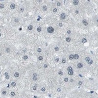
Strong staining in adipocytes or lung alveolar cells.Those cell types are reported as Not detected (HPA: Adipose tissue; HPA: Lung). Consider cross-reactivity or endogenous chromogenic activity, particularly if the signal also appears in a no-primary control (general IHC practice).
Broad, hazy color across cells and tissue spaces, without clear nuclear boundaries.A diffuse pattern cannot establish the reported nuclear location (UniProt Q5TB30; HPA: tissue IHC). Examine the no-primary control, reagent background and tissue morphology; interpret only cell-associated staining that can be localized confidently (general IHC practice).
No convincing nuclear signal in a testis section containing pachytene spermatocytes.Pachytene spermatocytes are a reported High population (HPA: Testis). An absent signal calls for review of tissue identification, antibody and detection steps, and a control section before calling the specimen negative (general IHC practice).
💡Expected DEPDC1 appearanceA convincing positive is predominantly nuclear staining in testis germ cells, strongest in pachytene spermatocytes (HPA: tissue IHC; HPA: Testis); dominant membrane staining or color in reported negative cell types warrants control checks (UniProt Q5TB30 topology; HPA: Adipose tissue; HPA: Lung; general IHC practice).
How each factor affects the staining
How should tissue choice affect interpretation?Testis pachytene spermatocytes provide a reported High reference; several other tissues have Medium staining in specified cells (HPA: tissue IHC). Match each result to its cell type: whole-section color alone does not reproduce the reported pattern (general IHC practice).
How much confidence does the antibody record provide?The listed antibody CAB017845 is IHC Approved, while the tissue profile has medium consistency with RNA and awaits external verification (HPA: antibody validation; HPA: tissue IHC reliability). Treat unexpected staining as provisional and compare it with appropriate controls (general IHC practice).
Can this stain identify a DEPDC1 isoform?UniProt lists 2 isoforms and a full-length 1–811 chain (UniProt Q5TB30). No epitope position or isoform-specific staining evidence is supplied (UniProt Q5TB30 record; HPA: antibody validation); this IHC pattern alone cannot assign an isoform.
What should IF/ICC show?HPA reports mainly nucleoplasmic signal, with additional nucleolar fibrillar center localization; both locations are Supported in its ICC-IF record (HPA: subcellular). These IF observations help interpret compartment, while the tissue IHC record supplies the paraffin-section cell pattern (HPA: tissue IHC).
Why is my staining missing, weak or wrong?
SituationLikely causeNext action
Testis positive control has no nuclear staining.The expected High pachytene spermatocyte signal is missing (HPA: Testis); a failed staining step or unsuitable section is possible (general IHC practice).Confirm pachytene spermatocytes are present, then review antibody incubation, detection and control-slide performance (general IHC practice).
No-primary control develops chromogenic color.Endogenous detection activity or reagent background can produce color without primary antibody (general IHC practice).Address endogenous activity and reagent background, then repeat the control before assigning cellular DEPDC1 staining (general IHC practice).
Color is diffuse or obscures nuclei.Excess background can conceal the reported nuclear pattern (HPA: tissue IHC; general IHC practice).Review blocking, washes and chromogen development; compare nuclear boundaries with the counterstain and no-primary control (general IHC practice).
A presumed negative section shows focal nuclear staining.HPA negatives refer to named cell types, such as adipocytes and alveolar cells, rather than every cell in those tissues (HPA: Adipose tissue; HPA: Lung).Identify the stained cells and compare their compartment with controls before treating the entire tissue as a contradiction (general IHC practice).
Bone marrow RNA suggests expression, but hematopoietic cells lack staining.HPA lists bone marrow among tissue-enhanced RNA sites yet reports hematopoietic-cell protein staining as Not detected (HPA: RNA specificity; HPA: Bone marrow).Record the protein-level observation by cell type; use testis pachytene spermatocytes to assess staining performance (HPA: Testis; general IHC practice).
Staining is mainly at cell borders or in cytoplasm.That distribution disagrees with the nuclear tissue profile and UniProt localization (HPA: tissue IHC; UniProt Q5TB30).Check morphology and no-primary staining, then compare with a testis control before scoring it as DEPDC1 (HPA: Testis; general IHC practice).

Sample controls for DEPDC1 IHC & IF

🧪Run testis first: pachytene spermatocytes should stain (HPA: High in pachytene spermatocytes). Run adipose tissue as a negative comparator (HPA: Not detected in adipocytes); on the testis slide, compare non-pachytene cells with the stained spermatocytes and record which remain unstained.
Positive control tissue: Testis (Pachytene spermatocytes, HPA High)
Negative control tissue: Adipose tissue (HPA Not detected)
ICC-IF cell lines (HPA subcellular resource): HPA ICC-IF images show DEPDC1 in A-431, U-251MG, U2OS, with annotated localisation: Nucleoplasm (supported) (HPA subcellular).
Technical controls: Include no-primary (secondary-only) and isotype controls matched to the primary antibody’s host species, immunoglobulin class, and concentration. Use a DEPDC1 knockout specimen processed identically, or immunizing-peptide competition if the peptide is available; check the testis section for endogenous peroxidase activity before DAB detection (selected-SKU caption: peroxidase secondary and DAB staining).
⚠️Feasibility: A formalin-fixed, paraffin-embedded human testis section is shown with chromogenic detection (selected-SKU caption: formalin-fixed paraffin-embedded testis, peroxidase secondary, DAB), but a target-specific fixation window and antigen-retrieval requirement are unreported. The supplied evidence does not establish whether frozen sections or IF are easier; IF/ICC images support nucleoplasmic localization, with additional signal in the nucleoli fibrillar center (HPA subcellular: supported locations). In testis, check whether endogenous peroxidase produces background that could be mistaken for DAB signal (selected-SKU caption: DAB staining).

HPA tissue IHC evidence for DEPDC1

Comprehensive Human Protein Atlas IHC scoring per tissue (reliability: Approved — Medium consistency between antibody staining and RNA expression data. Pending external verification.). Rows are taken directly from the HPA tissue chart — click any row's HPA link to view the source.

Positive expression · recommended positive controls

TissueCell typeLevelEvidenceSource
Testis Pachytene spermatocytes High Protein (IHC) HPA →
Caudate Neuronal cells Medium Protein (IHC) HPA →
Cerebral cortex Endothelial cells Medium Protein (IHC) HPA →
Duodenum Glandular cells Medium Protein (IHC) HPA →
Esophagus Squamous epithelial cells Medium Protein (IHC) HPA →

Undetected expression · recommended negative controls

TissueCell typeLevelEvidenceSource
Adipose tissue Adipocytes Not detected Protein (IHC) HPA →
Adrenal gland Glandular cells Not detected Protein (IHC) HPA →
Bone marrow Hematopoietic cells Not detected Protein (IHC) HPA →
Breast Adipocytes Not detected Protein (IHC) HPA →
Endometrium Cells in endometrial stroma Not detected Protein (IHC) HPA →
Section 3

Advanced DEPDC1 IHC Tips

Troubleshoot DEPDC1 staining in paraffin sections by checking nuclear localisation, cell identity and controls alongside the chromogenic signal.

What retrieval should I try first if DEPDC1 nuclear staining is weak?
Start with Tris-EDTA at pH 9.0, heated to 95–98 °C for 20 min (page retrieval setting). Allow sections to cool in the retrieval buffer before washing, and run a known positive testis section beside the study sections (standard IHC practice; UniProt: testis expression). If staining remains weak, compare a shorter heating interval or citrate at pH 6.0 on adjacent sections as fallback conditions, keeping detection and exposure to DAB constant (standard IHC practice). Judge improvement by nuclear signal in spermatocytes and by tissue integrity, since nuclear expression is expected and pachytene spermatocytes stain strongly in the reference profile (HPA tissue IHC).
How can I separate fixation problems from low DEPDC1 expression?
The catalog antibody has been shown on formalin fixed, paraffin embedded human testis with peroxidase and DAB detection (A08594 tissue-IHC caption). No comparison of fixatives or fixation times is supplied, so DEPDC1 sensitivity to fixation remains unknown (A08594 tissue-IHC caption). Process a known positive testis control with the study sections and compare nuclear signal after the same pH 9.0, 20 min retrieval (UniProt: testis expression; page retrieval setting; standard IHC practice). If the control stains but study sections do not, examine tissue preservation and sample history before assigning a biological negative; if both fail, check the staining run and retrieval (standard IHC practice).
Should cytoplasmic or nucleolar staining count as DEPDC1 positive?
Score nucleoplasmic staining as the primary pattern: DEPDC1 is nuclear and colocalizes with ZNF224, while the reference subcellular profile places it mainly in the nucleoplasm (UniProt: subcellular location; HPA subcellular). A nucleolar fibrillar-center component is also reported, so a discrete nuclear substructure need not be discarded automatically (HPA subcellular). Diffuse cytoplasmic DAB alone deserves scrutiny because DEPDC1 has no transmembrane segment and its supported main location is nucleoplasmic (UniProt: topology; HPA subcellular). Compare the pattern with a positive testis section and a no-primary control, and record nuclear and cytoplasmic staining separately rather than combining them into one positive score (UniProt: testis expression; standard IHC practice).
Could isoforms or epitope masking explain discordant DEPDC1 staining?
DEPDC1 has 2 listed isoforms, but the precise residues recognized by the N-terminal catalog antibody are not supplied (UniProt: isoforms; A08594 tissue-IHC caption). The protein contains a DEP domain at residues 24–108 and a Rho-GAP domain at 281–321; neither location establishes which sequence this antibody detects (UniProt: domains; A08594 tissue-IHC caption). A reported phosphoserine at residue 512 likewise does not establish an effect on staining without epitope evidence (UniProt: modified residues). If sections disagree, compare adjacent sections under the same pH 9.0 retrieval and seek epitope mapping or an independently characterized antibody before attributing the difference to an isoform (page retrieval setting; standard IHC practice).
How should I check a multiplex IF result against chromogenic DEPDC1 IHC?
Use the chromogenic testis pattern as an anatomical reference: the catalog antibody was demonstrated by DAB IHC in formalin fixed, paraffin embedded testis, while the reference profile highlights nuclear staining in spermatocytes (A08594 tissue-IHC caption; HPA tissue IHC). In IF, pair DEPDC1 with a separately validated marker of the expected cell population and a nuclear counterstain, then inspect colocalisation cell by cell (HPA tissue IHC; standard IF practice). Choose a far-red fluorophore and include unstained tissue to assess autofluorescence before interpreting weak signal (standard IF practice). Because DEPDC1 is nuclear and has no transmembrane segment, assess permeabilisation for access to the nuclear epitope; optimize IF fixation and antibody conditions independently (UniProt: subcellular location and topology; standard IF practice).
What causes diffuse brown staining despite weak nuclear DEPDC1 signal?
Check a no-primary section and inspect whether brown deposits follow tissue edges, damaged areas or cells rather than nuclei (standard IHC practice; UniProt: nuclear location). The catalog tissue image used a peroxidase-linked secondary antibody and DAB, so endogenous peroxidase activity and excessive chromogen development are relevant controls for this detection format (A08594 tissue-IHC caption; standard IHC practice). Apply a peroxidase block and an appropriate protein block according to their reagent instructions, then compare DAB development across matched sections (standard IHC practice). Retain the pH 9.0, 20 min retrieval setting initially; alter one step at a time and accept signal only when nuclear contrast improves without parallel background (page retrieval setting; standard IHC practice).
How should I quantify DEPDC1 across paraffin sections? ⚠ ANSWER MARKED FOR VERIFICATION
Define the cell population and tissue region before scoring, since the reference profile reports strongest staining in pachytene spermatocytes and nuclear expression across several tissues (HPA tissue IHC). For each region, report the percentage of cells with nuclear staining and, when intensity grading is reproducible, an H-score calculated from the percentages in intensity categories 0–3 (standard IHC practice). Alternatively, report positive nuclei per mm² when cell counts vary substantially across regions (standard IHC practice). Normalize to the number of evaluable nuclei or the measured viable tissue area, keep thresholds and DAB development consistent, and exclude folds and necrosis before comparing samples (standard IHC practice).
When is a DEPDC1-positive IHC result convincing rather than artefactual?
A convincing result shows nuclear staining in morphologically intact cells and a compatible positive control, particularly testis spermatocytes where the reference profile reports strong signal (UniProt: nuclear location; HPA tissue IHC). Treat isolated cytoplasmic deposits cautiously because the supported main location is nucleoplasmic, although a nucleolar fibrillar-center component is reported (HPA subcellular). Exclude edge staining, necrotic areas and signal reproduced in a no-primary control; with DAB detection, also consider endogenous peroxidase before calling cells positive (A08594 tissue-IHC caption; standard IHC practice). Interpret a negative study section against a successfully stained control under the same pH 9.0, 20 min retrieval, and do not infer clinical significance from the catalog image (page retrieval setting; A08594 tissue-IHC caption).
Boster reagents

Best DEPDC1 / DEP domain-containing protein 1A IHC Antibodies

A08594 has real chromogenic IHC data from formalin-fixed, paraffin-embedded human testis (A08594 IHC image caption). No IF image is provided (catalog: IF images absent).

Real IHC data DEPDC1 Antibody (N-term) (Cat. #A08594) immunohistochemistry analysis in formalin fixed and paraffin embedded human testis tissue followed by peroxidase conjugation of the secondary antibody and DAB staining. This data demonstrates the use of the DEPDC1 Antibody (N-term) for immunohistochemistry. Clinical relevance has not been evaluated.
Anti-DEPDC1 Antibody (N-term)
Cat # A08594

A08594 is listed for human IHC-P and WB (catalog: A08594 applications and reactivity). Its IHC image shows DAB staining of formalin-fixed, paraffin-embedded human testis (A08594 IHC image caption).

Which to pick: For tissue IHC, choose A08594: it is a rabbit polyclonal antibody listed for human IHC-P, with a starting dilution of 1:50–1:100 (catalog: A08594 host, clonality, applications, reactivity and IHC dilution). Its image supports use on formalin-fixed, paraffin-embedded human testis (A08594 IHC image caption). No SKU in this payload is listed for IF/ICC or nonhuman reactivity, so there is no supported IF/ICC or cross-species pick (catalog: A08594 applications and reactivity).

Each figure is that product's own IHC / IF validation image from its datasheet.

References

  1. UniProt Consortium. UniProt entry Q5TB30 (DEP1A_HUMAN, DEP domain-containing protein 1A).
  2. Human Protein Atlas. DEPDC1 tissue IHC expression (reliability: Approved).
  3. Human Protein Atlas. DEPDC1 subcellular location (ICC-IF): Mainly localized to the nucleoplasm. In addition localized to the nucleoli fibrillar center..
  4. Human Protein Atlas. DEPDC1 antibody validation summary (1 antibodies).
  5. DEP domain containing 1 as a biomarker for poor prognosis in lung adenocarcinoma. Heliyon 2024 — PMC11101781.
  6. High Expression of DEPDC1 Promotes Malignant Phenotypes of Breast Cancer Cells and Predicts Poor Prognosis in Patients With Breast Cancer. Frontiers in oncology 2019 — PMC6473048.
  7. DEPDC1 is required for cell cycle progression and motility in nasopharyngeal carcinoma. Oncotarget 2017 — PMC5609947.
  8. DEPDC1 as a crucial factor in the progression of human osteosarcoma. Cancer medicine 2023 — PMC10028160.
  9. PubMed PMID:17452976 — UniProt-cited evidence.
  10. PubMed PMID:16710414 — UniProt-cited evidence.
  11. PubMed PMID:15489334 — UniProt-cited evidence.