This website uses cookies to ensure you get the best experience on our website.


- Table of Contents
Plan chromogenic DEPTOR IHC-P around variable cytoplasmic staining, with cardiomyocytes as a high-staining reference (HPA tissue IHC). This guide covers consistent fixation, the catalog antibody’s documented IHC-P workflow (datasheet A03811-2), and interpretation of tissue-dependent intensity (HPA tissue IHC).

Expected localisation, validated protocols, controls and antibodies — the at-a-glance facts below, then the full design guide.
| Expected localisation | Cytoplasmic IHC (HPA tissue IHC); lysosomal membrane when mTOR-associated (UniProt) | |
| Staining pattern | Variable cytoplasmic staining; high in cardiomyocytes (HPA tissue IHC) | |
| Antigen retrieval | EDTA pH 8.0 HIER, heat-mediated (datasheet A03811-2) | |
| Positive control | Heart muscle+4 more · see all | |
| Negative control | Caudate+4 more · see all |
| Fixation | Keep fixation consistent across sections (standard IHC practice; not target-specific) | |
| Caveat | Tissue staining varies; RNA concordance is medium (HPA tissue IHC) | |
| Regulation | Expression regulation not annotated (UniProt) | |
| Isoform / epitope | 2 isoforms; map the antibody epitope to each (UniProt) |
The catalog antibody uses EDTA pH 8.0 heat retrieval (datasheet: A03811-2). Four published DEPTOR IHC methods provide paraffin-section conditions (PMC7471900; PMC5598754; PMC4657798; PMC4924707).
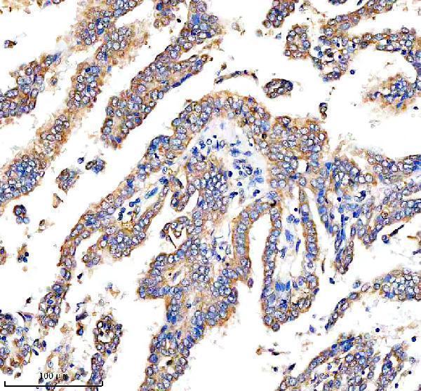
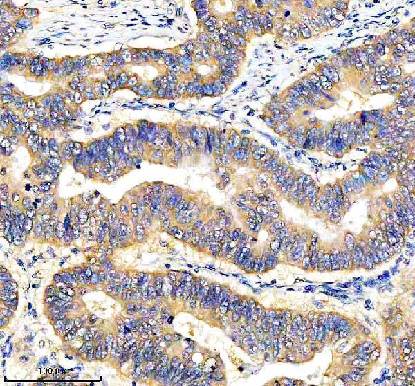
| Sample | Paraffin-embedded human lung cancer tissue; fixative not specified (datasheet A03811-2) |
| Fixation | Image fixative and duration unreported (datasheet A03811-2); verify before use. |
| Sectioning | 4–5 µm sections on charged slides (standard) |
| Deparaffinisation | Xylene, graded ethanol series to water (standard) |
| Antigen retrieval | Heat retrieval: EDTA pH 8.0 (datasheet A03811-2); 20 min, 95–100 °C (standard) |
| Peroxidase block | 3% H2O2, 10 min, room temperature (standard) |
| Blocking | 10% goat serum (datasheet A03811-2) |
| Primary antibody | Rabbit anti-DEPTOR, 2-5 μg/ml (datasheet A03811-2) |
| Primary incubation | Overnight at 4 °C (datasheet A03811-2) |
| Detection | HRP-conjugated secondary, DAB chromogen (datasheet A03811-2) |
| Counterstain | Hematoxylin, blue, dehydrate and mount (standard) |
| Expected result | DEPTOR-positive staining in cardiomyocytes of heart muscle (HPA tissue IHC: High). HPA tissue profile: Cytoplasmic expression at variable levels in most tissues. No signal in the no-primary control. |
In paraffin-section IHC, expect variable cytoplasmic DEPTOR staining across tissues, with strong signal in cardiomyocytes, skeletal myocytes, placental syncytiotrophoblast cell bodies, and salivary glandular cells (HPA: tissue IHC). DEPTOR associates with lysosomal membranes through mTOR complexes and has no transmembrane segment (UniProt Q8TB45: localization and topology). HPA rates the tissue profile Enhanced, while noting medium staining–RNA consistency and pending external verification (HPA: tissue IHC).
| Clear cytoplasmic signal in the expected cells, strongest in heart muscle cardiomyocytes or skeletal muscle myocytes (HPA: High). | This fits the reported IHC distribution. Compare cells within the same section before scoring: HPA reports variable cytoplasmic expression across most tissues, so uniform intensity is not required (HPA: tissue IHC). |
| Predominantly nuclear or sharply surface-restricted chromogenic staining, with little cytoplasmic signal. | Treat this as discordant with the HPA tissue-IHC profile and check specificity and staining controls (HPA: cytoplasmic profile). UniProt places complex-associated DEPTOR at lysosomal membranes but reports no transmembrane segment; that annotation does not require visible lysosomal puncta in routine IHC (UniProt Q8TB45: localization and topology). |
| Strong staining in a cell population listed as Not detected, such as esophageal squamous epithelium (HPA: Not detected). | Consider cross-reactivity or endogenous detection activity; inspect a no-primary control and compare another IHC-validated antibody if available (standard IHC practice). A single positive field does not overturn the HPA cell-specific observation, especially given its pending external verification (HPA: tissue IHC). |
| Diffuse color over cells and surrounding section, without clear cellular boundaries. | This is background rather than a scorable DEPTOR pattern. Check blocking, washes, primary-antibody concentration, and the detection-only control (standard IHC practice). HPA describes cellular cytoplasmic staining, not a diffuse section-wide deposit (HPA: tissue IHC). |
| No signal in heart muscle cardiomyocytes despite interpretable tissue morphology (HPA: High). | The run needs review before a negative call. Confirm the IHC-validated antibody, tissue processing and retrieval used for that assay, reagent activity, and the positive-control slide (standard IHC practice). HPA's High category supports a useful comparison tissue; it does not guarantee every specimen will stain (HPA: tissue IHC). |
| Cell type and tissue | Choose a reported High population for a positive comparison and a specified Not detected population for contrast; score the named cells, not an entire tissue as uniformly positive or negative (HPA: tissue IHC). |
| Localization and topology | The IHC readout is cytoplasmic (HPA: tissue IHC). Lysosomal-membrane association depends on mTOR-complex association, and DEPTOR lacks a transmembrane segment; neither fact predicts a crisp membrane outline in chromogenic sections (UniProt Q8TB45: localization and topology). |
| Antibody evidence | HPA lists Enhanced IHC evidence for HPA023938, HPA023945, and CAB020841, and Supported IHC evidence for HPA024011 (HPA: antibodies). The tissue profile still has medium staining–RNA consistency and awaits external verification (HPA: tissue IHC). |
| Isoforms and modified residues | UniProt lists two isoforms and multiple modified residues, including phosphorylation sites (UniProt Q8TB45: isoforms and modified residues). Their effects on a particular antibody's IHC signal cannot be assigned without its epitope and validation data; no target-specific fixation sensitivity is supplied. |
| IF/ICC Q&A: should it match the IHC compartment exactly? | No direct match is established. HPA reports mainly mitochondrial ICC-IF signal, with additional nucleoplasm and nuclear-body signal, while its tissue IHC profile is cytoplasmic (HPA: subcellular ICC-IF; HPA: tissue IHC). Interpret each assay with its own validation and controls (standard IHC/IF practice). |
| Situation | Likely cause | Next action |
|---|---|---|
| Positive comparison tissue is blank. | A failed staining run, inactive detection reagents, or an unsuitable assay setup is possible (standard IHC practice). | Check the IHC-validated antibody's instructions, retrieval and detection steps, and a known working control before interpreting DEPTOR absence (standard IHC practice; HPA: High in cardiomyocytes). |
| All cells show similar brown color. | Excess background or endogenous detection activity may obscure the expected cell-specific pattern (standard IHC practice; HPA: variable cytoplasmic expression). | Run no-primary and detection-only controls; review blocking, washes, and primary concentration for the chosen detection system (standard IHC practice). |
| A reported Not detected cell population stains strongly. | Cross-reactivity, endogenous activity, or a specimen-specific difference may explain discordance (standard IHC practice; HPA: Not detected categories). | Verify cell identity and controls, then compare staining with an independent IHC-validated antibody where feasible; record the discrepancy rather than calling it established DEPTOR expression (HPA: antibodies; standard IHC practice). |
| Signal is nuclear with little cytoplasmic staining. | This differs from the HPA tissue-IHC profile; ICC-IF nuclear annotations alone do not validate that chromogenic IHC result (HPA: tissue IHC; HPA: subcellular ICC-IF). | Review controls, tissue morphology, and antibody-specific IHC evidence; score the nuclear pattern separately until its specificity is established (standard IHC practice). |
| Staining appears weak in a low-expression comparison tissue. | HPA already reports Low staining in populations such as cerebral-cortex endothelial cells and duodenal glandular cells (HPA: tissue IHC). | Compare with a reported High population in the same validated workflow before changing retrieval or dilution; document cell type and intensity separately (HPA: tissue IHC; standard IHC practice). |
| IHC looks diffuse, but an ICC-IF image looks punctate. | The reported readouts differ: cytoplasmic tissue IHC versus mainly mitochondrial ICC-IF; morphology and assay context also affect what can be resolved (HPA: tissue IHC; HPA: subcellular ICC-IF; standard microscopy practice). | Judge the paraffin-section result against tissue-IHC controls and the correct cells. Use the ICC-IF annotation as context, not as a required punctate IHC scoring rule (HPA: tissue IHC; HPA: subcellular ICC-IF). |


Comprehensive Human Protein Atlas IHC scoring per tissue (reliability: Enhanced — Medium consistency between antibody staining and RNA expression data. Pending external verification.). Rows are taken directly from the HPA tissue chart — click any row's HPA link to view the source.
| Tissue | Cell type | Level | Evidence | Source |
|---|---|---|---|---|
| Heart muscle | Cardiomyocytes | High | Protein (IHC) | HPA → |
| Placenta | Syncytiotrophoblasts - cell body | High | Protein (IHC) | HPA → |
| Salivary gland | Glandular cells | High | Protein (IHC) | HPA → |
| Skeletal muscle | Myocytes | High | Protein (IHC) | HPA → |
| Adrenal gland | Glandular cells | Medium | Protein (IHC) | HPA → |
| Tissue | Cell type | Level | Evidence | Source |
|---|---|---|---|---|
| Caudate | Glial cells | Not detected | Protein (IHC) | HPA → |
| Esophagus | Squamous epithelial cells | Not detected | Protein (IHC) | HPA → |
| Gallbladder | Glandular cells | Not detected | Protein (IHC) | HPA → |
| Hippocampus | Glial cells | Not detected | Protein (IHC) | HPA → |
| Nasopharynx | Respiratory epithelial cells | Not detected | Protein (IHC) | HPA → |
Use the catalog antibody’s paraffin-section IHC conditions as a starting point, then judge DEPTOR staining by cell type, compartment and appropriate controls.
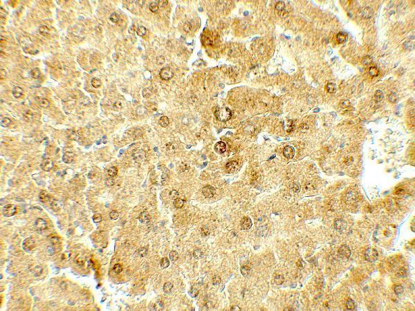
Two anti-DEPTOR antibodies have real IHC images: human paraffin sections for A03811-2 and mouse liver for A03811 (catalog image captions). Neither has IF data (catalog application lists and images).
A03811-2 shows IHC in human lung and stomach cancer paraffin sections (catalog image captions). A03811 shows IHC in mouse liver (catalog image caption).
Which to pick: Choose A03811-2 for human paraffin-section IHC; its caption reports EDTA retrieval and 2 μg/ml primary antibody, but does not report the fixative (A03811-2 image caption). Choose A03811 for mouse liver IHC at 5 μg/mL (A03811 image caption). Both list human, mouse and rat reactivity, but their pictured IHC results cover only the samples above; neither lists IF/ICC or provides an IF image, so neither is established here for IF/ICC (catalog reactivity, application lists and images).