DHODH / Dihydroorotate dehydrogenase (quinone), mitochondrial · IHC design guide

Design Immunohistochemistry for DHODH

Plan chromogenic DHODH IHC in paraffin sections using the IHC-validated antibody M04035-1 (datasheet M04035-1). Start with 2–5 µg/mL (datasheet M04035-1) and assess cytoplasmic staining in glandular epithelia (HPA tissue IHC).

Evidence assembled Oct 2026 · For research use; verify linked source records and product datasheet before use
Immunohistochemistry protocol sheet for DHODH (IHC for DHODH): expected localisation Cytoplasmic tissue staining (HPA tissue IHC); inner mitochondrial membrane expected (UniProt), antibody M04035-1, validated IHC image, and IHC protocol steps
Printable DHODH IHC protocol sheet — expected localisation Cytoplasmic tissue staining (HPA tissue IHC); inner mitochondrial membrane expected (UniProt), antibody M04035-1, controls and protocol steps. Open the full DHODH IHC guide →

DHODH Immunohistochemistry Experimental Design Guide

Expected localisation, validated protocols, controls and antibodies — the at-a-glance facts below, then the full design guide.

Must know before staining
Expected localisation Cytoplasmic tissue staining (HPA tissue IHC); inner mitochondrial membrane expected (UniProt)
Staining pattern General cytoplasmic staining, high in glandular epithelia (HPA tissue IHC)
Antigen retrieval EDTA pH 8.0 HIER, heat-mediated (datasheet M04035-1)
Positive control ⓘ Appendix+4 more · see all
Negative control ⓘ Caudate
Important caveats
Reasons your staining may differ from the expected pattern.
Fixation Matched tissue-IHC evidence does not establish the fixation claim. Validate the specimen-specific method before use. (selected-SKU IHC image M04035-1); optimize empirically.
Caveat IHC staining has low consistency with RNA expression (HPA tissue IHC)
Regulation Expression regulation unreported (UniProt)
Isoform / epitope No annotated isoforms; chain spans residues 1–395 (UniProt)
Section 1

Recommended DHODH IHC & IF Protocols

The catalog antibody’s IHC-P protocol is paired with two published DHODH staining protocols for human brain and paraffin-embedded tissue (PMC6814620; PMC11910144).

Recommended immunohistochemistry (IHC-P) protocol parameters
SampleParaffin-embedded human breast cancer tissue; fixative not specified (datasheet M04035-1)
FixationImage fixative and duration unreported (datasheet M04035-1); verify before use.
Sectioning4–5 µm sections on charged slides (standard)
DeparaffinisationXylene, graded ethanol series to water (standard)
Antigen retrievalHeat retrieval: EDTA pH 8.0 (datasheet M04035-1); 20 min, 95–100 °C (standard)
Peroxidase block3% H2O2, 10 min, room temperature (standard)
Blocking10% goat serum (datasheet M04035-1)
Primary antibodyMouse monoclonal (clone 2G7) anti-DHODH, 2 μg/ml (datasheet M04035-1)
Primary incubationOvernight at 4 °C (datasheet M04035-1)
DetectionStreptavidin-biotin complex (SABC), DAB chromogen (datasheet M04035-1)
CounterstainHematoxylin, blue, dehydrate and mount (standard)
Expected resultDHODH-positive staining in glandular cells of appendix (HPA tissue IHC: High). HPA tissue profile: General cytoplasmic expression. High levels in glandular epithelia, endocrine tissues and in lymphoid tissues. No signal in the no-primary control.
💡Decision noteStart with EDTA pH 8.0 heat retrieval for the catalog antibody (datasheet M04035-1). Published retrieval conditions differ or leave the buffer choice open (PMC6814620; PMC11910144).
Section 2

What Is the Expected DHODH Staining Pattern?

DHODH is anchored in the mitochondrial inner membrane by residues 11–30, with most of the protein facing the intermembrane space (UniProt Q02127 topology). In paraffin IHC, expect predominantly cytoplasmic staining, especially in glandular cells and kidney tubules (HPA tissue IHC). HPA rates its tissue staining Approved but reports low agreement with RNA expression and pending external verification (HPA tissue IHC).

What am I looking at on my slide?
Granular or uneven cytoplasmic signal in glandular cells or kidney tubules.This fits the expected cellular compartment and high-staining cell groups (UniProt Q02127 topology; HPA tissue IHC). Chromogenic IHC cannot resolve the inner membrane, so judge the cellular pattern rather than individual mitochondria (standard IHC practice).
Predominantly nuclear staining, with little cytoplasmic signal.Question a nuclear-dominant IHC result because mitochondria are the main location (UniProt Q02127; HPA subcellular IF). HPA also reports nucleoplasmic signal in ICC-IF, so nuclear signal alone does not prove an artefact (HPA subcellular IF). Check controls and repeat the localisation assessment (standard IHC practice).
Strong staining in caudate glial cells instead of the expected positive cells.HPA reports DHODH as undetected in caudate glial cells (HPA tissue IHC). An unexpected strong signal there warrants checks for cross-reactivity or endogenous detection activity; it does not, by itself, establish either cause (standard IHC practice).
Diffuse colour across cells and surrounding tissue, without a discernible cellular pattern.Treat this as background until controls establish a specific signal (standard IHC practice). HPA describes general cytoplasmic expression, with high levels in selected cell groups; it does not describe uniform staining of every tissue component (HPA tissue IHC).
No signal in appendix or colon glandular cells, or in kidney tubules.These are high-staining examples in HPA tissue IHC, so a blank result calls for a run-level check before biological interpretation (HPA tissue IHC; standard IHC practice). Review the positive control, antibody application and detection steps (standard IHC practice). HPA's tissue assessment remains pending external verification (HPA tissue IHC).
💡Expected DHODH appearanceCall a positive result when cytoplasmic staining is clear and relatively strong in HPA high-staining glandular cells or kidney tubules (HPA tissue IHC), consistent with mitochondrial DHODH (UniProt Q02127); widespread uniform colour or isolated nuclear-dominant staining should trigger control review (standard IHC practice; HPA subcellular IF).
How each factor affects the staining
Topology and IHC resolutionResidues 11–30 form the inner-membrane anchor; residues 31–395 face the intermembrane space (UniProt Q02127 topology). Interpret a cellular cytoplasmic pattern in chromogenic IHC, since this method cannot pinpoint the membrane face (standard IHC practice).
Tissue choice and evidence strengthAppendix, breast, colon, duodenum and other listed glandular cells, plus kidney tubules, stain High (HPA tissue IHC). HPA rates tissue IHC Approved while reporting low staining–RNA consistency and pending external verification; use its examples as comparisons, not absolute cutoffs (HPA tissue IHC).
Antibody validationHPA010123 is Approved for IHC and Enhanced for ICC; HPA011942 has Enhanced ICC evidence but no IHC status listed (HPA antibodies). ICC validation does not establish paraffin-IHC performance for an antibody without an IHC rating (standard IHC practice).
IF/ICC Q&A: Where should DHODH appear?Mainly in mitochondria, with additional nucleoplasmic and cytosolic localisation reported in MCF-7, U2OS and A-549 ICC-IF images (HPA subcellular IF). This IF observation helps assess compartment claims; it does not set an IHC staining intensity threshold (standard IHC practice).
Why is my staining missing, weak or wrong?
SituationLikely causeNext action
Expected positive glandular cells or kidney tubules are blank.A failed staining run is possible; the HPA high-staining examples make a technical check useful (HPA tissue IHC; standard IHC practice).Confirm the positive control stained, then review antibody application, retrieval and detection using the validated IHC workflow (standard IHC practice). Do not infer DHODH-specific retrieval sensitivity from HPA or UniProt.
The whole section is uniformly brown.Background or endogenous detection activity can obscure cell-level interpretation (standard IHC practice).Compare reagent-omission controls, inspect blocking and washing, and assess whether signal follows cellular boundaries (standard IHC practice). Reassess the result against HPA's cytoplasmic cell-group pattern (HPA tissue IHC).
Strong signal is confined to nuclei.The dominant compartment conflicts with mitochondrial localisation, although HPA reports additional nucleoplasmic ICC-IF signal (UniProt Q02127; HPA subcellular IF).Check controls and the presence of cytoplasmic staining before calling it specific IHC signal (standard IHC practice). Avoid treating the reported ICC-IF location as proof of a nuclear-dominant paraffin-IHC pattern.
Caudate glial cells stain strongly.That differs from HPA's Not detected glial-cell result; cross-reactivity or detection background is possible (HPA tissue IHC; standard IHC practice).Review control staining and compare with a positive tissue on the same run (standard IHC practice). Record the discrepancy rather than assigning DHODH positivity from this observation alone.
A candidate antibody gives a convincing ICC image but uncertain IHC staining.HPA lists Enhanced ICC evidence for both antibodies, but only HPA010123 has an IHC rating (HPA antibodies).Use an antibody with documented IHC validation for paraffin-section interpretation, and assess its tissue controls in the same workflow (HPA antibodies; standard IHC practice).
IHC and an RNA-based expectation disagree.HPA explicitly reports low consistency between antibody staining and RNA expression data (HPA tissue IHC).Score the observed cellular staining and document controls; keep the discordance visible in the interpretation (standard IHC practice). Do not use the liver-enriched RNA label to invent a liver IHC intensity (HPA tissue IHC).

Sample controls for DHODH IHC & IF

🧪Run breast first and score glandular cells for DHODH staining (High; HPA: breast glandular cells). Run caudate as the negative tissue and score glial cells (Not detected; HPA: caudate glial cells); on the breast slide, use adjacent nonglandular cells to assess background without assuming they are DHODH-negative (HPA: breast glandular cells).
Positive control tissue: Appendix (Glandular cells, HPA High)
Negative control tissue: Caudate (HPA Not detected)
ICC-IF cell lines (HPA subcellular resource): HPA ICC-IF images show DHODH in MCF-7, U2OS, A-549, with annotated localisation: Mitochondria (enhanced) (HPA subcellular).
Technical controls: Include a no-primary, secondary-only control; a concentration-matched mouse IgG isotype control for the captioned mouse primary; and DHODH knockout material as a biological negative (M04035-1 caption: mouse primary; standard IHC practice). For the captioned SABC/DAB detection in breast tissue, block endogenous peroxidase and assess endogenous biotin background (M04035-1 caption: SABC/DAB; standard IHC practice).
⚠️Feasibility: A target-specific fixation window or fixation effect is unreported, and the selected M04035-1 paraffin-section caption does not report a fixative (M04035-1 caption). Heat retrieval in EDTA at pH 8.0 is a documented starting condition, but retrieval dependence has not been established (M04035-1 caption). The supplied evidence does not establish that frozen sections or IF are easier than paraffin IHC; with SABC/DAB detection, endogenous biotin background is an artefact to assess in the breast section (M04035-1 caption; standard IHC practice).

HPA tissue IHC evidence for DHODH

Comprehensive Human Protein Atlas IHC scoring per tissue (reliability: Approved — Low consistency between antibody staining and RNA expression data. Pending external verification.). Rows are taken directly from the HPA tissue chart — click any row's HPA link to view the source.

Positive expression · recommended positive controls

TissueCell typeLevelEvidenceSource
Appendix Glandular cells High Protein (IHC) HPA →
Breast Glandular cells High Protein (IHC) HPA →
Cerebellum Purkinje cells High Protein (IHC) HPA →
Colon Glandular cells High Protein (IHC) HPA →
Duodenum Glandular cells High Protein (IHC) HPA →

Undetected expression · recommended negative controls

TissueCell typeLevelEvidenceSource
Caudate Glial cells Not detected Protein (IHC) HPA →
Section 3

Advanced DHODH IHC Tips

Troubleshoot DHODH staining in paraffin sections by checking retrieval, tissue controls, subcellular pattern, and detection chemistry together (datasheet M04035-1; UniProt Q02127; standard IHC practice).

What retrieval should I use when DHODH staining is weak in paraffin sections?
Start with heat-mediated EDTA retrieval at pH 8.0 for paraffin-section DHODH IHC (datasheet M04035-1). The selected breast cancer staining example used that retrieval before incubation with 2 μg/ml primary antibody overnight at 4°C (datasheet M04035-1). If staining is weak, check that sections experienced uniform heating and that a tissue control stains before changing the buffer (standard IHC practice). If those checks pass, a citrate buffer at pH 6.0 can be tested as a fallback on adjacent sections, with morphology and background assessed alongside signal (standard IHC practice). Keep detection conditions matched so the retrieval comparison is interpretable (standard IHC practice).
Could fixation explain variable DHODH staining across my paraffin sections?
Target-specific DHODH sensitivity to fixation is unknown from the supplied evidence (datasheet M04035-1). The selected image documents a paraffin-embedded breast cancer section but does not report its fixative, so it cannot establish how DHODH responds to formalin or another fixative (datasheet M04035-1). Record each specimen’s fixative and processing history, then compare sections processed together using the same EDTA pH 8.0 retrieval and detection conditions (datasheet M04035-1; standard IHC practice). If staining differs, inspect morphology and a concurrent control before attributing the difference to fixation (standard IHC practice). Avoid drawing a fixation conclusion from tissue expression patterns alone (standard IHC practice).
Should DHODH appear diffuse or punctate in chromogenic tissue staining?
Expect predominantly cytoplasmic staining at light-microscope resolution, potentially with a granular distribution consistent with mitochondria (HPA tissue IHC: general cytoplasmic expression; UniProt Q02127: mitochondrial inner membrane). DHODH has a membrane-spanning segment at residues 11–30, while residues 31–395 face the mitochondrial intermembrane space (UniProt Q02127 topology). DAB deposits do not resolve individual mitochondrial membranes, so score the cellular pattern without claiming membrane-level localisation from chromogenic IHC alone (standard IHC practice). HPA also reports nucleoplasmic and cytosolic signals in subcellular imaging; an isolated nuclear DAB pattern therefore needs corroboration rather than automatic acceptance (HPA subcellular; standard IHC practice). Compare matched sections for morphology and reproducibility (standard IHC practice).
Can topology or isoforms explain inconsistent DHODH antibody staining?
The supplied UniProt record lists 0 isoforms, so an isoform switch is unsupported as an explanation for discrepant IHC patterns (UniProt Q02127). It places the transmembrane segment at residues 11–30 and the larger 31–395 region on the intermembrane-space side (UniProt Q02127 topology). The antibody’s epitope is unspecified here, so topology alone cannot establish whether retrieval exposes its binding site (datasheet M04035-1; UniProt Q02127 topology). Compare retrieval conditions on adjacent sections while holding antibody concentration and DAB development constant (standard IHC practice). If two antibodies with documented, distinct epitopes are available, agreement in cellular staining can strengthen an interpretation (standard IHC practice).
How can IF help assess a questionable DHODH IHC pattern?
Use IF as a separate localisation check, with a mitochondrial marker and, in glandular tissue, an epithelial marker such as pan-cytokeratin in distinct channels (UniProt Q02127: mitochondrial inner membrane; HPA tissue IHC: high glandular-cell staining; standard IF practice). Choose fluorophores whose emission can be distinguished from tissue autofluorescence, and include single-stain and no-primary controls when evaluating overlap (standard IF practice). Because residues 31–395 face the intermembrane space, test permeabilisation conditions against marker preservation if the antibody binds that region; its epitope is unspecified here (UniProt Q02127 topology; datasheet M04035-1). HPA reports mitochondrial staining in ICC/IF, with additional nucleoplasmic and cytosolic localisation (HPA subcellular). Interpret IF overlap as supporting evidence for an IHC pattern, while accounting for the different preparations (standard IF/IHC practice).
How do I troubleshoot widespread brown signal in DHODH IHC?
Check no-primary and secondary-only sections to separate primary-dependent signal from detection-system background (standard IHC practice). The selected method used a biotinylated secondary, streptavidin–biotin detection, and DAB; assess endogenous biotin and peroxidase contributions with appropriate controls when using that workflow (datasheet M04035-1; standard IHC practice). Its tissue block was 10% goat serum, and the primary was 2 μg/ml overnight at 4°C (datasheet M04035-1). If background persists, compare blocking, washing, and primary concentration on adjacent sections while keeping DAB development matched (standard IHC practice). Review whether signal follows intact cellular cytoplasm or instead pools in damaged regions and tissue edges (standard IHC practice).
What should I score when comparing DHODH staining between tissue groups? ⚠ ANSWER MARKED FOR VERIFICATION
Define the cell population before scoring, because DHODH IHC varies by tissue and cell type (HPA tissue IHC). For glandular samples, an H-score can combine staining intensity and percentage of positive glandular cells; alternatively report percentage positive and intensity separately (HPA tissue IHC: high glandular-cell staining; standard IHC practice). Normalise cell counts to the number of eligible cells, or express positive-cell density per mm² of viable tissue when comparing areas (standard IHC practice). Keep retrieval, antibody concentration, development, and image acquisition comparable across groups, and exclude folds, necrosis, and edge artefacts by a prespecified rule (standard IHC practice). Report the scoring compartment and controls alongside the summary statistic (standard IHC practice).
When is a DHODH-positive DAB pattern convincing rather than artefactual?
A convincing pattern is reproducible in intact cells and broadly compatible with cytoplasmic mitochondrial distribution (UniProt Q02127: mitochondrial inner membrane; HPA tissue IHC: general cytoplasmic expression; standard IHC practice). High staining in glandular cells can provide context, but HPA marks its tissue IHC profile as Approved with low consistency against RNA data and pending external verification (HPA tissue IHC). Treat isolated nuclear staining, tissue-edge accentuation, necrotic deposits, or signal retained in no-primary controls as reasons to investigate before assigning DHODH positivity (HPA subcellular; standard IHC practice). With DAB detection, exclude endogenous peroxidase and, for the selected streptavidin–biotin method, assess endogenous biotin background (datasheet M04035-1; standard IHC practice). Interpret differences using matched controls and morphology, rather than staining intensity alone (standard IHC practice).
Boster reagents

Best DHODH / Dihydroorotate dehydrogenase (quinone), mitochondrial IHC Antibodies

Anti-DHODH antibodies have IHC examples in human and mouse paraffin sections and an IF/ICC example in human MCF-7 cells (catalog image captions: M04035-1, PB10056, M04035).

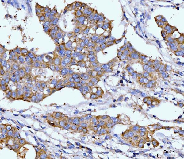
Real IHC data IHC analysis of DHODH using anti-DHODH antibody (M04035-1). DHODH was detected in a paraffin-embedded section of human breast cancer tissue. Heat mediated antigen retrieval was performed in EDTA buffer (pH 8.0, epitope retrieval solution). The tissue section was blocked with 10% goat serum. The tissue section was then incubated with 2 μg/ml mouse anti-DHODH Antibody (M04035-1) overnight at 4°C. Biotinylated goat anti-mouse IgG was used as secondary antibody and incubated for 30 minutes at 37°C. The tissue section was developed using Strepavidin-Biotin-Complex (SABC) (Catalog # SA1021) with DAB as the chromogen.
Anti-DHODH Antibody ® (monoclonal, 2G7)
Cat # M04035-1
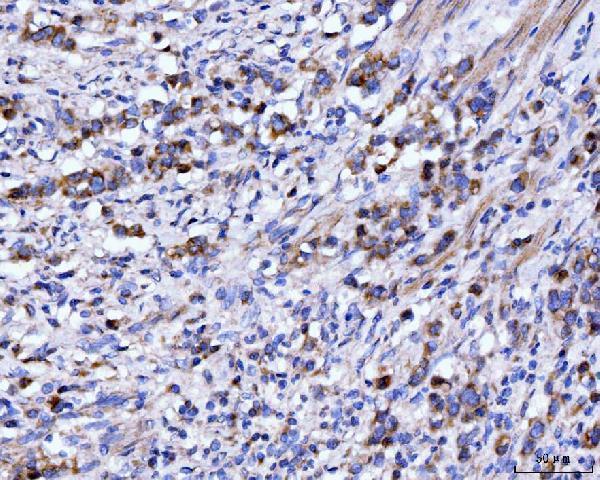
Real IHC data IHC analysis of DHODH using anti-DHODH antibody (PB10056). DHODH was detected in a paraffin-embedded section of mouse intestine tissue. Heat mediated antigen retrieval was performed in EDTA buffer (pH 8.0, epitope retrieval solution). The tissue section was blocked with 10% goat serum. The tissue section was then incubated with 1 μg/ml rabbit anti-DHODH Antibody (PB10056) overnight at 4°C. Biotinylated goat anti-rabbit IgG was used as secondary antibody and incubated for 30 minutes at 37°C. The tissue section was developed using Strepavidin-Biotin-Complex (SABC) (Catalog # SA1022) with DAB as the chromogen.
Anti-DHODH Antibody ®
Cat # PB10056
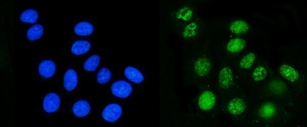
Real IF data IF analysis of DHODH using anti-DHODH antibody (M04035). DHODH was detected in an immunocytochemical section of MCF-7 cells. Enzyme antigen retrieval was performed using IHC enzyme antigen retrieval reagent (AR0022) for 15 mins. The cells were blocked with 10% goat serum. And then incubated with 5 μg/mL mouse anti-DHODH Antibody (M04035) overnight at 4°C. DyLight®488 Conjugated Goat Anti-Mouse IgG (BA1126) was used as secondary antibody at 1:100 dilution and incubated for 30 minutes at 37°C. The section was counterstained with DAPI. Visualize using a fluorescence microscope and filter sets appropriate for the label used.
Anti-DHODH Antibody ® (monoclonal, 4E3)
Cat # M04035

M04035-1 was shown on human breast cancer paraffin sections; PB10056 was shown on mouse intestine paraffin sections (catalog IHC image captions: M04035-1, PB10056). M04035 was shown by IF/ICC in MCF-7 cells (catalog IF image caption: M04035).

Which to pick: For human paraffin-section IHC, choose mouse monoclonal M04035-1, shown at 2 μg/ml on human breast cancer tissue (catalog applications and IHC image caption: M04035-1). For IHC across human, mouse, and rat samples, choose rabbit PB10056: its IHC application lists all three species, and its captions show human lung cancer, mouse intestine, and rat intestine paraffin sections (catalog application, reactivity, and IHC image captions: PB10056). For IF/ICC, choose mouse monoclonal M04035, shown at 5 μg/mL in MCF-7 cells; the fixative is unreported in these image captions (catalog applications and IF image caption: M04035; catalog IHC image captions: M04035-1, PB10056).

Each figure is that product's own IHC / IF validation image from its datasheet.

References

  1. UniProt Consortium. UniProt entry Q02127 (PYRD_HUMAN, Dihydroorotate dehydrogenase (quinone), mitochondrial).
  2. Human Protein Atlas. DHODH tissue IHC expression (reliability: Approved).
  3. Human Protein Atlas. DHODH subcellular location (ICC-IF): Mainly localized to the mitochondria. In addition localized to the nucleoplasm and cytosol..
  4. Human Protein Atlas. DHODH antibody validation summary (2 antibodies).
  5. Metabolomics profiles delineate uridine deficiency contributes to mitochondria-mediated apoptosis induced by celastrol in human acute promyelocytic leukemia cells. Oncotarget 2016 — PMC5216817.
  6. Brain pyrimidine nucleotide synthesis and Alzheimer disease. Aging 2019 — PMC6814620.
  7. Designer Exosomes for Targeted Delivery of a Novel Therapeutic Cargo to Enhance Sorafenib-Mediated Ferroptosis in Hepatocellular Carcinoma. Frontiers in oncology 2022 — PMC9263838.
  8. Targeting YBX1-m5C mediates RNF115 mRNA circularisation and translation to enhance vulnerability of ferroptosis in hepatocellular carcinoma. Clinical and translational medicine 2025 — PMC11910144.
  9. PubMed PMID:1446837 — UniProt-cited evidence.
  10. PubMed PMID:7487077 — UniProt-cited evidence.
  11. PubMed PMID:14702039 — UniProt-cited evidence.