This website uses cookies to ensure you get the best experience on our website.


- Table of Contents
Plan paraffin-section DMTN IHC around cytoplasmic staining in several tissues and red blood cell positivity (HPA tissue IHC). Start the catalog antibody at 1:100–1:300 (datasheet: A07422), and interpret results cautiously because antibody staining and RNA show low consistency (HPA tissue IHC).

Expected localisation, validated protocols, controls and antibodies — the at-a-glance facts below, then the full design guide.
| Expected localisation | Cytoplasmic staining in tissue (HPA tissue IHC) | |
| Staining pattern | Cytoplasmic signal in several tissues; red blood cells positive (HPA tissue IHC) | |
| Antigen retrieval | Citrate pH 6.0 HIER, 95–98 °C, 20 min (rule: cytoplasmic / membrane antigen) | |
| Positive control | Adipose tissue+4 more · see all | |
| Negative control | Appendix+4 more · see all |
| Fixation | Keep paraffin-section fixation consistent (standard IHC practice; not target-specific) | |
| Caveat | Antibody staining and RNA show low consistency (HPA tissue IHC) | |
| Regulation | Brain-enhanced RNA expression (HPA tissue RNA) | |
| Isoform / epitope | Four isoforms; isoform-specific staining is unresolved (UniProt) |
Compare the catalog antibody’s IHC-P protocol with one published DMTN IHC protocol (PMC6277997).
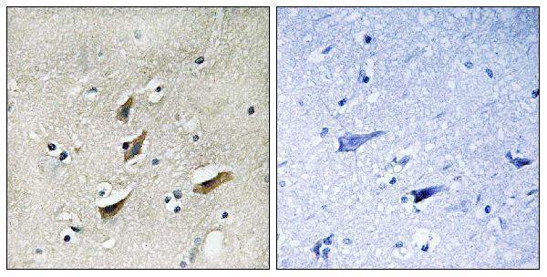
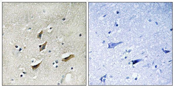
| Sample | Paraffin-embedded human brain tissue; fixative not specified (datasheet A07422) |
| Fixation | Image fixative and duration unreported (datasheet A07422); verify before use. |
| Sectioning | 4–5 µm sections on charged slides (standard) |
| Deparaffinisation | Xylene, graded ethanol series to water (standard) |
| Antigen retrieval | Heat-induced epitope retrieval in citrate buffer, pH 6.0, 20 min at 95–98 °C (standard rule: cytoplasmic / membrane antigen) |
| Peroxidase block | 3% H2O2, 10 min, room temperature (standard) |
| Blocking | 10% normal serum of the secondary host, 30 min, room temperature (standard) |
| Primary antibody | Rabbit anti-DMTN, 1:100-1:300 (datasheet A07422) |
| Primary incubation | Overnight at 4 °C (standard) |
| Detection | HRP-polymer secondary, DAB chromogen 5–10 min (standard) |
| Counterstain | Hematoxylin, blue, dehydrate and mount (standard) |
| Expected result | DMTN-positive staining in adipocytes of adipose tissue (HPA tissue IHC: Medium). HPA tissue profile: Cytoplasmic expression in several tissue. Additional positivity in red blood cells. No signal in the no-primary control. |
DMTN is a cytoplasmic and cytoskeletal protein that also associates with membranes, including the erythrocyte spectrin–actin junction; it has no transmembrane segment (UniProt Q08495 topology/localization). In tissue IHC, expect cytoplasmic staining in selected cells and possible red blood cell positivity (HPA: tissue IHC profile). Treat the pattern as provisional: HPA rates tissue staining Uncertain because antibody staining and RNA expression show low consistency (HPA: reliability).
| Cytoplasmic staining in bronchial respiratory epithelium, with red blood cell positivity. | This fits the reported tissue profile: bronchial respiratory epithelial cells show Medium staining, and HPA notes additional red blood cell positivity (HPA: bronchus; tissue IHC profile). Assess epithelial cytoplasm separately from blood-filled spaces; erythrocyte-associated DMTN is biologically plausible (UniProt Q08495 localization). |
| Signal follows cell borders or processes as well as cytoplasm, without a dominant nuclear pattern. | A membrane-associated component can fit DMTN’s cytoskeletal and erythrocyte membrane localization, although HPA describes the tissue pattern broadly as cytoplasmic (UniProt Q08495 localization; HPA: tissue IHC profile). A predominantly nuclear result lacks support from either supplied localization record and warrants comparison with controls (UniProt Q08495 localization; HPA: tissue IHC profile). |
| Strong staining appears in appendix glandular cells while the expected comparison tissue stains weakly or not at all. | HPA reports appendix glandular cells as Not detected; this contrast raises concern for cross-reactivity or detection-system activity (HPA: appendix; general IHC practice). It is a warning, not proof of antibody failure, because the HPA tissue IHC profile itself is rated Uncertain (HPA: reliability). |
| Weak, diffuse chromogen covers stroma, lumina and many unrelated cells without clear boundaries. | A widespread haze does not resemble the reported cytoplasmic, cell-associated pattern (HPA: tissue IHC profile). Compare the no-primary control and examine blocking, washes and detection conditions before assigning that signal to DMTN (general IHC practice). |
| No convincing signal appears in bronchial respiratory epithelial cells or kidney glomerular cells. | Both are reported at Medium staining and can serve as comparison areas (HPA: bronchus; kidney). Check tissue integrity, the positive control and the detection run before interpreting absence as biological; the HPA result is Uncertain and does not guarantee positivity in every specimen (HPA: reliability; general IHC practice). |
| Cell type and compartment | Compare like cells: HPA reports Medium staining in bronchial respiratory epithelial cells and kidney glomerular cells, but Not detected in appendix glandular cells (HPA: bronchus; kidney; appendix). UniProt places DMTN in cytosol, cytoskeleton and membrane-associated sites, with no transmembrane segment (UniProt Q08495 topology/localization). |
| Confidence in the tissue pattern | HPA rates tissue IHC Uncertain owing to low consistency between antibody staining and RNA expression; both listed rabbit polyclonal antibodies have Uncertain IHC validation (HPA: reliability; antibodies HPA024290/HPA058487). Use morphology and controls when interpreting a deviation (general IHC practice). |
| Isoforms and epitope interpretation | UniProt lists 4 DMTN isoforms and an HP domain at residues 337–405 (UniProt Q08495 isoforms/domains). The supplied sources do not map the antibody epitope, so staining cannot establish which isoform was detected or whether the domain is represented. |
| Modification and processing | UniProt lists 19 modified residues, including phosphoserines, and a chain spanning residues 1–405, with no signal peptide or propeptide (UniProt Q08495 modifications/processing). These facts alone do not predict staining strength, antigen retrieval needs or epitope accessibility. |
| Chromogenic detection in blood-rich areas | Red blood cells may contain target-associated signal (HPA: tissue IHC profile; UniProt Q08495 localization). In peroxidase-based IHC, endogenous activity can also produce chromogen, so judge blood-rich areas against an appropriate no-primary control (general IHC practice). |
| Situation | Likely cause | Next action |
|---|---|---|
| Bronchial respiratory epithelium is negative. | The run may have a staining or detection problem, or this specimen may differ from the reported Medium pattern (HPA: bronchus; reliability; general IHC practice). | Review morphology and a concurrent positive control, then check the antibody and detection steps before calling the sample negative (general IHC practice). |
| Appendix glandular cells stain more strongly than the expected positive comparison cells. | That reverses HPA’s Not detected appendix glandular pattern and may reflect cross-reactivity or background (HPA: appendix; general IHC practice). | Compare the same cell types across sections, inspect a no-primary control and avoid treating this contrast alone as proof of DMTN expression (general IHC practice; HPA: reliability). |
| Red blood cell-rich spaces dominate the slide. | HPA reports red blood cell positivity, and erythrocyte membrane association is described by UniProt; peroxidase-based detection can add endogenous chromogen (HPA: tissue IHC profile; UniProt Q08495 localization; general IHC practice). | Score tissue cells separately from blood and compare with a no-primary control; assess endogenous peroxidase blocking if the control also develops chromogen (general IHC practice). |
| Most nuclei stain while cytoplasm is faint. | Predominantly nuclear staining is unsupported by the supplied DMTN localization and tissue IHC profile (UniProt Q08495 localization; HPA: tissue IHC profile). | Check the no-primary control, counterstain and detection conditions; require a convincing cell-associated cytoplasmic pattern before attributing nuclear color to DMTN (general IHC practice). |
| Diffuse background obscures cell boundaries. | Non-specific binding or insufficient washing can create a broad chromogenic haze (general IHC practice). | Compare the no-primary control and review blocking, washes and antibody concentration; reassess only interpretable, cell-associated signal (general IHC practice). |
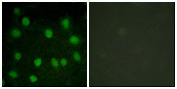
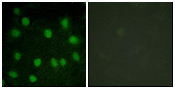
| Can IF/ICC localization be inferred from these images? | HPA provides no main ICC-IF location and lists 0 cell lines with ICC-IF images (HPA: subcellular record). | Use UniProt’s cytoplasmic, cytoskeletal and membrane-associated localization only as a hypothesis; confirm any IF/ICC pattern with controls in the separate IF/ICC design (UniProt Q08495 localization; HPA: subcellular record; general IF practice). |


Comprehensive Human Protein Atlas IHC scoring per tissue (reliability: Uncertain — Low consistency between antibody staining and RNA expression data.). Rows are taken directly from the HPA tissue chart — click any row's HPA link to view the source.
| Tissue | Cell type | Level | Evidence | Source |
|---|---|---|---|---|
| Adipose tissue | Adipocytes | Medium | Protein (IHC) | HPA → |
| Bone marrow | Hematopoietic cells | Medium | Protein (IHC) | HPA → |
| Bronchus | Respiratory epithelial cells | Medium | Protein (IHC) | HPA → |
| Cerebral cortex | Neuropil | Medium | Protein (IHC) | HPA → |
| Colon | Endothelial cells | Medium | Protein (IHC) | HPA → |
| Tissue | Cell type | Level | Evidence | Source |
|---|---|---|---|---|
| Appendix | Glandular cells | Not detected | Protein (IHC) | HPA → |
| Duodenum | Glandular cells | Not detected | Protein (IHC) | HPA → |
| Endometrium | Cells in endometrial stroma | Not detected | Protein (IHC) | HPA → |
| Fallopian tube | Glandular cells | Not detected | Protein (IHC) | HPA → |
| Heart muscle | Cardiomyocytes | Not detected | Protein (IHC) | HPA → |
Troubleshoot DMTN staining in paraffin sections by checking retrieval, compartment, cell identity, controls, and scoring before interpreting intensity.
Anti-DMTN antibodies have paraffin-section IHC images from human brain and IF images from HUVEC cells (catalog image captions); both list human and mouse reactivity (catalog applications/reactivity).
A07422 lists IHC and IF, with human brain paraffin-section IHC and HUVEC IF images (A07422 applications and image captions). A30444 lists IHC, IF and ICC, with human brain paraffin-section IHC and HUVEC IF images (A30444 applications and image captions).
Which to pick: For tissue IHC, either SKU has its own human brain paraffin-section image (A07422 and A30444 IHC image captions); the fixative is unreported in both captions. For IF, either lists IF and has its own HUVEC image; choose A30444 if ICC is required because ICC appears in its application list (A07422 and A30444 applications and IF image captions). For human or mouse samples, both are listed as rabbit polyclonal antibodies reactive with those species (A07422 and A30444 catalog payloads).