DOCK1 / Dedicator of cytokinesis protein 1 · IHC design guide

Design Immunohistochemistry for DOCK1

Plan DOCK1 chromogenic IHC on paraffin sections with 2–5 μg/ml primary antibody (datasheet A03440-2). Compare cytoplasmic staining in breast glandular cells, where signal is high, with adrenal glandular cells, where it is undetected (HPA tissue IHC).

Evidence assembled Oct 2026 · For research use; verify linked source records and product datasheet before use
Immunohistochemistry protocol sheet for DOCK1 (IHC for DOCK1): expected localisation Cytoplasmic staining in several cell types (HPA tissue IHC), antibody A03440-2, validated IHC image, and IHC protocol steps
Printable DOCK1 IHC protocol sheet — expected localisation Cytoplasmic staining in several cell types (HPA tissue IHC), antibody A03440-2, controls and protocol steps. Open the full DOCK1 IHC guide →

DOCK1 Immunohistochemistry Experimental Design Guide

Expected localisation, validated protocols, controls and antibodies — the at-a-glance facts below, then the full design guide.

Must know before staining
Expected localisation Cytoplasmic staining in several cell types (HPA tissue IHC)
Staining pattern Cytoplasmic signal across several cell types (HPA tissue IHC)
Antigen retrieval EDTA pH 8.0 HIER, heat-mediated (datasheet A03440-2)
Positive control ⓘ Breast+4 more · see all
Negative control ⓘ Adrenal gland+4 more · see all
Important caveats
Reasons your staining may differ from the expected pattern.
Fixation Keep fixation consistent across sections (standard IHC practice; not target-specific)
Caveat Signal varies by cell type; some are undetected (HPA tissue IHC)
Regulation No specific regulator reported (UniProt)
Isoform / epitope No listed isoforms; one 1–1865 chain (UniProt)
Section 1

Recommended DOCK1 IHC & IF Protocols

The catalog antibody’s EDTA retrieval protocol (datasheet A03440-2) and a published endometrial tissue IHC protocol (PMC10913561) provide starting conditions for chromogenic DOCK1 staining.

Recommended immunohistochemistry (IHC-P) protocol parameters
SampleParaffin-embedded human colorectal adenocarcinoma tissue; fixative not specified (datasheet A03440-2)
FixationImage fixative and duration unreported (datasheet A03440-2); verify before use.
Sectioning4–5 µm sections on charged slides (standard)
DeparaffinisationXylene, graded ethanol series to water (standard)
Antigen retrievalHeat retrieval: EDTA pH 8.0 (datasheet A03440-2); 20 min, 95–100 °C (standard)
Peroxidase block3% H2O2, 10 min, room temperature (standard)
Blocking10% goat serum (datasheet A03440-2)
Primary antibodyRabbit anti-DOCK1, 2-5 μg/ml (datasheet A03440-2)
Primary incubationOvernight at 4 °C (datasheet A03440-2)
DetectionHRP-conjugated secondary, DAB chromogen (datasheet A03440-2)
CounterstainHematoxylin, blue, dehydrate and mount (standard)
Expected resultDOCK1-positive staining in glandular cells of breast (HPA tissue IHC: High). HPA tissue profile: Cytoplasmic expression in several cell types. No signal in the no-primary control.
💡Decision noteStart with heat-mediated EDTA retrieval at pH 8.0 (datasheet A03440-2); microwave citrate retrieval is the published alternative (PMC10913561).
Section 2

What Is the Expected DOCK1 Staining Pattern?

In paraffin-section IHC, expect DOCK1 chiefly in the cytoplasm of several cell types, including breast and thyroid glandular cells and tonsillar squamous epithelial cells (HPA tissue IHC: Approved; pending external verification). UniProt places DOCK1 in the cytoplasm and at membranes, with recruitment linked to phosphatidylinositol 3,4,5-trisphosphate; it has no transmembrane segment (UniProt Q14185: subcellular location and topology).

What am I looking at on my slide?
Cytoplasmic staining in breast or thyroid glandular cells, or tonsillar squamous epithelial cells.This matches cell types scored High by HPA tissue IHC (HPA: High in those cells). Compare stained cells with the section's morphology; tissue-wide colour alone cannot establish the expected cell pattern (general IHC interpretation).
Predominantly nuclear staining in an IHC section, with little cytoplasmic signal.Check for artefact or nonspecific staining against the cytoplasmic tissue IHC profile (HPA tissue IHC). Nuclear signal is not inherently impossible: HPA reports approved nucleoplasmic localisation in ICC-IF, a different application (HPA subcellular ICC-IF).
Strong staining in a cell population reported as not detected, such as lymph-node germinal center cells.This conflicts with the reported IHC pattern for that cell population (HPA: Not detected in lymph-node germinal center cells). Consider cross-reactivity or endogenous chromogen-generating activity; neither can be assigned from the slide alone (general IHC practice).
Diffuse colour across cells and extracellular spaces, obscuring boundaries.Treat the pattern as background until controls separate specific cellular staining from nonspecific antibody binding or endogenous detection activity (general IHC practice). HPA describes cytoplasmic expression in several cell types, rather than a diffuse tissue-level readout (HPA tissue IHC).
No staining in a breast, thyroid or tonsil section with suitable target cells.A missing signal warrants a technical check because HPA scores the specified cell populations High (HPA tissue IHC). HPA's IHC status is Approved with external verification pending, so a negative result alone does not establish absence of DOCK1 (HPA: reliability).
💡Expected DOCK1 appearanceCall an IHC section positive when identifiable breast or thyroid glandular cells, or tonsillar squamous epithelial cells, show clear cytoplasmic staining consistent with HPA's High scores (HPA tissue IHC); isolated nuclear colour or diffuse colour outside cells calls for control-based review, although nucleoplasmic DOCK1 is reported in ICC-IF (HPA subcellular ICC-IF; general IHC practice).
How each factor affects the staining
Compartment and membrane recruitmentThe tissue IHC profile is cytoplasmic (HPA tissue IHC). UniProt also lists membrane localisation through phosphatidylinositol 3,4,5-trisphosphate interaction and no transmembrane segment (UniProt Q14185). Assess any edge staining in the context of a cellular pattern; membrane staining is not a required positive-call criterion from these IHC data.
Cell-specific tissue distributionHPA scores bronchial respiratory epithelial cells, caudate glial cells and glandular cells in cervix, duodenum and endometrium Medium, while several other populations are Low or Not detected (HPA tissue IHC). Select and score the named cell population, since a tissue label alone does not predict uniform staining.
Antibody validation scopeHPA lists IHC as Approved for HPA048692 and CAB004378; neither entry is labelled Enhanced in the supplied record (HPA antibodies). The tissue profile's reliability is Approved with external verification pending (HPA tissue IHC). Treat a conflicting slide as a result to investigate, rather than as definitive proof that either profile is wrong.
IF/ICC Q: Should its localisation match paraffin IHC?A: HPA reports approved nucleoplasm and cytosol as main ICC-IF locations, plus nucleoli, plasma membrane, actin filaments and focal adhesions as additional locations (HPA subcellular ICC-IF). These observations inform interpretation; the supplied IHC tissue profile remains cytoplasmic (HPA tissue IHC).
Processing and epitope scopeUniProt lists one chain spanning residues 1–1865, no signal peptide or propeptide, and no annotated isoforms (UniProt Q14185). These facts do not identify the epitope of a chosen antibody or establish a DOCK1-specific antigen-retrieval or fixation response.
Why is my staining missing, weak or wrong?
SituationLikely causeNext action
A High-scored target cell population is unstained.Possible failure in staining or detection; the absence conflicts with HPA's breast, thyroid or tonsil scores (HPA tissue IHC; general IHC practice).Check that the expected cells are present, then review the antibody's IHC validation, retrieval, primary incubation and detection controls (HPA antibodies; general IHC practice). Do not infer DOCK1-specific fixation sensitivity from these sources.
Nuclei dominate the IHC signal.This departs from HPA's cytoplasmic tissue IHC profile, although nucleoplasm is an approved ICC-IF location (HPA tissue IHC; HPA subcellular ICC-IF).Review morphology and a primary-omission control for detection background; assess whether cytoplasmic staining remains in the named positive cells (general IHC practice; HPA tissue IHC).
A reported Not detected cell population stains strongly.The result differs from its HPA IHC score; antibody cross-reactivity or endogenous detection activity are possible technical causes (HPA tissue IHC; general IHC practice).Confirm cell identity on the counterstained section and compare a primary-omission control; interpret a persistent primary-dependent signal cautiously (general IHC practice).
Colour spreads beyond cell outlines.Diffuse chromogen can reflect nonspecific binding or endogenous detection activity rather than the reported cellular pattern (general IHC practice; HPA tissue IHC).Compare a primary-omission control, review blocking and washing, and score only signal assignable to identifiable cells (general IHC practice).
Only faint staining appears in an HPA Medium or Low population.A weaker signal may accord with that population's reported level; HPA scores vary by cell type (HPA tissue IHC).Compare the correct cell population with a High-scored control section processed in the same run before calling the faint result a failed stain (HPA tissue IHC; general IHC practice).
An IHC result is being compared with an ICC-IF image.HPA reports a cytoplasmic tissue IHC profile and approved nucleoplasmic and cytosolic main ICC-IF locations (HPA tissue IHC; HPA subcellular ICC-IF).Interpret each result within its application and cell context; use the tissue IHC cell scores for the paraffin-section positive call (HPA tissue IHC; general IHC interpretation).

Sample controls for DOCK1 IHC & IF

🧪Run breast first and require staining in glandular cells (HPA: High in breast glandular cells). Use adrenal gland glandular cells as the negative tissue (HPA: Not detected in adrenal gland glandular cells); on the breast slide, assess morphologically distinct, unstained cells as internal background without assuming a specific cell type is negative (standard IHC practice).
Positive control tissue: Breast (Glandular cells, HPA High)
Negative control tissue: Adrenal gland (HPA Not detected)
ICC-IF cell lines (HPA subcellular resource): HPA ICC-IF images show DOCK1 in AF22, SuSa, U2OS, RPTEC/TERT1, hTERT-RPE1 (serum starved), with annotated localisation: Nucleoplasm (approved), Cytosol (approved) (HPA subcellular).
Technical controls: Include a no-primary, secondary-only control; a concentration-matched rabbit IgG isotype control matched to the primary antibody’s clonality where known; and DOCK1-knockout material as a biological negative (selected IHC caption: rabbit primary; standard IHC practice). Quench endogenous peroxidase and inspect the breast section for residual background before HRP/DAB scoring (selected IHC caption: HRP/DAB; standard IHC practice).
⚠️Feasibility: A target-specific fixation window or fixation effect is unreported, and the selected A03440-2 paraffin-section caption does not state the fixative (selected IHC caption). The caption uses heat-mediated EDTA retrieval at pH 8.0, but does not establish that retrieval is required; whether frozen sections or IF are easier is unreported (selected IHC caption). In breast sections, endogenous peroxidase can confound DAB scoring if it is not quenched (standard IHC practice).

HPA tissue IHC evidence for DOCK1

Comprehensive Human Protein Atlas IHC scoring per tissue (reliability: Approved — Pending external verification.). Rows are taken directly from the HPA tissue chart — click any row's HPA link to view the source.

Positive expression · recommended positive controls

TissueCell typeLevelEvidenceSource
Breast Glandular cells High Protein (IHC) HPA →
Thyroid gland Glandular cells High Protein (IHC) HPA →
Tonsil Squamous epithelial cells High Protein (IHC) HPA →
Bronchus Respiratory epithelial cells Medium Protein (IHC) HPA →
Caudate Glial cells Medium Protein (IHC) HPA →

Undetected expression · recommended negative controls

TissueCell typeLevelEvidenceSource
Adrenal gland Glandular cells Not detected Protein (IHC) HPA →
Gallbladder Glandular cells Not detected Protein (IHC) HPA →
Lymph node Germinal center cells Not detected Protein (IHC) HPA →
Rectum Glandular cells Not detected Protein (IHC) HPA →
Skeletal muscle Myocytes Not detected Protein (IHC) HPA →
Section 3

Advanced DOCK1 IHC Tips

Troubleshoot DOCK1 staining in paraffin section IHC using the catalog antibody’s tissue example and compartment and cell type controls.

Which retrieval condition should I use when DOCK1 staining is weak?
Start with heat mediated antigen retrieval in EDTA at pH 8.0 for paraffin sections (datasheet A03440-2). The selected tissue example used this condition before staining human colorectal adenocarcinoma with 2 μg/ml primary antibody overnight at 4°C (datasheet A03440-2). If staining is weak, first check that sections received consistent heating and cooling and that the retrieval solution maintained its intended pH (standard IHC practice). Compare any retrieval adjustment on adjacent sections with the same detection and counterstain, since altered treatment can change tissue morphology and background (standard IHC practice). Score signal in intact cells rather than damaged edges (standard IHC practice).
Could fixation explain inconsistent DOCK1 staining between paraffin blocks?
Target specific fixation sensitivity is unknown: the selected paraffin section caption does not state a fixative (datasheet A03440-2). Record each block’s fixative and processing history, then compare sections processed and stained together so those variables can be assessed (standard IHC practice). Keep retrieval at EDTA pH 8.0 and the documented 2 μg/ml overnight primary incubation while investigating block differences (datasheet A03440-2). Include a previously staining control section in the same run to distinguish a block issue from a staining run issue (standard IHC practice). Do not assign a DOCK1 specific fixation effect without a controlled comparison (standard IHC practice).
Where should I look for convincing DOCK1 signal in a tissue section?
Evaluate cytoplasmic staining first, because tissue IHC reports cytoplasmic expression in several cell types (HPA tissue IHC). Membrane associated signal can also fit DOCK1, which is recruited to membranes through interaction with phosphatidylinositol 3,4,5 trisphosphate and has no transmembrane segment (UniProt Q14185). Cellular IF data additionally report nucleoplasm and cytosol as main locations, with plasma membrane, actin filaments and focal adhesion sites as additional locations (HPA subcellular). Assess these compartments within intact, identifiable cells and compare the pattern with neighbouring cells on the same section (standard IHC practice). Treat isolated nuclear chromogen as a finding requiring corroboration, rather than as sufficient tissue IHC evidence (HPA tissue IHC; standard IHC practice).
Can isoforms or epitope masking explain a changed DOCK1 staining pattern?
The supplied record lists 0 isoforms, so it provides no isoform specific explanation for a changed stain (UniProt Q14185). It describes an SH3 domain at residues 9–70, a C2 DOCK type domain at 425–609, and a DOCKER domain at 1207–1617 (UniProt Q14185). The antibody epitope is not supplied, so do not assign altered staining to one of those domains or to the recorded phosphoserines without further evidence (supplied antibody evidence; UniProt Q14185). Compare adjacent sections under the documented EDTA pH 8.0 retrieval and identical detection before interpreting an intensity change as biological (datasheet A03440-2; standard IHC practice). Record the cellular compartment alongside intensity when reviewing the result (standard IHC practice).
How can I check a DOCK1 IHC pattern with multiplex immunofluorescence?
Treat IF as a separate assay: the selected antibody example establishes staining in a paraffin tissue section with chromogenic detection (datasheet A03440-2). Pair DOCK1 with a marker of the cell type under study, then check whether the two signals occupy the expected cells and compartments (standard IF practice; HPA subcellular). Select fluorophores whose emission is distinct from the section’s autofluorescence, and inspect unstained tissue and single stain controls before interpreting overlap (standard IF practice). Because DOCK1 has no transmembrane segment and is reported in cytosol and nucleoplasm, choose permeabilisation that permits access to the compartment being tested (UniProt Q14185; HPA subcellular; standard IF practice). Optimise that IF condition directly rather than importing the chromogenic incubation as an IF validation (standard IF practice).
How do I reduce diffuse brown background without losing DOCK1 staining?
The selected tissue example used 10% goat serum blocking, 2 μg/ml primary antibody overnight at 4°C, and a peroxidase conjugated secondary for 30 minutes at 37°C (datasheet A03440-2). Keep those documented conditions as a comparison point while checking wash consistency and whether the primary concentration needs optimisation in your sections (datasheet A03440-2; standard IHC practice). Include a no primary control and block endogenous peroxidase before DAB development to identify enzyme driven colour (standard IHC practice). Examine tissue folds, damaged margins and extracellular deposits separately from intact cell staining (standard IHC practice). Use matched development time and counterstain when comparing a background adjustment with the reference section (standard IHC practice).
How should I quantify DOCK1 staining across different tissue regions? ⚠ ANSWER MARKED FOR VERIFICATION
Define the cell population and region of interest before scoring, because tissue IHC reports different DOCK1 levels across cell types (HPA tissue IHC; standard IHC practice). For identifiable cells, record percent positive and intensity or calculate an H score on the usual 0–300 scale (standard IHC practice). If cells cannot be separated reliably, measure positive area or cell density per mm² in consistently selected tissue regions (standard IHC practice). Normalise to the number of evaluable target cells or intact tissue area, as appropriate, and exclude folds, necrosis and empty space by a stated rule (standard IHC practice). Keep retrieval, imaging and DAB development consistent across compared sections (standard IHC practice).
When is an apparent DOCK1 positive stain likely to be artefact?
A credible result should show staining in intact cells with a coherent cytoplasmic pattern; tissue IHC describes cytoplasmic expression in several cell types (HPA tissue IHC). As context for cell identity, HPA reports high staining in breast glandular cells and no detected staining in rectal glandular cells, while its tissue IHC reliability is approved pending external verification (HPA tissue IHC). Scrutinise signal confined to section edges, folds or necrotic areas, and compare it with a no primary control for nonspecific colour (standard IHC practice). Check endogenous peroxidase blocking when DAB appears without a plausible cellular pattern (standard IHC practice). Interpret isolated nuclear colour cautiously in tissue IHC, even though nucleoplasm is reported in cellular IF data (HPA tissue IHC; HPA subcellular).
Boster reagents

Best DOCK1 / Dedicator of cytokinesis protein 1 IHC Antibodies

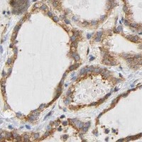
A03440-2 has IHC images from human paraffin sections of colorectal and lung adenocarcinoma, plus IF images from human paraffin sections of esophageal squamous cell carcinoma and intestinal cancer (catalog image captions).

Real IHC data IHC analysis of DOCK180/DOCK1 using anti-DOCK180/DOCK1 antibody (A03440-2). DOCK180/DOCK1 was detected in a paraffin-embedded section of human colorectal adenocarcinoma tissue. Heat mediated antigen retrieval was performed in EDTA buffer (pH 8.0, epitope retrieval solution). The tissue section was blocked with 10% goat serum. The tissue section was then incubated with 2 μg/ml rabbit anti-DOCK180/DOCK1 Antibody (A03440-2) overnight at 4°C. Peroxidase Conjugated Goat Anti-rabbit IgG was used as secondary antibody and incubated for 30 minutes at 37°C. The tissue section was developed using HRP Conjugated Rabbit IgG Super Vision Assay Kit (Catalog # SV0002) with DAB as the chromogen.
Anti-DOCK180/DOCK1 Antibody ®
Cat # A03440-2

A03440-2 is listed for human IHC and IF (catalog applications and reactivity). Its IHC images show colorectal and lung adenocarcinoma sections; its IF images show esophageal squamous cell carcinoma and intestinal cancer sections (catalog image captions).

Which to pick: Choose A03440-2 for human paraffin-section IHC; its captions document EDTA retrieval at pH 8.0 and primary antibody at 2 μg/ml, but do not report the fixative (catalog IHC image captions). For IF, A03440-2 has human paraffin-section images at 5 μg/ml; ICC validation and clonality are unreported (catalog IF image captions; catalog applications and clone field). No cross-species option is documented here because A03440-2 lists human reactivity only (catalog reactivity).

Each figure is that product's own IHC / IF validation image from its datasheet.

References

  1. UniProt Consortium. UniProt entry Q14185 (DOCK1_HUMAN, Dedicator of cytokinesis protein 1).
  2. Human Protein Atlas. DOCK1 tissue IHC expression (reliability: Approved).
  3. Human Protein Atlas. DOCK1 subcellular location (ICC-IF): Mainly localized to the nucleoplasm and cytosol. In addition localized to the nucleoli, plasma membrane, actin filaments and focal adhesion sites..
  4. Human Protein Atlas. DOCK1 antibody validation summary (3 antibodies).
  5. DOCK1 regulates the malignant biological behavior of endometrial cancer through c-Raf/ERK pathway. BMC cancer 2024 — PMC10913561.
  6. MiR-486-5p inhibits IL-22-induced epithelial-mesenchymal transition of breast cancer cell by repressing Dock1. Journal of Cancer 2019 — PMC6746125.
  7. TBOPP, a DOCK1 Inhibitor, Potentiates Cisplatin Efficacy in Breast Cancer by Regulating Twist-mediated EMT. Current cancer drug targets 2025 — PMC11826914.
  8. Impaired cell death and mammary gland involution in the absence of Dock1 and Rac1 signaling. Cell death & disease 2014 — PMC4454313.
  9. PubMed PMID:8657152 — UniProt-cited evidence.
  10. PubMed PMID:15164054 — UniProt-cited evidence.
  11. PubMed PMID:8662907 — UniProt-cited evidence.