This website uses cookies to ensure you get the best experience on our website.


- Table of Contents
Plan DPAGT1 IHC-P around granular cytoplasmic staining, with colon glandular cells as a high-staining control (HPA tissue IHC). This guide covers fixation, retrieval, chromogenic detection and scoring, starting the IHC-validated antibody at 2.5 μg/mL (datasheet).

Expected localisation, validated protocols, controls and antibodies — the at-a-glance facts below, then the full design guide.
| Expected localisation | Granular cytoplasm (HPA tissue IHC); ER membrane (UniProt) | |
| Staining pattern | Often granular cytoplasm in glandular and epithelial cells (HPA tissue IHC) | |
| Antigen retrieval | Citrate pH 6.0 HIER, 95–98 °C, 20 min (rule: cytoplasmic / membrane antigen) | |
| Positive control | Adrenal gland+4 more · see all | |
| Negative control | Adipose tissue+3 more · see all |
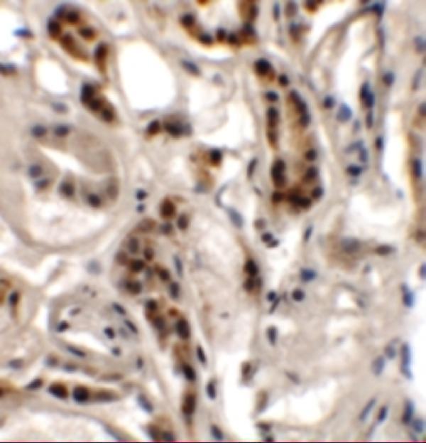
| Fixation | Keep paraffin tissue fixation consistent (standard IHC practice; not target-specific); Matched tissue-IHC evidence does not establish the fixation claim. Validate the specimen-specific method before use. (selected-SKU IHC image A08142) | |
| Caveat | Staining and RNA show only medium consistency (HPA tissue IHC) | |
| Regulation | No specific regulator annotated (UniProt) | |
| Isoform / epitope | 3 isoforms; lumenal vs cytoplasmic epitope matters (UniProt) |
The catalog antibody’s IHC-P protocol is accompanied by a published paraffin-section protocol for human tongue squamous cell carcinoma (PMC7758185).
| Sample | Tissue sections; selected-image fixative not specified (standard IHC workflow) |
| Fixation | Image fixative and duration unreported (datasheet A08142); verify before use. |
| Sectioning | 4–5 µm sections on charged slides (standard) |
| Deparaffinisation | Xylene, graded ethanol series to water (standard) |
| Antigen retrieval | Heat-induced epitope retrieval in citrate buffer, pH 6.0, 20 min at 95–98 °C (standard rule: cytoplasmic / membrane antigen) |
| Peroxidase block | 3% H2O2, 10 min, room temperature (standard) |
| Blocking | 10% normal serum of the secondary host, 30 min, room temperature (standard) |
| Primary antibody | Rabbit anti-DPAGT1, 2.5 μg/mL (datasheet A08142) |
| Primary incubation | Overnight at 4 °C (standard) |
| Detection | HRP-polymer secondary, DAB chromogen 5–10 min (standard) |
| Counterstain | Hematoxylin, blue, dehydrate and mount (standard) |
| Expected result | DPAGT1-positive staining in glandular cells of adrenal gland (HPA tissue IHC: High). HPA tissue profile: General cytoplasmic expression, often in a granular pattern. No signal in the no-primary control. |
DPAGT1 is a 10-pass endoplasmic reticulum membrane protein (UniProt Q9H3H5 topology). In paraffin-section IHC, expect cytoplasmic staining, often granular, in cells such as colon and duodenal glandular cells, bronchial ciliated-cell bodies, and adrenal glandular cells (HPA tissue IHC: High; general cytoplasmic, often granular profile). HPA rates its tissue staining Approved, with medium consistency between antibody staining and RNA expression (HPA tissue IHC: reliability).
| Granular cytoplasmic signal in colon or duodenal glandular cells, with little signal in nearby unstained structures. | This matches HPA's general cytoplasmic, often granular profile and its High staining in these glandular cells (HPA tissue IHC). The pattern is compatible with DPAGT1's ER membrane location; chromogenic IHC alone does not resolve individual ER membranes (UniProt Q9H3H5; general IHC practice). |
| Predominantly nuclear, cell-surface, or extracellular staining with little cytoplasmic signal. | Treat this as a compartment mismatch: UniProt places DPAGT1 in the ER membrane, while HPA reports a cytoplasmic tissue pattern (UniProt Q9H3H5; HPA tissue IHC). Review controls and staining distribution before assigning such signal to DPAGT1 (general IHC practice). |
| Strong staining in adipocytes, smooth muscle cells, ovarian follicle cells, or chondrocytes. | HPA reports DPAGT1 as Not detected in those cell types (HPA tissue IHC). Unexpected staining raises possible cross-reactivity or endogenous detection activity; a single discrepant section cannot establish either cause, and Not detected does not prove protein absence (general IHC interpretation). |
| Diffuse color across cells, stroma, and slide background without a recognizable granular cytoplasmic pattern. | This is less convincing than HPA's often granular cytoplasmic profile (HPA tissue IHC). Widespread nonspecific antibody binding, inadequate blocking, or endogenous detection activity are possible technical explanations to test with controls (general IHC practice). |
| No signal in a well-preserved colon or duodenal glandular-cell section. | HPA reports High staining in these cells, so absence of signal calls for a run-level check (HPA tissue IHC). Review the catalog antibody's IHC-P conditions, retrieval, detection reagents, and a positive control before interpreting the specimen as negative (general IHC practice). |
| Compartment and topology | DPAGT1 has 10 transmembrane segments and an ER membrane location (UniProt Q9H3H5 topology). Interpret its predicted location against HPA's cytoplasmic, often granular IHC profile; routine chromogenic resolution does not prove ER colocalization (HPA tissue IHC; general IHC practice). |
| Tissue choice and expression range | HPA reports High staining in several epithelial or glandular populations, Low staining in selected others, and Not detected in specific cell types; its RNA summary is low tissue specificity (HPA tissue IHC). Compare named cell populations rather than labeling a whole organ uniformly positive or negative (general IHC interpretation). |
| Antibody evidence | HPA lists HPA053878 as IHC Approved and describes tissue-level agreement with RNA as medium consistency (HPA antibodies; HPA tissue IHC). This supports cautious pattern comparison; the supplied record does not report IHC Enhanced validation or an epitope, so neither independent-antibody concordance nor epitope accessibility can be assumed (HPA antibodies; supplied record). |
| Isoforms and processing | UniProt lists 3 isoforms, one chain spanning residues 1–408, and no annotated signal peptide or propeptide (UniProt Q9H3H5). Because the supplied antibody record gives no epitope, this section cannot determine which isoforms its IHC signal represents (HPA antibodies; supplied record). |
| Can IF/ICC confirm this pattern? | UniProt's ER membrane assignment makes intracellular, ER-like fluorescence a plausible comparison, but HPA's ICC-IF entry says only Membrane, gives no main location, and lists no cell-line images (UniProt Q9H3H5; HPA subcellular). It therefore supplies no observed ER colocalization or IF/ICC validation here (HPA subcellular; HPA antibodies). |
| Situation | Likely cause | Next action |
|---|---|---|
| Expected cytoplasmic signal is absent in a High-staining control tissue (HPA tissue IHC). | An IHC run, retrieval, antibody, or detection failure is possible; the slide alone cannot identify which (general IHC practice). | Check the catalog antibody's IHC-P instructions and run a suitable positive control through the same retrieval and detection steps (general IHC practice). |
| Only nuclei or extracellular material stain. | The dominant compartment conflicts with UniProt's ER membrane location and HPA's cytoplasmic IHC profile (UniProt Q9H3H5; HPA tissue IHC). | Inspect staining morphology and controls, then reassess antibody specificity and detection background before scoring the result (general IHC practice). |
| Not-detected reference cell types show strong color (HPA tissue IHC). | Possible antibody cross-reactivity or endogenous detection activity; HPA's Not detected designation is an observed staining result, not proof of molecular absence (HPA tissue IHC; general IHC interpretation). | Compare a no-primary control and the expected positive-cell pattern; investigate background before calling those cells DPAGT1-positive (general IHC practice). |
| Color covers stroma and many unrelated cell populations. | Nonspecific binding or endogenous chromogenic activity may obscure HPA's often granular cytoplasmic profile (HPA tissue IHC; general IHC practice). | Review blocking and wash steps, use a no-primary control, and evaluate signal against cell boundaries and morphology (general IHC practice). |
| A weak tissue is negative while a High-staining control is positive (HPA tissue IHC). | HPA reports Low staining in some cell types, so a negative call in a weak sample may reflect limited assay sensitivity (HPA tissue IHC; general IHC interpretation). | Score the control and sample separately; record the weak sample as not detected under the tested conditions rather than inferring DPAGT1 is absent (general IHC interpretation). |
| An IF/ICC image seems to show a precise ER pattern. | The supplied HPA ICC-IF entry has no cell-line images or specified main location; ER localization comes from UniProt (HPA subcellular; UniProt Q9H3H5). | Describe the observed fluorescence separately from the UniProt prediction, and require independent colocalization evidence before claiming ER-specific IF staining (general IF practice). |


Comprehensive Human Protein Atlas IHC scoring per tissue (reliability: Approved — Medium consistency between antibody staining and RNA expression data.). Rows are taken directly from the HPA tissue chart — click any row's HPA link to view the source.
| Tissue | Cell type | Level | Evidence | Source |
|---|---|---|---|---|
| Adrenal gland | Glandular cells | High | Protein (IHC) | HPA → |
| Bronchus | Ciliated cells (cell body) | High | Protein (IHC) | HPA → |
| Cerebellum | Cells in granular layer | High | Protein (IHC) | HPA → |
| Colon | Glandular cells | High | Protein (IHC) | HPA → |
| Duodenum | Glandular cells | High | Protein (IHC) | HPA → |
| Tissue | Cell type | Level | Evidence | Source |
|---|---|---|---|---|
| Adipose tissue | Adipocytes | Not detected | Protein (IHC) | HPA → |
| Ovary | Follicle cells | Not detected | Protein (IHC) | HPA → |
| Smooth muscle | Smooth muscle cells | Not detected | Protein (IHC) | HPA → |
| Soft tissue | Chondrocytes | Not detected | Protein (IHC) | HPA → |
Troubleshoot DPAGT1 staining in paraffin section IHC using the specified retrieval method, membrane topology, and reported tissue staining patterns (UniProt Q9H3H5; HPA tissue IHC).
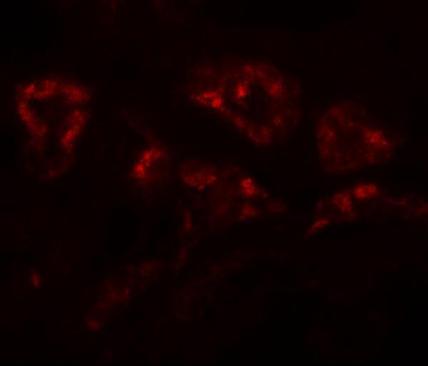
The catalog lists one anti-DPAGT1 antibody with IHC and IF images from human kidney tissue (catalog: image captions); human and mouse reactivity is listed (catalog: reactivity).
A08142 will render with a human kidney IHC image at 2.5 μg/mL (catalog: IHC image caption). A08142 also has a human kidney IF image at 20 μg/mL (catalog: IF image caption).
Which to pick: Choose A08142 for paraffin-section IHC (catalog: IHC-P application); its IHC caption shows human kidney tissue, but does not report the fixative (catalog: IHC image caption). Choose A08142 for IF; ICC validation is not listed (catalog: applications). For work involving both human and mouse samples, A08142 lists reactivity with both species, although the supplied IHC and IF images show human tissue only (catalog: reactivity and image captions); no clone is specified.