This website uses cookies to ensure you get the best experience on our website.


- Table of Contents
Plan chromogenic DPF3 IHC around the nuclear staining reported in neurons, ovary, testis and muscle (HPA tissue IHC). This guide covers tissue controls, consistent fixation and interpretation of the IHC-validated antibody’s staining, with HPA’s medium RNA concordance in mind (HPA tissue IHC).

Expected localisation, validated protocols, controls and antibodies — the at-a-glance facts below, then the full design guide.
| Expected localisation | Nuclear staining in positive tissues (HPA tissue IHC) | |
| Staining pattern | Nuclei of neurons, follicle cells, spermatocytes and myocytes (HPA tissue IHC) | |
| Antigen retrieval | Tris-EDTA pH 9.0 HIER, 95–98 °C, 20 min (rule: nuclear antigen) | |
| Positive control | Cerebellum+4 more · see all | |
| Negative control | Adipose tissue+4 more · see all |
| Fixation | Keep fixation consistent across sections. (standard IHC practice; not target-specific) | |
| Caveat | Staining has medium concordance with RNA data (HPA tissue IHC) | |
| Regulation | Staining varies by tissue and cell type (HPA tissue IHC) | |
| Isoform / epitope | Five isoforms; epitope coverage needs confirmation (UniProt) |
The catalog antibody protocol is paired with one published DPF3 IHC protocol reporting primary dilution and antigen retrieval without retrieval conditions (PMC8897700 methods).
| Sample | Tissue sections; selected-image fixative not specified (standard IHC workflow) |
| Fixation | Image fixative and duration unreported (datasheet A09485); verify before use. |
| Sectioning | 4–5 µm sections on charged slides (standard) |
| Deparaffinisation | Xylene, graded ethanol series to water (standard) |
| Antigen retrieval | Heat-induced epitope retrieval in Tris-EDTA buffer, pH 9.0, 20 min at 95–98 °C (standard rule: nuclear antigen) |
| Peroxidase block | 3% H2O2, 10 min, room temperature (standard) |
| Blocking | 10% normal serum of the secondary host, 30 min, room temperature (standard) |
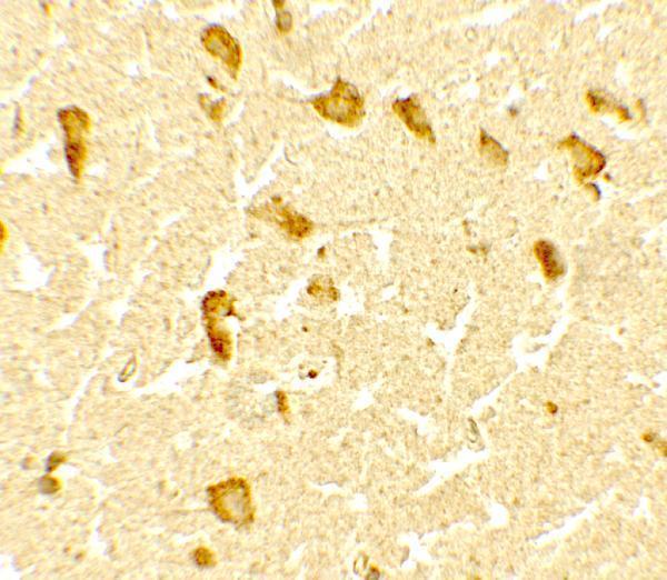
| Primary antibody | Rabbit anti-DPF3, 5 μg/mL (datasheet A09485) |
| Primary incubation | Overnight at 4 °C (standard) |
| Detection | HRP-polymer secondary, DAB chromogen 5–10 min (standard) |
| Counterstain | Hematoxylin, blue, dehydrate and mount (standard) |
| Expected result | DPF3-positive staining in gLUC cells - nucleus of cerebellum (HPA tissue IHC: High). HPA tissue profile: Nuclear expression in neurons, ovary, testis and heart and skeletal myocytes. No signal in the no-primary control. |
DPF3 is a nuclear chromatin regulator with no transmembrane segment (UniProt Q92784: subcellular location, topology). In paraffin-section IHC, expect staining chiefly in neuronal nuclei and in the nuclei of heart and skeletal myocytes; HPA also describes nuclear expression in ovary and testis (HPA: tissue profile). HPA rates the tissue IHC evidence Enhanced, with medium staining–RNA consistency and external verification pending (HPA: tissue reliability).
| High nuclear staining in cerebellar GLUC cells; neuronal staining in cerebral cortex (HPA: tissue IHC). | This fits the strongest supplied neural examples. Assess signal in the annotated cells and compartment: HPA explicitly calls cerebellar GLUC nuclei high, while its cortex entry calls neuronal cells high (HPA: tissue IHC). |
| Nuclear staining in pachytene spermatocytes, cardiomyocytes or ovarian follicle cells (HPA: tissue IHC). | These are supported cell populations, with high staining in pachytene spermatocytes and medium staining in cardiomyocytes and follicle cells (HPA: tissue IHC). Compare like cells; a whole-tissue average can hide their distribution (standard IHC interpretation). |
| Predominantly cytoplasmic, membrane or extracellular signal, without convincing nuclear staining. | Treat this as discordant with DPF3's nuclear annotation and HPA's nuclear tissue profile (UniProt Q92784: subcellular location; HPA: tissue profile). Recheck localisation against a nuclear counterstain before calling it DPF3 (standard IHC practice). |
| Staining in an HPA-undetected cell population, especially if it follows vessels, pigment or section edges. | For example, HPA reports adipocytes in adipose tissue and respiratory epithelial cells in bronchus as not detected (HPA: tissue IHC). Such signal warrants checks for cross-reactivity, endogenous chromogenic activity or deposition artefact (standard IHC practice). |
| Diffuse colour across cells and stroma, or no nuclear signal in cerebellar GLUC cells. | Diffuse colour obscures compartment scoring and suggests background (standard IHC interpretation). Absent signal in a supplied high nuclear reference raises a run or detection concern, though one negative slide cannot establish absent DPF3 expression (HPA: cerebellum tissue IHC; standard IHC practice). |
| Reference-cell selection | Use the annotated cell population, not an entire organ, to judge concordance. HPA reports high nuclear staining in cerebellar GLUC cells, but only low staining in hippocampal neuronal cells and skeletal myocytes (HPA: tissue IHC). |
| Tissue evidence and antibody validation | HPA's tissue pattern has Enhanced reliability but only medium staining–RNA consistency, with external verification pending (HPA: tissue reliability). HPA066790 has Enhanced IHC validation; HPA070601 has no listed IHC status, so its Enhanced ICC status should not be read as IHC validation (HPA: antibody validation). |
| Cellular location and processing | A predominantly nuclear result agrees with the UniProt nucleus annotation and HPA tissue profile (UniProt Q92784: subcellular location; HPA: tissue profile). UniProt lists no transmembrane segment, signal peptide or propeptide, and a single 1–378 chain (UniProt Q92784: topology and processing). |
| Isoforms and modified residue | UniProt lists 5 isoforms and phosphoserine at residue 323 (UniProt Q92784: isoforms, modified residues). The supplied evidence does not map an antibody epitope to an isoform or this residue; do not infer staining differences from them (supplied UniProt and HPA records). |
| Antigen retrieval and fixation | Retrieval conditions can be optimised as a general paraffin-IHC workflow step (standard IHC practice). No supplied target-specific evidence establishes that fixation or retrieval changes DPF3 staining, so report the tested condition without attributing a staining difference to DPF3-specific sensitivity (supplied UniProt and HPA records). |
| IF/ICC Q: where should fluorescence appear? | A: HPA reports DPF3 in the nucleoplasm, with an Enhanced main-location assessment and ICC-IF images from BJ, PC-3 and U2OS cells (HPA: subcellular). This is an IF/ICC localisation reference, not a paraffin-IHC protocol (HPA: subcellular). |
| Situation | Likely cause | Next action |
|---|---|---|
| No nuclear staining in a cerebellar reference section. | The reference is discordant with HPA's high nuclear GLUC-cell result; failed staining or detection is possible (HPA: cerebellum tissue IHC; standard IHC practice). | Confirm the annotated cells are present; review the run control, antibody dilution, retrieval and detection steps before interpreting the specimen as negative (standard IHC practice). |
| Only cytoplasmic or membrane colour is visible. | This conflicts with the nuclear location reported by UniProt and HPA; nonspecific binding or misplaced chromogen may explain the colour (UniProt Q92784: subcellular location; HPA: tissue profile; standard IHC interpretation). | Compare with the nuclear counterstain and a reagent control, then score nuclear and non-nuclear signal separately (standard IHC practice). |
| Broad staining appears in cells expected to be undetected. | HPA lists adipocytes in adipose tissue and respiratory epithelial cells in bronchus as not detected; cross-reactivity or endogenous detection activity is possible (HPA: tissue IHC; standard IHC practice). | Check a reagent control, endogenous-enzyme blocking and chromogen deposition; reassess the exact cell type rather than the organ label (standard chromogenic-IHC practice). |
| Diffuse colour makes nuclei hard to distinguish. | Nonspecific background or excessive detection can obscure compartment assignment (standard IHC interpretation). | Review blocking, antibody dilution, washes and detection development; use a nuclear counterstain to judge whether a discrete nuclear component remains (standard IHC practice). |
| Heart or skeletal muscle looks weaker than cerebellum. | HPA reports medium cardiomyocyte staining and low skeletal-myocyte staining, versus high cerebellar GLUC-cell staining (HPA: tissue IHC). | Score the relevant cells against their own HPA level; do not require muscle to match the cerebellar reference intensity (HPA: tissue IHC; standard IHC interpretation). |
| An ICC-validated antibody is being considered for paraffin IHC. | HPA070601 has Enhanced ICC validation but no listed IHC status; the validation entries cover different applications (HPA: antibody validation). | Use an IHC-validated antibody as the paraffin-section reference; HPA066790 has Enhanced IHC validation, while an ICC result alone does not establish IHC performance (HPA: antibody validation). |


![HPA ICC-IF BJ [Human fibroblast] — DPF3 (HPA066790)](https://images.proteinatlas.org/66790/1544_B12_2_blue_red_green.jpg)
![HPA ICC-IF BJ [Human fibroblast] — DPF3 (HPA066790)](https://images.proteinatlas.org/66790/1544_B12_3_blue_red_green.jpg)
Comprehensive Human Protein Atlas IHC scoring per tissue (reliability: Enhanced — Medium consistency between antibody staining and RNA expression data. Pending external verification.). Rows are taken directly from the HPA tissue chart — click any row's HPA link to view the source.
| Tissue | Cell type | Level | Evidence | Source |
|---|---|---|---|---|
| Cerebellum | GLUC cells - nucleus | High | Protein (IHC) | HPA → |
| Cerebral cortex | Neuronal cells | High | Protein (IHC) | HPA → |
| Kidney | Proximal tubules (cell body) | High | Protein (IHC) | HPA → |
| Testis | Pachytene spermatocytes | High | Protein (IHC) | HPA → |
| Adrenal gland | Glandular cells | Medium | Protein (IHC) | HPA → |
| Tissue | Cell type | Level | Evidence | Source |
|---|---|---|---|---|
| Adipose tissue | Adipocytes | Not detected | Protein (IHC) | HPA → |
| Breast | Adipocytes | Not detected | Protein (IHC) | HPA → |
| Bronchus | Respiratory epithelial cells | Not detected | Protein (IHC) | HPA → |
| Cervix | Glandular cells | Not detected | Protein (IHC) | HPA → |
| Colon | Endothelial cells | Not detected | Protein (IHC) | HPA → |
Troubleshoot DPF3 chromogenic IHC by checking nuclear staining, tissue context and matched controls before interpreting signal intensity.
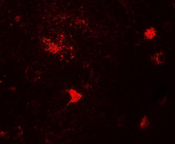
A09485 has human brain IHC and IF images (catalog image captions) and lists human and mouse reactivity (catalog reactivity).
A09485 is listed for IHC-P and has a human brain IHC image at 5 μg/mL (catalog applications; IHC image caption). A09485 is also listed for IF and has a human brain IF image at 20 μg/mL (catalog applications; IF image caption).
Which to pick: For paraffin-section tissue IHC, choose A09485: it lists IHC-P and shows human brain staining; the fixative is unreported (catalog applications; IHC image caption). For IF, choose A09485 based on its human brain IF image; ICC validation is unreported (IF image caption; catalog applications). For mouse samples, A09485 lists mouse reactivity, but both images show human brain; its clonality is unreported (catalog reactivity; image captions; catalog clone field).