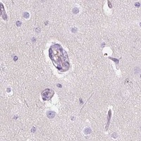
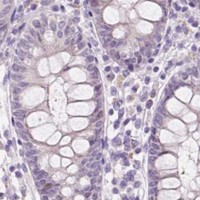
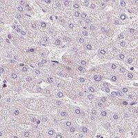
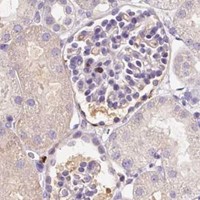
DPPA4 / Developmental pluripotency-associated protein 4 · IHC design guide

Design Immunohistochemistry for DPPA4

Plan chromogenic IHC-P around nuclear staining in testis spermatogonia and placental cytotrophoblasts (HPA tissue IHC). The catalog antibody was used at 1:25 (datasheet A10283-2); assess nuclear signal with controls for possible off-target binding (HPA tissue IHC).

Evidence assembled Oct 2026 · For research use; verify linked source records and product datasheet before use
Immunohistochemistry protocol sheet for DPPA4 (IHC for DPPA4): expected localisation Nuclear staining in testis and placenta (HPA tissue IHC), antibody A10283-2, validated IHC image, and IHC protocol steps
Printable DPPA4 IHC protocol sheet — expected localisation Nuclear staining in testis and placenta (HPA tissue IHC), antibody A10283-2, controls and protocol steps. Open the full DPPA4 IHC guide →

DPPA4 Immunohistochemistry Experimental Design Guide

Expected localisation, validated protocols, controls and antibodies — the at-a-glance facts below, then the full design guide.

Must know before staining
Expected localisation Nuclear staining in testis and placenta (HPA tissue IHC)
Staining pattern High nuclear spermatogonia; medium nuclear cytotrophoblasts (HPA tissue IHC)
Antigen retrieval Citrate pH 6 HIER, heat-mediated (datasheet A10283-2)
Positive control ⓘ Testis+1 more · see all
Negative control ⓘ Adipose tissue+4 more · see all
Important caveats
Reasons your staining may differ from the expected pattern.
Fixation Formaldehyde-fixed paraffin sections (selected-SKU IHC image A10283-2)
Caveat Presumed off-target binding may mimic DPPA4 staining (HPA tissue IHC)
Regulation Expression regulation unreported (UniProt)
Isoform / epitope No annotated isoforms; one 1–304 chain (UniProt)
Section 1

Recommended DPPA4 IHC & IF Protocols

The catalog antibody’s IHC-P protocol uses citrate retrieval (datasheet A10283-2). The published protocols below cover colon tissue microarrays (PMC4466839) and paraffin-embedded lung tumors (PMC6580026).

Recommended immunohistochemistry (IHC-P) protocol parameters
SampleFormaldehyde-fixed, paraffin-embedded human testis tissue (datasheet A10283-2)
FixationImage formalin-fixed; duration unreported (datasheet A10283-2); verify before use.
Sectioning4–5 µm sections on charged slides (standard)
DeparaffinisationXylene, graded ethanol series to water (standard)
Antigen retrievalHeat retrieval: Citrate pH 6 (datasheet A10283-2); 20 min, 95–100 °C (standard)
Peroxidase block3% H2O2, 10 min, room temperature (standard)
Blocking10% normal serum of the secondary host, 30 min, room temperature (standard)
Primary antibodyRabbit anti-DPPA4, 1:25 (datasheet A10283-2)
Primary incubation1 hours at 37°C (datasheet A10283-2)
DetectionHRP-polymer secondary, DAB chromogen 5–10 min (standard)
CounterstainHematoxylin, blue, dehydrate and mount (standard)
Expected resultDPPA4-positive staining in spermatogonia cells of testis (HPA tissue IHC: High). HPA tissue profile: Nuclear expression mainly in testis and placenta. No signal in the no-primary control.
💡Decision noteStart with heat-mediated citrate pH 6 retrieval for the catalog antibody (datasheet A10283-2). The lung protocol also uses heated citrate but does not state its pH (PMC6580026).
Section 2

What Is the Expected DPPA4 Staining Pattern?

DPPA4 is a nuclear protein associated with transcriptionally active chromatin (UniProt Q7L190). In paraffin-section IHC, expect nuclear staining mainly in testis spermatogonia and placenta cytotrophoblasts (HPA tissue IHC). HPA rates the tissue profile Enhanced, while noting medium agreement with RNA data and presumed off-target staining that was disregarded (HPA tissue IHC). DPPA4 has no transmembrane segment (UniProt Q7L190 topology).

What am I looking at on my slide?
Strong nuclear staining in spermatogonia, with other testicular cells less conspicuous.This matches the clearest supplied tissue positive: spermatogonia are High (HPA tissue IHC). Judge the result by cell identity and nuclear location together (HPA tissue IHC; UniProt Q7L190). A uniformly stained testis section deserves scrutiny because the reported pattern is cell specific (HPA tissue IHC).
Nuclear staining in placental cytotrophoblasts, weaker than the testis positive.Cytotrophoblasts are Medium, whereas spermatogonia are High (HPA tissue IHC). Compare staining within identified cells rather than expecting equal intensity across tissues (HPA tissue IHC). Placental signal provides a second supported tissue context, although its moderate reported level makes a faint result harder to judge alone (HPA tissue IHC).
Predominantly cytoplasmic, membrane, or extracellular color without convincing nuclear staining.That is discordant with the reported nuclear tissue profile and UniProt nuclear location (HPA tissue IHC; UniProt Q7L190). Consider nonspecific staining or detection artefact before scoring it as DPPA4 (general IHC practice). HPA also reports cytosol in ICC-IF, so an isolated cytosolic signal cannot settle antibody specificity across applications (HPA subcellular ICC-IF).
Staining concentrates in adipocytes or marrow hematopoietic cells instead of the expected nuclei.HPA reports these cell populations as Not detected by tissue IHC (HPA tissue IHC). Review morphology, antibody-dependent cross-reactivity, and endogenous chromogen activity before calling them positive (general IHC practice). Bone marrow is tissue enhanced at the RNA level, but its listed hematopoietic cells remain protein-negative by IHC (HPA tissue IHC).
Diffuse color across cells, stroma, or the whole section obscures nuclear boundaries.This distribution cannot support a cell-specific nuclear call (HPA tissue IHC; general IHC practice). Check whether a no-primary control retains color and whether background tracks the detection reagent (general IHC practice). HPA explicitly disregarded presumed off-target staining when assigning its tissue profile, so broad color should not be accepted as DPPA4 without controls (HPA tissue IHC).
💡Expected DPPA4 appearanceCall a positive when spermatogonial nuclei stain strongly or cytotrophoblast nuclei stain moderately (HPA tissue IHC); widespread nonnuclear color is suspect (HPA tissue IHC; general IHC practice).
How each factor affects the staining
Cell compartment and protein structureNuclear interpretation is supported by both tissue IHC and UniProt localization (HPA tissue IHC; UniProt Q7L190). The protein has no transmembrane segment, signal peptide, or propeptide; its annotated chain spans residues 1–304 (UniProt Q7L190). These annotations do not specify an antibody epitope or an antigen-retrieval condition (UniProt Q7L190).
Antibody evidenceThe HPA tissue profile is Enhanced, with medium antibody–RNA consistency and presumed off-target binding disregarded (HPA tissue IHC). Among listed antibodies, HPA035250 is IHC Enhanced; HPA035249 and CAB020678 are IHC Supported (HPA antibodies). These labels support confidence in the reported pattern, while control slides remain needed for a particular staining run (general IHC practice).
RNA versus stained cellsRNA is tissue enhanced in bone marrow, lymphoid tissue, and testis (HPA tissue IHC). Yet the supplied marrow hematopoietic-cell IHC entry is Not detected (HPA tissue IHC). Select IHC controls from the observed protein pattern rather than treating tissue-level RNA enrichment as a promise of staining in every cell (HPA tissue IHC).
IF/ICC Q&A: where may fluorescence appear?HPA reports mainly nucleoli and nucleoli rims, with additional nucleoplasm, mitotic-chromosome, and cytosol localization in ICC-IF (HPA subcellular ICC-IF). This answers the compartment question for cultured-cell imaging; it does not replace the nuclear, cell-specific tissue-IHC expectation or establish an IF protocol here (HPA subcellular ICC-IF; HPA tissue IHC).
Why is my staining missing, weak or wrong?
SituationLikely causeNext action
No staining in testis spermatogonia.The supplied high-positive population is missing (HPA tissue IHC); the cause cannot be assigned from the image alone (general IHC practice).Verify cell identification, reagent activity, retrieval and detection against the chosen antibody's validated IHC-P instructions; compare a known-positive section (HPA tissue IHC; general IHC practice).
Placenta looks negative while testis stains.Placental cytotrophoblast staining is Medium versus High in spermatogonia, so it may be less conspicuous (HPA tissue IHC).Inspect identified cytotrophoblast nuclei and compare sections under the same scoring criteria; avoid declaring a global assay failure from placenta alone (HPA tissue IHC; general IHC practice).
Strong cytoplasmic or membrane color dominates.The tissue pattern and UniProt location favor nuclei (HPA tissue IHC; UniProt Q7L190); nonspecific binding or detection background is possible (general IHC practice).Check a no-primary control and optimize blocking or antibody dilution using the antibody's IHC-P instructions before scoring nonnuclear color (general IHC practice).
Marrow hematopoietic cells stain despite a clean testis control.Those cells are Not detected by HPA IHC, although marrow RNA is tissue enhanced (HPA tissue IHC). Cross-reactivity is possible (general IHC practice).Recheck cell identity and compare with a no-primary control and another validated antibody where available; do not infer protein positivity from marrow RNA alone (HPA tissue IHC; general IHC practice).
Diffuse chromogen remains on the no-primary slide.Antibody-independent detection background, including endogenous enzyme activity, can color paraffin sections (general IHC practice).Review enzyme-blocking and detection steps, then repeat the no-primary control before interpreting DPPA4 staining (general IHC practice).
Different antibodies produce conflicting tissue patterns.HPA lists different IHC validation statuses and notes presumed off-target binding in its tissue assessment (HPA antibodies; HPA tissue IHC).Compare nuclear staining in spermatogonia and cytotrophoblasts with the reported High and Medium levels, and retain antibody-specific controls when judging discordant signal (HPA tissue IHC; general IHC practice).

Sample controls for DPPA4 IHC & IF

🧪Run testis first: spermatogonia should show DPPA4 staining (HPA: High in testis spermatogonia). Use adipose tissue adipocytes as an external negative (HPA: Not detected in adipocytes); on the testis slide, assess cells without nuclear signal as internal background references rather than assuming every other cell type is negative (UniProt Q7L190: nuclear localization).
Positive control tissue: Testis (Spermatogonia cells, HPA High)
Negative control tissue: Adipose tissue (HPA Not detected)
ICC-IF cell lines (HPA subcellular resource): HPA ICC-IF images show DPPA4 in A-431, U-251MG, U2OS, AF22, PC-3, with annotated localisation: Nucleoli (approved), Nucleoli rim (approved) (HPA subcellular).
Technical controls: Include a no-primary, secondary-only control and an isotype control matched to the primary antibody’s host species and subclass; use DPPA4 knockout material as a biological negative (standard IHC practice). For testis sections, check endogenous biotin when using the caption’s biotinylated secondary antibody, and block endogenous peroxidase if using peroxidase detection (A10283-2 caption: biotinylated secondary; standard IHC practice).
⚠️Feasibility: A target-specific fixation window or fixation effect is unreported, and the exact selected-SKU tissue-IHC record leaves the fixative unreported (A10283-2 fixative field: not stated). The selected IHC caption uses heat-mediated citrate retrieval at pH 6, but does not establish that retrieval is required; it provides no basis to call frozen sections or IF easier (A10283-2 caption: citrate retrieval in paraffin sections). HPA has ICC-IF images and reports mainly nucleolar and nucleolar-rim localization, so assess that pattern separately from the tissue IHC result; a biotin-based detection system warrants an endogenous-biotin check in the testis section (HPA: subcellular localization; A10283-2 caption: biotinylated secondary; standard IHC practice).

HPA tissue IHC evidence for DPPA4

Comprehensive Human Protein Atlas IHC scoring per tissue (reliability: Enhanced — Medium consistency between antibody staining and RNA expression data. Presumed off target binding observed and disregarded.). Rows are taken directly from the HPA tissue chart — click any row's HPA link to view the source.

Positive expression · recommended positive controls

TissueCell typeLevelEvidenceSource
Testis Spermatogonia cells High Protein (IHC) HPA →
Placenta Cytotrophoblasts Medium Protein (IHC) HPA →

Undetected expression · recommended negative controls

TissueCell typeLevelEvidenceSource
Adipose tissue Adipocytes Not detected Protein (IHC) HPA →
Adrenal gland Glandular cells Not detected Protein (IHC) HPA →
Appendix Glandular cells Not detected Protein (IHC) HPA →
Bone marrow Hematopoietic cells Not detected Protein (IHC) HPA →
Breast Adipocytes Not detected Protein (IHC) HPA →
Section 3

Advanced DPPA4 IHC Tips

Troubleshoot DPPA4 staining in paraffin sections by checking nuclear localisation, cell identity and staining controls before comparing signal intensity.

How should I adjust retrieval when DPPA4 staining is weak in paraffin sections?
Start with heat-mediated citrate retrieval at pH 6 for the catalog antibody (datasheet A10283-2). Keep heating and cooling conditions consistent across slides, and compare a testis control with a no-primary control in the same run (HPA: high staining in spermatogonia; standard IHC practice). If nuclear staining remains weak, vary retrieval duration on adjacent sections before changing buffer chemistry, while watching for tissue damage (standard IHC practice). Compare signal in intact spermatogonia rather than scoring diffuse color across the section (HPA: high staining in spermatogonia; UniProt Q7L190: nuclear localisation). Record each retrieval condition alongside primary dilution and detection settings so the source of any improvement is clear (standard IHC practice).
Could fixation explain weak or uneven DPPA4 staining?
Target-specific DPPA4 sensitivity to fixation is unknown from the supplied evidence; do not assign weak staining to a particular fixative without a controlled comparison. For paraffin sections, compare matched tissue processed with documented fixation durations, then hold retrieval, primary incubation and chromogen development constant (standard IHC practice). Examine morphology and staining at section edges as well as the center, because processing differences can produce uneven IHC signal (standard IHC practice). Use a testis section to judge whether the expected cells remain assessable, with spermatogonia as the relevant reference population (HPA: high staining in spermatogonia). Report fixation conditions with the result rather than treating an optimized condition as established DPPA4 biology.
What staining pattern should count as DPPA4 positive?
Score convincing nuclear staining in the expected cells as the primary IHC pattern (UniProt Q7L190: nucleus; HPA tissue IHC: nuclear expression mainly in testis and placenta). In testis, assess spermatogonia; in placenta, assess cytotrophoblasts rather than averaging all cells in either section (HPA: high in spermatogonia; medium in cytotrophoblasts). DPPA4 is associated with transcriptionally active chromatin, which supports a nuclear expectation without specifying a uniform nuclear pattern (UniProt Q7L190). The HPA subcellular IF record additionally places signal at nucleoli and their rim, with other approved locations, but those images do not establish a paraffin IHC scoring threshold (HPA subcellular). Treat isolated diffuse cytoplasmic chromogen cautiously and check its persistence in negative controls (standard IHC practice).
How can I troubleshoot an uncertain DPPA4 epitope in paraffin sections?
Check the antibody's documented immunogen or epitope before attributing a staining difference to DPPA4 sequence variation; none is specified in the supplied product evidence. The reference record describes a 304 amino acid chain with no annotated isoforms or domains, so it does not identify an alternative isoform-specific IHC pattern (UniProt Q7L190). Phosphorylation is annotated at residues 215, 221 and 226, but any effect on this antibody's binding remains unestablished (UniProt Q7L190). Compare retrieval conditions and nuclear cell patterns on serial sections while holding detection constant (standard IHC practice). If epitope identity remains unknown, report the reagent and staining conditions without claiming phospho-specificity or isoform discrimination.
How should I cross-check DPPA4 IHC with an IF experiment?
Use IF as a complementary localisation check, selecting a validated marker for the expected cell population when multiplexing with DPPA4 (HPA: spermatogonia in testis and cytotrophoblasts in placenta; standard IF practice). Choose spectrally separated fluorophores after inspecting tissue autofluorescence in unstained and single-stained controls (standard IF practice). DPPA4 has no annotated transmembrane segment and is primarily nuclear, so use permeabilisation suitable for antibody access to nuclear epitopes and verify that cell morphology remains interpretable (UniProt Q7L190; standard IF practice). HPA IF images show approved nucleolar and nucleolar-rim localisation, plus additional nucleoplasmic, mitotic-chromosome and cytosolic localisation (HPA subcellular). Compare compartment and cell identity across methods without assuming that IF intensity predicts chromogenic IHC intensity.
What should I check when DPPA4 IHC has widespread background?
Run no-primary and reagent controls to distinguish primary-dependent staining from secondary or detection background (standard IHC practice). The selected testis IHC example used 3% BSA for 0.5 hour and primary antibody at 1:25 for 1 hour at 37°C (caption: A10283-2). If background persists, titrate the primary and review blocking, washes and chromogen development on adjacent sections (standard IHC practice). With a biotin-based detection workflow, assess endogenous biotin; with peroxidase detection, include an appropriate endogenous peroxidase block (standard IHC practice; caption A10283-2: biotinylated secondary). Reject widespread color that obscures nuclear assessment in spermatogonia or cytotrophoblasts (HPA: DPPA4 tissue IHC pattern).
How should I quantify DPPA4 staining across paraffin sections? ⚠ ANSWER MARKED FOR VERIFICATION
Define the relevant cell population before scoring: spermatogonia in testis or cytotrophoblasts in placenta (HPA: high and medium staining, respectively). Record the percentage of cells with nuclear staining and, when intensity categories are reproducible, calculate a nuclear H-score from the same annotated regions (standard IHC practice). Normalize positive counts to the number of eligible cells, or report positive-cell density per mm² of eligible tissue when cell counts cannot be made reliably (standard IHC practice). Keep retrieval, detection, exposure to chromogen and scoring thresholds consistent across compared sections (standard IHC practice). Report excluded damaged or necrotic areas and retain a matched negative control for each scoring run (standard IHC practice).
How can I separate true DPPA4 staining from artefact?
A credible result aligns nuclear signal with the expected cells: spermatogonia in testis or cytotrophoblasts in placenta (UniProt Q7L190: nucleus; HPA tissue IHC: high and medium, respectively). Review unexpected cytoplasmic or widespread staining against no-primary controls before assigning it to DPPA4 (standard IHC practice). Exclude section-edge deposits, necrotic regions and endogenous enzyme signal from biological interpretation (standard IHC practice). HPA rates its tissue staining reliability as Enhanced while noting presumed off-target binding that was disregarded, so concordance alone does not establish antibody specificity (HPA tissue IHC). Document which cells and nuclear regions stained, and resolve discordant patterns with an independent antibody or orthogonal assay (standard IHC practice).
Boster reagents

Best DPPA4 / Developmental pluripotency-associated protein 4 IHC Antibodies

The catalog contains one human-reactive anti-DPPA4 antibody with IHC-P data from human testis paraffin sections (catalog: A10283-2 image caption). No IF/ICC validation is listed (catalog: applications).

Real IHC data A10283-2 staining DPPA4 in human testis tissue sections by Immunohistochemistry (IHC-P -paraformaldehyde-fixed, paraffin-embedded sections). Tissue was fixed with formaldehyde and blocked with 3% BSA for 0. 5 hour at room temperature; antigen retrieval was by heat mediation with a citrate buffer (pH6). Samples were incubated with primary antibody (1/25) for 1 hours at 37°C. A undiluted biotinylated goat polyvalent antibody was used as the secondary antibody.
Anti-DPPA4 Antibody (N-term)
Cat # A10283-2

A10283-2 is listed for human IHC-P (catalog: applications/reactivity). Its own IHC image caption documents staining in human testis paraffin sections (catalog: A10283-2 image caption).

Which to pick: For tissue IHC, choose A10283-2, a rabbit polyclonal antibody listed for human IHC-P at 1:25 (catalog: host/dilution/applications/reactivity); its own caption reports formaldehyde fixation of human testis paraffin sections (catalog: A10283-2 image caption). No IF/ICC or cross-species choice is validated here: A10283-2 lists human reactivity and IHC-P/WB applications, with no IF figure (catalog: reactivity/applications/IF image alts).

Each figure is that product's own IHC / IF validation image from its datasheet.

References

  1. UniProt Consortium. UniProt entry Q7L190 (DPPA4_HUMAN, Developmental pluripotency-associated protein 4).
  2. Human Protein Atlas. DPPA4 tissue IHC expression (reliability: Enhanced).
  3. Human Protein Atlas. DPPA4 subcellular location (ICC-IF): Mainly localized to the nucleoli and nucleoli rim. In addition localized to the nucleoplasm, mitotic chromosome and cytosol..
  4. Human Protein Atlas. DPPA4 antibody validation summary (3 antibodies).
  5. Developmental pluripotency-associated 4: a novel predictor for prognosis and a potential therapeutic target for colon cancer. Journal of experimental & clinical cancer research : CR 2015 — PMC4466839.
  6. High developmental pluripotency‑associated 4 expression promotes cell proliferation and glycolysis, and predicts poor prognosis in non‑small‑cell lung cancer. Molecular medicine reports 2019 — PMC6580026.
  7. PubMed PMID:14702039 — UniProt-cited evidence.
  8. PubMed PMID:16641997 — UniProt-cited evidence.
  9. PubMed PMID:15489334 — UniProt-cited evidence.