DSG2 / Desmoglein-2 · IHC design guide

Design Immunohistochemistry for DSG2

Plan DSG2 chromogenic IHC on paraffin sections using 2–5 µg/ml catalog antibody (datasheet: M02035-2). Compare breast glandular cells with lymph node germinal center cells, then score cytoplasmic and membranous staining (HPA tissue IHC).

Evidence assembled Oct 2026 · For research use; verify linked source records and product datasheet before use
Immunohistochemistry protocol sheet for DSG2 (IHC for DSG2): expected localisation Membranous and cytoplasmic tissue staining (HPA tissue IHC), antibody M02035-2, validated IHC image, and IHC protocol steps
Printable DSG2 IHC protocol sheet — expected localisation Membranous and cytoplasmic tissue staining (HPA tissue IHC), antibody M02035-2, controls and protocol steps. Open the full DSG2 IHC guide →

DSG2 Immunohistochemistry Experimental Design Guide

Expected localisation, validated protocols, controls and antibodies — the at-a-glance facts below, then the full design guide.

Must know before staining
Expected localisation Membranous and cytoplasmic tissue staining (HPA tissue IHC)
Staining pattern Most cells show cytoplasmic and membranous staining (HPA tissue IHC)
Antigen retrieval EDTA pH 8.0 HIER, heat-mediated (datasheet M02035-2)
Positive control ⓘ Appendix+4 more · see all
Negative control ⓘ Adipose tissue+4 more · see all
Important caveats
Reasons your staining may differ from the expected pattern.
Fixation Keep fixation consistent across sections. (standard IHC practice; not target-specific)
Caveat Apoptotic cleavage can shift DSG2 into cytoplasm (UniProt)
Regulation Expression declines with stem-cell differentiation (UniProt)
Isoform / epitope 0 isoforms listed; processing and epitope side matter (UniProt)
Section 1

Recommended DSG2 IHC & IF Protocols

The catalog antibody has an IHC-P protocol (datasheet M02035-2). Published protocols cover colon tissue microarrays (PMC7789404: methods) and biliary tissues (PMC7436288: methods).

Recommended immunohistochemistry (IHC-P) protocol parameters
SampleParaffin-embedded human breast cancer tissue; fixative not specified (datasheet M02035-2)
FixationImage fixative and duration unreported (datasheet M02035-2); verify before use.
Sectioning4–5 µm sections on charged slides (standard)
DeparaffinisationXylene, graded ethanol series to water (standard)
Antigen retrievalHeat retrieval: EDTA pH 8.0 (datasheet M02035-2); 20 min, 95–100 °C (standard)
Peroxidase block3% H2O2, 10 min, room temperature (standard)
Blocking10% goat serum (datasheet M02035-2)
Primary antibodyMouse monoclonal (clone 2B4D1) anti-DSG2, 2-5 µg/ml (datasheet M02035-2)
Primary incubationOvernight at 4 °C (datasheet M02035-2)
DetectionHRP-conjugated secondary, DAB chromogen (datasheet M02035-2)
CounterstainHematoxylin, blue, dehydrate and mount (standard)
Expected resultDSG2-positive staining in endocrine cells of appendix (HPA tissue IHC: High). HPA tissue profile: Cytoplasmic and membranous expression in essentially all cells except in lymphoid tissue and the CNS. No signal in the no-primary control.
💡Decision noteStart with heat-mediated EDTA at pH 8.0 for the catalog antibody (datasheet M02035-2). Citrate at pH 6.0 was used for biliary sections (PMC7436288: methods).
Section 2

What Is the Expected DSG2 Staining Pattern?

DSG2 is a desmosomal cell-junction protein with extracellular and cytoplasmic regions flanking one transmembrane segment (UniProt Q14126 topology). Expect membranous, often junctional staining, with possible cytoplasmic signal (HPA tissue IHC; HPA subcellular ICC-IF). HPA reports high staining in several endocrine, glandular, and respiratory epithelial cell populations, and rates its tissue IHC profile Enhanced for consistency with RNA expression (HPA tissue IHC).

What am I looking at on my slide?
Strong cell-border staining in breast glandular cells or bronchial respiratory epithelial cells, with some cytoplasmic signal (HPA tissue IHC: High).This fits DSG2 at desmosomal junctions and the broader membranous and cytoplasmic tissue profile (UniProt Q14126 subcellular; HPA tissue IHC). Score the named cell population and compartment, since adjacent cells need not share its staining level (HPA tissue IHC).
Predominantly nuclear staining, without a convincing cell-border component.Nuclear-only staining is outside the reported DSG2 locations and warrants suspicion of artefact (UniProt Q14126 subcellular; HPA subcellular ICC-IF). Cytoplasmic staining alone needs context: UniProt reports cytoplasmic relocation after CASP3 cleavage during apoptosis (UniProt Q14126 subcellular).
Strong staining in adipocytes or lymph-node germinal center cells, with little signal in expected positive cells.Those populations are reported as Not detected (HPA tissue IHC). Check for antibody cross-reactivity or endogenous chromogenic activity (general IHC practice); do not infer which caused the signal from its location alone.
Uniform colour across cells and empty spaces, obscuring cell borders.A diffuse field is hard to reconcile with the reported junctional location (UniProt Q14126 subcellular; HPA subcellular ICC-IF). Background from detection or blocking is a possibility; a no-primary control helps test that possibility (general IHC practice).
No staining in breast glandular or bronchial respiratory epithelial cells.Both are High in the HPA tissue IHC record, so an all-negative slide warrants a technical check (HPA tissue IHC). First confirm the relevant cells are present, then review antibody suitability, retrieval, detection, and controls (general IHC practice).
💡Expected DSG2 appearanceCall a convincing positive when the expected glandular or respiratory epithelial cells show clear membranous, preferably junctional colour with possible cytoplasmic staining; isolated nuclear colour or equally strong staining in HPA Not detected cells is suspect (HPA tissue IHC; UniProt Q14126 subcellular).
How each factor affects the staining
Membrane topologyDSG2 spans residues 610–634, with residues 50–609 extracellular and 635–1118 cytoplasmic (UniProt Q14126 topology). Interpret border staining against this architecture; the supplied record gives no epitope for the catalog antibody, so it cannot predict which side that antibody detects.
Processing and redistributionThe signal peptide and propeptide precede the mature chain, and cytoplasmic relocation is reported after CASP3 cleavage in apoptosis (UniProt Q14126 processing; UniProt Q14126 subcellular). Thus, cytoplasmic signal is possible, but its presence on a slide does not by itself establish cleavage.
Glycosylation and localisationUniProt lists five glycosylation sites and reports that glycosylation promotes plasma-membrane localisation (UniProt Q14126 glycosylation; UniProt Q14126 subcellular). These facts do not establish an IHC retrieval requirement or predict staining intensity for this antibody.
Tissue and cell selectionHPA calls endocrine cells in appendix, colon, and duodenum High, while adipocytes and lymph-node germinal center cells are Not detected (HPA tissue IHC). Use the specified cell populations for comparison; a tissue name alone does not define every cell as positive or negative.
Evidence by applicationHPA rates tissue IHC reliability Enhanced and lists HPA004896 and CAB025122 as IHC Enhanced; each is ICC Supported (HPA tissue IHC; HPA antibodies). Those ratings support the reported patterns, but do not supply an IHC-P dilution or retrieval condition for an unspecified catalog antibody.
Why is my staining missing, weak or wrong?
SituationLikely causeNext action
Expected positive cells are blank.The result conflicts with HPA High staining in breast glandular or bronchial respiratory epithelial cells (HPA tissue IHC); the cause remains undetermined.Verify cell identity and section quality, then inspect the catalog antibody's IHC-P instructions, retrieval, reagent activity, and a known-positive control (general IHC practice).
Borders are faint but the whole section is coloured.Diffuse background can hide junctional signal expected from DSG2 localisation (UniProt Q14126 subcellular).Compare a no-primary control; check blocking, washing, and chromogen development before changing the staining conditions (general IHC practice).
A nominal negative population stains strongly.Adipocytes or lymph-node germinal center cells are Not detected in HPA tissue IHC; cross-reactivity or endogenous detection activity are possible (HPA tissue IHC; general IHC practice).Inspect a no-primary control and the positive population on the same run; if using peroxidase detection, review the endogenous-enzyme control (general IHC practice).
Colour is mainly nuclear.The reported DSG2 locations are junctional, membranous, and sometimes cytoplasmic, rather than nuclear (UniProt Q14126 subcellular; HPA subcellular ICC-IF).Check the counterstain and no-primary control, then compare a known-positive cell-border pattern before scoring nuclei as specific (general IHC practice).
Cytoplasmic colour appears without crisp borders.HPA reports cytoplasmic as well as membranous tissue expression; UniProt also reports cytoplasmic relocation after apoptotic CASP3 cleavage (HPA tissue IHC; UniProt Q14126 subcellular).Review cell morphology and a matched positive control; record cytoplasmic and border staining separately without assigning a cleavage mechanism from IHC alone (general IHC practice).
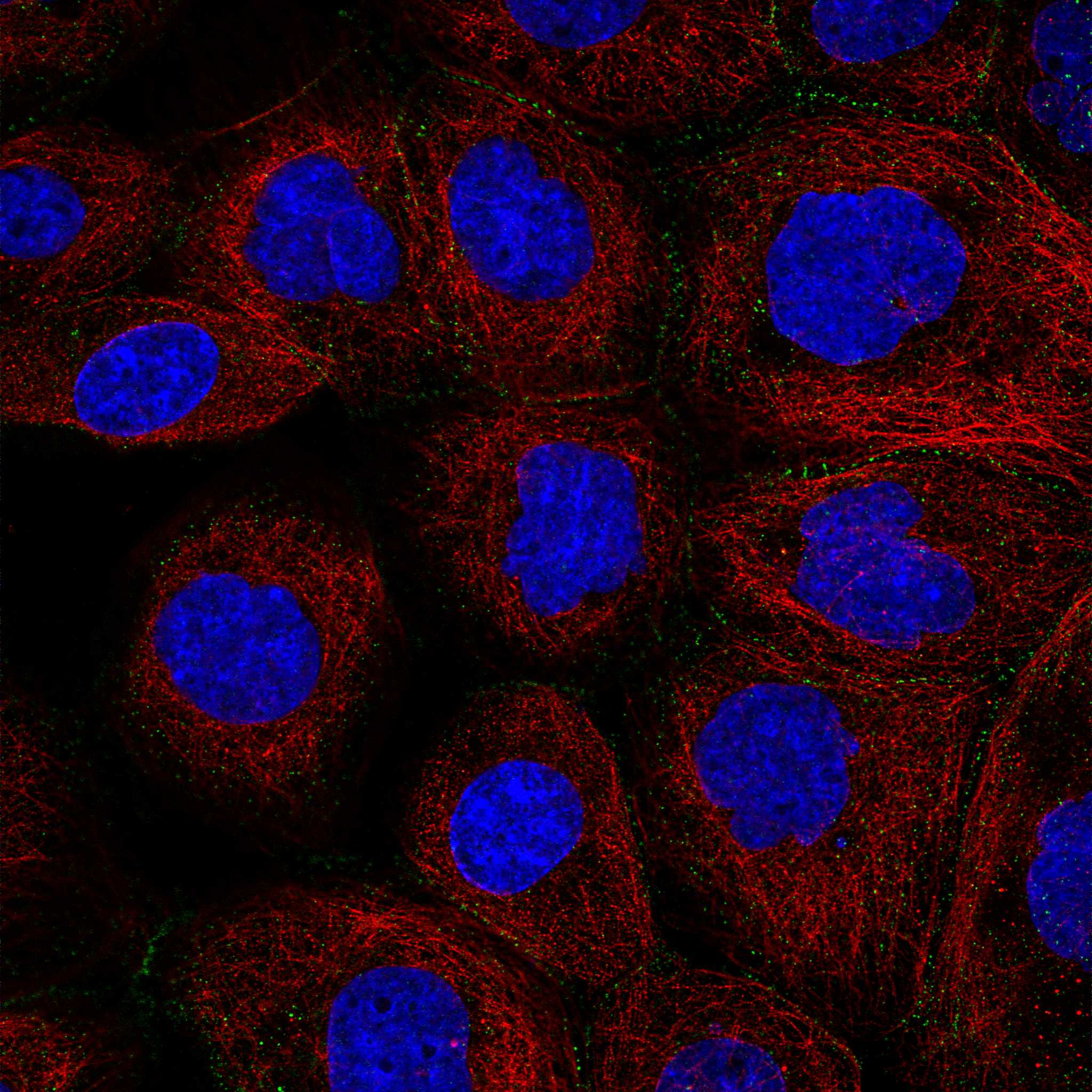
Q: What should an IF/ICC image show?A: HPA reports cell junctions as the main location, with plasma membrane and vesicles also observed (HPA subcellular ICC-IF).Compare the image with those locations and assess cell identity; consult the separate IF/ICC guide for experimental conditions (HPA subcellular ICC-IF).

Sample controls for DSG2 IHC & IF

🧪Run breast first; glandular cells should stain (HPA: High in breast glandular cells). Use adipose tissue as the negative comparator because adipocytes are not detected (HPA: Not detected in adipocytes); assess non-glandular cells on the breast slide for background staining without assuming they are DSG2-negative.
Positive control tissue: Appendix (Endocrine cells, HPA High)
Negative control tissue: Adipose tissue (HPA Not detected)
ICC-IF cell lines (HPA subcellular resource): HPA ICC-IF images show DSG2 in A-431, U-251MG, U2OS, KOLF2.1J, with annotated localisation: Cell Junctions (supported) (HPA subcellular).
Technical controls: Include a no-primary, secondary-only control and a concentration-matched mouse IgG isotype control for the mouse catalog antibody (caption: mouse primary antibody; standard IHC practice). Use DSG2 knockout tissue or a validated peptide-block control as a biological negative, and quench endogenous peroxidase before HRP/DAB detection (standard IHC practice; caption: HRP/DAB detection).
⚠️Feasibility: The selected M02035-2 paraffin-section caption does not report a fixative, and no target-specific fixation window or fixation effect is reported in the supplied evidence (caption: fixative not stated). The caption uses heat retrieval in EDTA at pH 8.0, but does not establish that retrieval is required (caption: EDTA retrieval, pH 8.0). The evidence does not establish that frozen sections or IF are easier; check breast sections for endogenous peroxidase background under the reported HRP/DAB method (HPA: breast glandular cells High; caption: HRP/DAB detection; standard IHC practice).

HPA tissue IHC evidence for DSG2

Comprehensive Human Protein Atlas IHC scoring per tissue (reliability: Enhanced — High consistency between antibody staining and RNA expression data.). Rows are taken directly from the HPA tissue chart — click any row's HPA link to view the source.

Positive expression · recommended positive controls

TissueCell typeLevelEvidenceSource
Appendix Endocrine cells High Protein (IHC) HPA →
Breast Glandular cells High Protein (IHC) HPA →
Bronchus Respiratory epithelial cells High Protein (IHC) HPA →
Cervix Glandular cells High Protein (IHC) HPA →
Colon Endocrine cells High Protein (IHC) HPA →

Undetected expression · recommended negative controls

TissueCell typeLevelEvidenceSource
Adipose tissue Adipocytes Not detected Protein (IHC) HPA →
Caudate Glial cells Not detected Protein (IHC) HPA →
Cerebellum Cells in granular layer Not detected Protein (IHC) HPA →
Cerebral cortex Endothelial cells Not detected Protein (IHC) HPA →
Hippocampus Glial cells Not detected Protein (IHC) HPA →
Section 3

Advanced DSG2 IHC Tips

Troubleshoot DSG2 staining in paraffin sections by checking retrieval, junctional pattern, controls and scoring before interpreting chromogenic signal.

Which retrieval condition should I try first for weak DSG2 staining?
Start with heat-mediated retrieval in EDTA at pH 8.0 for paraffin sections (datasheet M02035-2). The selected antibody stained a paraffin-embedded human breast cancer section after that retrieval, followed by 2 μg/ml primary antibody overnight at 4°C (datasheet M02035-2). If staining remains weak, compare a modestly adjusted heating duration on adjacent sections while keeping buffer and detection conditions constant (standard IHC practice). Include a known positive epithelial region and examine whether signal sharpens at cell borders, the expected major location for DSG2 (HPA subcellular; HPA tissue IHC). Excessively harsh retrieval can damage section morphology and complicate junctional scoring (standard IHC practice).
Could fixation explain loss of DSG2 staining in paraffin sections?
The selected tissue caption identifies a paraffin-embedded section but does not report its fixative, so DSG2-specific fixation sensitivity is unknown for this antibody (datasheet M02035-2). Record the specimen fixative and fixation interval, then compare sections processed under documented conditions with the same EDTA pH 8.0 retrieval (datasheet M02035-2; standard IHC practice). Review tissue preservation and whether cell borders remain intact before attributing faint staining to antigen loss (standard IHC practice). Keep the 2 μg/ml primary concentration and overnight 4°C incubation fixed during that comparison (datasheet M02035-2). Do not treat tissue expression patterns as a fixation experiment (HPA tissue IHC).
Where should convincing DSG2 chromogenic staining appear?
Prioritise crisp staining at contacts between adjacent cells: DSG2 is a desmosomal membrane protein, and junctions are its principal observed location (UniProt Q14126; HPA subcellular). Membranous and cytoplasmic staining occur across many tissues in the tissue atlas, so assess the cell type and morphology alongside compartment (HPA tissue IHC). In heart, the reported junctional site is the intercalated disk (UniProt Q14126, PubMed:31845994). Cytoplasmic signal can follow CASP3 cleavage during apoptosis, but diffuse staining alone does not establish that mechanism in a paraffin section (UniProt Q14126, PubMed:17559062; standard IHC interpretation). Compare the pattern with an appropriate negative control on the same staining run (standard IHC practice).
How should epitope location affect DSG2 staining interpretation?
Check the antibody's documented immunogen or epitope before assigning a missing or cytoplasmic signal to DSG2 processing (standard IHC practice). The supplied record lists an extracellular region at residues 50–609, a transmembrane segment at 610–634, and a cytoplasmic region at 635–1118 (UniProt Q14126 topology). It also lists a signal peptide at 1–23, a propeptide at 24–49, and extracellular glycosylation sites, all relevant to epitope accessibility or processing hypotheses (UniProt Q14126). No isoforms are listed in the supplied record, and the selected caption does not identify an epitope (UniProt Q14126; datasheet M02035-2). Test any proposed epitope-dependent explanation with an independently characterised antibody or control (standard IHC practice).
How can I adapt DSG2 localisation checks to multiplex IF?
Use IF as a separate localisation check: DSG2 is mainly junctional, with additional plasma membrane and vesicle localisation reported (HPA subcellular). Pair DSG2 with a marker identifying the expected cell population, such as an epithelial cell marker when examining glandular epithelium, and confirm marker specificity for the specimen (HPA tissue IHC; standard IF practice). Choose fluorophores after inspecting tissue autofluorescence and reserve a channel with low background for the weaker signal (standard IF practice). Set permeabilisation according to the antibody's documented epitope: an intracellular epitope requires access through the membrane, whereas excessive permeabilisation may compromise border definition (UniProt Q14126 topology; standard IF practice). Establish IF fixation and antibody performance independently; the paraffin IHC caption reports neither an IF method nor a fixative (datasheet M02035-2).
How do I reduce widespread brown signal without losing junctions?
First distinguish diffuse chromogen from cell-border staining by reviewing a negative control and the tissue architecture at matched magnification (standard IHC practice; HPA subcellular). For the selected paraffin example, the reported conditions were 10% goat serum blocking, 2 μg/ml primary overnight at 4°C, and a peroxidase-linked secondary for 30 minutes at 37°C (datasheet M02035-2). A peroxidase block, suitable washing, and controlled DAB development address common chromogenic background sources (standard IHC practice). If background persists, titrate primary concentration or detection exposure on adjacent sections while monitoring a positive epithelial region (standard IHC practice; HPA tissue IHC). Interpret residual cytoplasmic colour cautiously because DSG2 can also show cytoplasmic staining (HPA tissue IHC).
What should I score when DSG2 is both membranous and cytoplasmic? ⚠ ANSWER MARKED FOR VERIFICATION
Define the compartment before scoring: report junctional or membranous staining separately from cytoplasmic staining because both occur in tissue images (HPA tissue IHC; HPA subcellular). Within a specified cell population, record the percentage of positive cells and an intensity-based H-score using the same thresholds across sections (standard IHC practice). Normalise counts to the number of evaluable target cells; if measuring stained junction length or positive area, also report the analysed tissue area in mm² (standard IHC practice). Exclude folds, detached edges, and necrotic regions by a prespecified rule, and retain the same retrieval and DAB development settings across cases (standard IHC practice). Report cell type and compartment with every score because tissue expression is heterogeneous (HPA tissue IHC).
When is DSG2 staining more likely artefact than true signal?
A convincing result follows intact cell contacts in an expected population and is reproducible in a positive control stained in the same run (HPA subcellular; HPA tissue IHC; standard IHC practice). High staining is reported in breast glandular cells, whereas lymph-node germinal-center cells are reported as undetected; identify the actual cell type before judging a brown region (HPA tissue IHC). Treat isolated nuclear staining, section-edge colour, necrotic deposits, and signal retained in a negative control as reasons to investigate artefact (UniProt Q14126 localisation; standard IHC practice). Diffuse cytoplasmic colour needs care: cytoplasmic tissue staining is reported, and apoptosis-associated cleavage can redistribute DSG2, but morphology alone cannot prove cleavage (HPA tissue IHC; UniProt Q14126, PubMed:17559062). Check endogenous peroxidase control and DAB timing when background is widespread (standard IHC practice).
Boster reagents

Best DSG2 / Desmoglein-2 IHC Antibodies

Anti-DSG2 antibodies have IHC images from human and rat paraffin sections and IF/ICC images from MCF-7 cells (catalog image captions: M02035-2, A02035, PA1559).

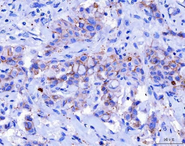
Real IHC data IHC analysis of Desmoglein 2/DSG2 using anti-Desmoglein 2/DSG2 antibody (M02035-2). Desmoglein 2/DSG2 was detected in a paraffin-embedded section of human breast cancer tissue. Heat mediated antigen retrieval was performed in EDTA buffer (pH 8.0, epitope retrieval solution). The tissue section was blocked with 10% goat serum. The tissue section was then incubated with 2 μg/ml mouse anti-Desmoglein 2/DSG2 Antibody (M02035-2) overnight at 4°C. Peroxidase Conjugated Goat Anti-mouse IgG was used as secondary antibody and incubated for 30 minutes at 37°C. The tissue section was developed using HRP Conjugated Mouse IgG Super Vision Assay Kit (Catalog # SV0001) with DAB as the chromogen.
Anti-Desmoglein 2/DSG2 Antibody ® (monoclonal, 2B4D1)
Cat # M02035-2
Real IHC data IHC analysis of DSG2 using anti-DSG2 antibody (A02035). DSG2 was detected in paraffin-embedded section of human appendicitis tissue. Heat mediated antigen retrieval was performed in EDTA buffer (pH8.0, epitope retrieval solution). The tissue section was blocked with 10% goat serum. The tissue section was then incubated with 1μg/ml rabbit anti-DSG2 Antibody (A02035) overnight at 4°C. Biotinylated goat anti-rabbit IgG was used as secondary antibody and incubated for 30 minutes at 37°C. The tissue section was developed using Strepavidin-Biotin-Complex (SABC) (Catalog # SA1022) with DAB as the chromogen.
Anti-Desmoglein 2/DSG2 Antibody ®
Cat # A02035
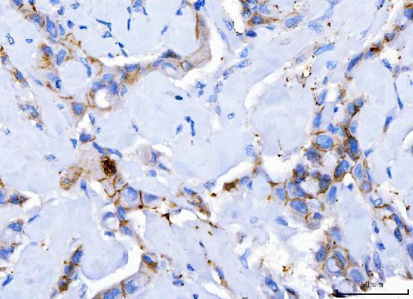
Real IHC data IHC analysis of DSG2 using anti-DSG2 antibody (PA1559). DSG2 was detected in paraffin-embedded section of human breast carcinoma tissue. Heat mediated antigen retrieval was performed in EDTA buffer (pH8.0, epitope retrieval solution). The tissue section was blocked with 10% goat serum. The tissue section was then incubated with 1μg/ml rabbit anti-DSG2 Antibody (PA1559) overnight at 4°C. Biotinylated goat anti-rabbit IgG was used as secondary antibody and incubated for 30 minutes at 37°C. The tissue section was developed using Strepavidin-Biotin-Complex (SABC) (Catalog # SA1022) with DAB as the chromogen.
Anti-Desmoglein 2/DSG2 Antibody ®
Cat # PA1559

M02035-2 has IHC images from human breast cancer, hepatocellular carcinoma, and laryngeal squamous cell carcinoma paraffin sections (M02035-2 image captions). A02035 has IHC images from human appendicitis, prostatic cancer, and rectal cancer paraffin sections plus IF/ICC data from MCF-7 cells; PA1559 has IHC images from human breast carcinoma, lung cancer, and rat intestine paraffin sections plus IF/ICC data from MCF-7 cells (A02035 and PA1559 image captions).

Which to pick: For human tissue IHC, choose mouse monoclonal M02035-2 at 2–5 µg/ml; its own paraffin-section images used EDTA retrieval at pH 8.0, and the fixative is unreported (M02035-2 catalog entry and image captions). For IF/ICC, choose A02035 or PA1559 at 2 µg/ml; both have MCF-7 IF/ICC images (A02035 and PA1559 catalog entries and IF image captions). For work across species, consider PA1559: its catalog lists human, mouse, and rat reactivity, while its own paraffin-section IHC images show human and rat tissue; the fixative is unreported (PA1559 catalog entry and IHC image captions).

Each figure is that product's own IHC / IF validation image from its datasheet.

References

  1. UniProt Consortium. UniProt entry Q14126 (DSG2_HUMAN, Desmoglein-2).
  2. Human Protein Atlas. DSG2 tissue IHC expression (reliability: Enhanced).
  3. Human Protein Atlas. DSG2 subcellular location (ICC-IF): Mainly localized to the cell junctions. In addition localized to the plasma membrane and vesicles..
  4. Human Protein Atlas. DSG2 antibody validation summary (2 antibodies).
  5. DSG2 expression is low in colon cancer and correlates with poor survival. BMC gastroenterology 2021 — PMC7789404.
  6. Negative Expression of DSG1 and DSG2, as Prognostic Biomarkers, Impacts on the Overall Survival in Patients with Extrahepatic Cholangiocarcinoma. Analytical cellular pathology (Amsterdam) 2020 — PMC7436288.
  7. Desmoglein-2 dysregulation impairs epithelial barrier integrity in eosinophilic asthma. Respiratory research 2026 — PMC12908299.
  8. Desmoglein 2 is associated with dihydroartemisinin resistance in HeLa cervical cancer cells: a potential link to the PI3K/AKT signaling pathway. Discover oncology 2026 — PMC13481938.
  9. PubMed PMID:8143788 — UniProt-cited evidence.
  10. PubMed PMID:15489334 — UniProt-cited evidence.
  11. PubMed PMID:1935985 — UniProt-cited evidence.