This website uses cookies to ensure you get the best experience on our website.


- Table of Contents
Use high-staining glandular tissue to plan a positive control for DYRK1A paraffin IHC (HPA tissue IHC). Evaluate the cytoplasmic tissue pattern (HPA tissue IHC) alongside the annotated nuclear localisation (UniProt), and check epitope coverage across 5 isoforms (UniProt).

Expected localisation, validated protocols, controls and antibodies — the at-a-glance facts below, then the full design guide.
| Expected localisation | Cytoplasmic tissue staining (HPA tissue IHC); nuclear localisation (UniProt) | |
| Staining pattern | Cytoplasmic staining across cell types (HPA tissue IHC) | |
| Antigen retrieval | Tris-EDTA pH 9.0 HIER, heat-mediated (datasheet A30487) | |
| Positive control | Appendix+4 more · see all | |
| Negative control | Liver |
| Fixation | Keep fixation consistent across sections (standard IHC practice; not target-specific) | |
| Caveat | Nuclear annotation differs from cytoplasmic tissue staining (UniProt; HPA tissue IHC) | |
| Regulation | Ubiquitous; highest in skeletal muscle and testis (UniProt) | |
| Isoform / epitope | 5 isoforms; check epitope coverage across sequences (UniProt) |
The catalog antibody’s paraffin-section protocol is followed by published DYRK1A chromogenic IHC methods for HNSCC, colon biopsies, brain cryosections, and mouse brainstem sections (PMC5086852; PMC13065994; PMC6421685; PMC3546979).
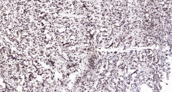
| Sample | Paraffin-embedded human Colon cancer tissue; fixative not specified (datasheet A30487) |
| Fixation | Image fixative and duration unreported (datasheet A30487); verify before use. |
| Sectioning | 4–5 µm sections on charged slides (standard) |
| Deparaffinisation | Xylene, graded ethanol series to water (standard) |
| Antigen retrieval | Heat retrieval: Tris-EDTA pH 9.0 (datasheet A30487); 20 min, 95–100 °C (standard) |
| Peroxidase block | 3% H2O2, 10 min, room temperature (standard) |
| Blocking | 10% normal serum of the secondary host, 30 min, room temperature (standard) |
| Primary antibody | Rabbit anti-DYRK1A, 1:100 - 1:300 (datasheet A30487) |
| Primary incubation | Overnight at 4 °C (standard) |
| Detection | HRP-polymer secondary, DAB chromogen 5–10 min (standard) |
| Counterstain | Hematoxylin, blue, dehydrate and mount (standard) |
| Expected result | DYRK1A-positive staining in glandular cells of appendix (HPA tissue IHC: High). HPA tissue profile: Cytoplasmic expression in all cells types. No signal in the no-primary control. |
DYRK1A is a nuclear protein with nuclear speckle localization and no transmembrane segment (UniProt Q13627: subcellular location; topology). In tissue IHC, expect cytoplasmic staining across cell types, with strong staining in several glandular cell populations and placental decidual cells (HPA: tissue IHC). Interpret this compartment difference cautiously: HPA rates its tissue IHC profile Approved, with medium consistency between antibody staining and RNA expression (HPA: tissue IHC reliability).
| Strong cytoplasmic staining in glandular cells of appendix, breast, cervix, fallopian tube, gallbladder, salivary gland or small intestine. | This matches reported High staining in those cell and tissue combinations (HPA: tissue IHC). Judge intensity within the identified cells; the profile describes cytoplasmic expression across cell types, while reported levels vary by cell and tissue (HPA: tissue IHC). |
| Nuclear staining, including a punctate nuclear pattern, accompanies or differs from the cytoplasmic IHC profile. | Nuclear and nuclear speckle localization is biologically plausible (UniProt Q13627: subcellular location; HPA: subcellular ICC-IF). The HPA tissue IHC summary instead reports cytoplasmic expression (HPA: tissue IHC). Treat this as a source-dependent compartment difference; check morphology and controls before assigning specificity. |
| A crisp cell-surface rim or staining outside cells dominates the section. | This is a compartment mismatch: DYRK1A has no transmembrane segment or signal peptide (UniProt Q13627: topology; processing), and the reported tissue pattern is cytoplasmic (HPA: tissue IHC). Consider an artefact or nonspecific deposition, especially when expected intracellular staining is absent. |
| Cholangiocytes stain strongly, or a uniform deposit covers cells and surrounding tissue. | Cholangiocytes were not detected in the HPA liver image set (HPA: liver tissue IHC), so strong staining there warrants a specificity check. A diffuse deposit that ignores cell boundaries is background; in chromogenic IHC, nonspecific binding or endogenous detection activity can produce it (general IHC practice). |
| No signal appears in a reported high-staining glandular population. | Compare with the specified cell population, since HPA reports High staining in several glandular tissues (HPA: tissue IHC). An absent signal suggests a failed or insensitive run only after confirming that the relevant cells are present; HPA's Approved rating and medium RNA agreement do not guarantee positivity in every specimen (HPA: tissue IHC reliability). |
| Tissue and cell selection | Use the named glandular populations or placental decidual cells as reported high-staining references (HPA: tissue IHC). Liver cholangiocytes provide a reported not detected comparison (HPA: liver tissue IHC); that observation is cell-specific, not a claim that the whole liver is negative. |
| Compartment evidence | The tissue IHC profile is cytoplasmic (HPA: tissue IHC), whereas UniProt places DYRK1A in the nucleus and nuclear speckles (UniProt Q13627: subcellular location). Record the actual compartment rather than treating either source alone as a universal rule for every preparation. |
| IHC evidence strength | The tissue IHC profile is Approved but has medium consistency with RNA data (HPA: tissue IHC reliability). HPA lists two IHC Approved antibodies, HPA015323 and HPA015810 (HPA: antibodies); this does not make every unexpected pattern target-specific. |
| Protein form and topology | UniProt lists five isoforms, one chain spanning residues 1–763, no signal peptide and no transmembrane segment (UniProt Q13627: isoforms; processing; topology). These facts support intracellular interpretation; without an epitope map, they do not predict which isoforms an antibody detects. |
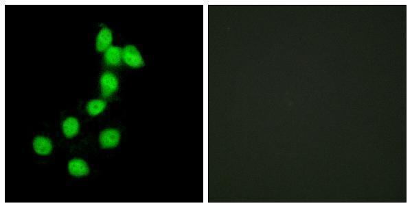
| IF/ICC Q&A: Where should fluorescence appear? | Mainly in nuclear speckles, with supported nucleoplasmic localization (HPA: subcellular ICC-IF). HPA also lists cytosol and centrosome as uncertain locations (HPA: subcellular ICC-IF). This IF observation is a comparison for interpretation, not an IHC protocol choice. |
| Situation | Likely cause | Next action |
|---|---|---|
| A reported high-staining tissue gives no chromogenic signal. | The sampled section may lack the named high-staining cells (HPA: tissue IHC), or the staining run may have failed (general IHC practice). | Confirm cell identity on the counterstain, then review the run's positive control, detection reagents and antigen retrieval record (general IHC practice). Do not infer a DYRK1A-specific retrieval requirement from HPA staining levels. |
| Staining is faint in hippocampal or caudate glial cells. | Those populations are reported at Low staining levels (HPA: tissue IHC). | Compare the same run with a named high-staining cell population and assess the faint signal against the negative control (HPA: tissue IHC; general IHC practice). Avoid scoring weak glial staining by the threshold used for high-staining glandular cells. |
| Most structures, including areas outside cells, show brown deposit. | Nonspecific binding, endogenous detection activity or excess chromogen development can create diffuse background (general chromogenic IHC practice). | Inspect the reagent control and review blocking, washes and development time (general chromogenic IHC practice). Score intracellular staining only where cell boundaries and morphology remain interpretable. |
| A membrane-like rim is the dominant result. | That pattern conflicts with the lack of a transmembrane segment or signal peptide (UniProt Q13627: topology; processing) and with HPA's cytoplasmic tissue profile (HPA: tissue IHC). | Check a negative reagent control and an HPA high-staining reference tissue in the same run; reassess whether the deposit follows cell membranes or an intracellular compartment (HPA: tissue IHC; general IHC practice). |
| Nuclear staining appears without the expected cytoplasmic IHC pattern. | DYRK1A has nuclear localization evidence (UniProt Q13627: subcellular location; HPA: subcellular ICC-IF), while HPA summarizes tissue IHC as cytoplasmic (HPA: tissue IHC). | Document the compartment and cell type, compare control sections, and avoid calling nuclear signal either definitive specificity or definitive failure from localization alone (general IHC interpretation). |
| Cholangiocytes show strong staining in a liver section. | HPA reports cholangiocytes as not detected (HPA: liver tissue IHC); unexpected signal may reflect background or cross-reactivity, though one reference observation cannot settle specificity. | Confirm the cells are cholangiocytes, examine the negative control, and compare staining with a reported high-staining population in the same run (HPA: tissue IHC; general IHC practice). |


Comprehensive Human Protein Atlas IHC scoring per tissue (reliability: Approved — Medium consistency between antibody staining and RNA expression data.). Rows are taken directly from the HPA tissue chart — click any row's HPA link to view the source.
| Tissue | Cell type | Level | Evidence | Source |
|---|---|---|---|---|
| Appendix | Glandular cells | High | Protein (IHC) | HPA → |
| Breast | Glandular cells | High | Protein (IHC) | HPA → |
| Cervix | Glandular cells | High | Protein (IHC) | HPA → |
| Fallopian tube | Glandular cells | High | Protein (IHC) | HPA → |
| Gallbladder | Glandular cells | High | Protein (IHC) | HPA → |
| Tissue | Cell type | Level | Evidence | Source |
|---|---|---|---|---|
| Liver | Cholangiocytes | Not detected | Protein (IHC) | HPA → |
Troubleshoot DYRK1A staining in paraffin sections by checking retrieval, compartment, cell type and controls before interpreting chromogenic signal.
A30487 has IHC data from paraffin-embedded human colon cancer and IF data from HepG2 cells (catalog image captions); listed reactivity covers human, mouse, and rat (catalog: reactivity).
A30487 is listed for IHC and IF/ICC (catalog: applications). Its images show IHC on paraffin-embedded human colon cancer and IF in HepG2 cells (catalog image captions).
Which to pick: For tissue IHC, choose A30487: its image shows paraffin-embedded human colon cancer with Tris-EDTA retrieval at pH 9.0 and primary antibody at 1:200 overnight; the fixative is unreported (A30487 IHC image caption). For IF/ICC, A30487 is listed for both applications and has an IF image in HepG2 cells (catalog: applications; A30487 IF image caption). For human, mouse, or rat samples, A30487 is the listed rabbit polyclonal option, though the supplied images show IHC only in human tissue and IF only in HepG2 cells (catalog: host, clonality, reactivity; A30487 image captions).