This website uses cookies to ensure you get the best experience on our website.


- Table of Contents
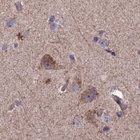
Plan ECSIT staining in paraffin sections using the observed cytoplasmic tissue pattern (HPA tissue IHC). The catalog antibody was used at 2 μg/mL in human tissue sections (datasheet A07966-3); compare matched sections with appropriate controls (standard IHC practice).

Expected localisation, validated protocols, controls and antibodies — the at-a-glance facts below, then the full design guide.
| Expected localisation | General cytoplasmic staining (HPA tissue IHC) | |
| Staining pattern | Cytoplasmic staining; high in colon glandular cells (HPA tissue IHC) | |
| Antigen retrieval | EDTA pH 8.0 HIER, heat-mediated (datasheet A07966-3) | |
| Positive control | Adrenal gland+4 more · see all | |
| Negative control | None in HPA (detected in all 45 tissues); use no-primary + isotype controls |
| Fixation | Matched tissue-IHC evidence does not establish the fixation claim. Validate the specimen-specific method before use. (selected-SKU IHC image A07966-3) | |
| Caveat | Low tissue specificity can limit tissue contrast (HPA tissue IHC) | |
| Regulation | Staining-intensity regulation unreported (UniProt) | |
| Isoform / epitope | 4 isoforms; epitope coverage unreported (UniProt) |
The catalog antibody’s IHC-P protocol is accompanied by three published ECSIT IHC methods using paraffin sections or reported IHC antibody conditions.
| Sample | Paraffin-embedded human bladder adenosquamous carcinoma tissue; fixative not specified (datasheet A07966-3) |
| Fixation | Image fixative and duration unreported (datasheet A07966-3); verify before use. |
| Sectioning | 4–5 µm sections on charged slides (standard) |
| Deparaffinisation | Xylene, graded ethanol series to water (standard) |
| Antigen retrieval | Heat retrieval: EDTA pH 8.0 (datasheet A07966-3); 20 min, 95–100 °C (standard) |
| Peroxidase block | 3% H2O2, 10 min, room temperature (standard) |
| Blocking | 10% goat serum (datasheet A07966-3) |
| Primary antibody | Rabbit anti-ECSIT, 2-5 μg/ml (datasheet A07966-3) |
| Primary incubation | Overnight at 4 °C (datasheet A07966-3) |
| Detection | Streptavidin-biotin complex (SABC), DAB chromogen (datasheet A07966-3) |
| Counterstain | Hematoxylin, blue, dehydrate and mount (standard) |
| Expected result | ECSIT-positive staining in glandular cells of adrenal gland (HPA tissue IHC: High). HPA tissue profile: General cytoplasmic expression. No signal in the no-primary control. |
ECSIT is reported in cytoplasm, nucleus and mitochondria, with no transmembrane segment (UniProt Q9BQ95). In paraffin-section IHC, expect chiefly cytoplasmic staining in the relevant cells; HPA describes general cytoplasmic expression and high staining in several glandular and neuronal cell populations (HPA tissue IHC). HPA rates its tissue IHC profile Approved, with medium consistency between staining and RNA data (HPA tissue IHC).
| Cytoplasmic staining in adrenal glandular cells, bronchial basal cells or cerebellar Purkinje cells. | This matches cell populations scored High by HPA (HPA tissue IHC). Assess staining within the named cells, alongside section morphology and controls; HPA’s overall IHC description is general cytoplasmic expression (HPA tissue IHC). |
| Predominant membrane-edge or extracellular staining, with little intracellular signal. | Treat this as a suspect pattern: ECSIT has no transmembrane segment, and HPA reports general cytoplasmic tissue staining (UniProt Q9BQ95 topology; HPA tissue IHC). Check morphology and detection controls before assigning the deposit to ECSIT. Nuclear signal alone needs separate review because UniProt also lists the nucleus (UniProt Q9BQ95). |
| Strong deposit in a cell population outside the expected stained cells, or in acellular material. | Possible explanations include cross-reactivity or endogenous chromogenic activity; appearance alone cannot distinguish them (general IHC practice). Compare the named HPA-positive cell population on the same or a parallel section and run an appropriate detection control (HPA tissue IHC; general IHC practice). |
| Diffuse colour across tissue, including areas without a clear cellular pattern. | A uniform haze makes cell-level scoring unreliable (general IHC practice). Inspect the detection-only control, washing and reagent concentration before treating the colour as ECSIT signal (general IHC practice). HPA’s reported pattern is cellular and generally cytoplasmic (HPA tissue IHC). |
| No staining in a selected HPA High population. | A blank result in adrenal glandular cells, bronchial basal cells or cerebellar Purkinje cells conflicts with the reported HPA pattern (HPA tissue IHC). First verify tissue identity and the control run, then review retrieval and detection conditions as general IHC troubleshooting steps. |
| Choice of reference cells | HPA scores adrenal and intestinal glandular cells, bronchial basal cells and selected neurons High, but oral squamous cells and adipocytes Low (HPA tissue IHC). Use a named High population to assess a weak run; Low does not mean a verified negative control. |
| Compartment and protein features | UniProt lists cytoplasm, nucleus and mitochondrion and reports no transmembrane segment or glycosylation sites (UniProt Q9BQ95). HPA’s tissue-level summary is general cytoplasmic expression (HPA tissue IHC). These records do not predict a fixation response. |
| Antibody evidence | HPA lists HPA042979 as IHC Approved and ICC Supported; its overall tissue IHC reliability is Approved with medium staining–RNA consistency (HPA antibodies; HPA tissue IHC). These ratings support comparison with the reported pattern, not automatic assignment of every deposit to ECSIT. |
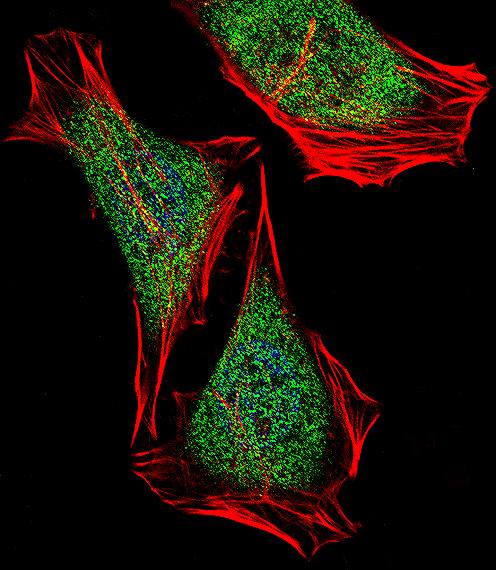
| IF/ICC Q&A: where is ECSIT seen? | HPA reports mainly nucleoplasmic signal with additional cytosol in ICC-IF, supported for both locations (HPA subcellular). This is an IF/ICC observation; paraffin-section IHC is described separately as generally cytoplasmic (HPA tissue IHC). |
| Situation | Likely cause | Next action |
|---|---|---|
| HPA High cells are blank. | Tissue selection, retrieval or detection may be at issue; the blank slide does not identify which step failed (HPA tissue IHC; general IHC practice). | Confirm the named cell population, inspect a positive tissue control, and review retrieval and detection settings under the IHC workflow (general IHC practice). |
| Signal is weak but cell-restricted. | The chosen population may be HPA Low, or staining conditions may give limited contrast (HPA tissue IHC; general IHC practice). | Compare with an HPA High population, such as bronchial basal cells, before changing staining conditions; keep comparisons within the same run where possible (HPA tissue IHC; general IHC practice). |
| Colour covers most of the section. | Diffuse background can result from nonspecific reagent binding or excess chromogen development (general IHC practice). | Check the detection-only control, blocking, washes and development time; score ECSIT only where a discernible cellular pattern remains (general IHC practice). |
| Deposit appears along membranes or in acellular spaces. | The location conflicts with the reported intracellular locations and lack of a transmembrane segment (UniProt Q9BQ95; HPA tissue IHC). | Review section morphology and a detection control, then compare intracellular staining in HPA High cells before interpreting the deposit (HPA tissue IHC; general IHC practice). |
| Unexpected cells stain more strongly than the intended reference cells. | Cross-reactivity or endogenous detection activity is possible; HPA’s Low populations are not established negatives (HPA tissue IHC; general IHC practice). | Use the named HPA High cells for pattern comparison and check the detection-only control; assess any endogenous enzyme activity relevant to the chromogen system (HPA tissue IHC; general IHC practice). |
| Nuclear staining dominates an IHC section. | Nuclear localisation is plausible from UniProt and HPA ICC-IF, while HPA summarises tissue IHC as generally cytoplasmic (UniProt Q9BQ95; HPA subcellular; HPA tissue IHC). | Check whether cytoplasmic staining is also present in HPA High cells and whether controls are clean; report the compartment observed without treating ICC-IF as an IHC protocol or identical tissue pattern (HPA tissue IHC; HPA subcellular; general IHC practice). |
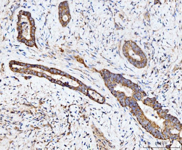
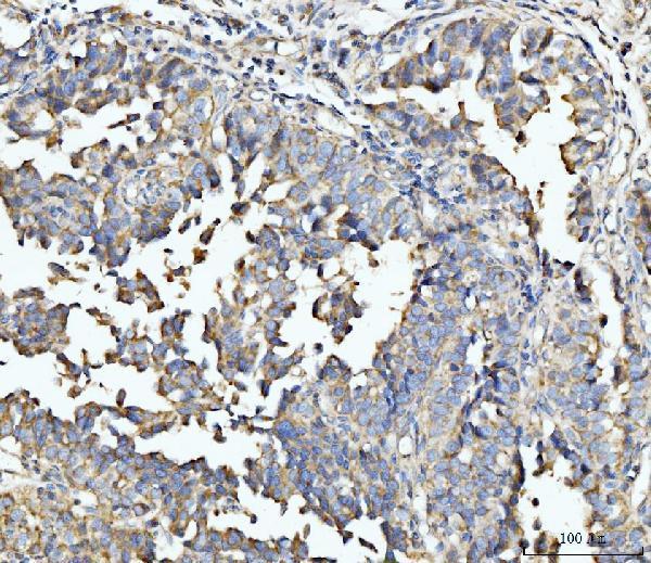
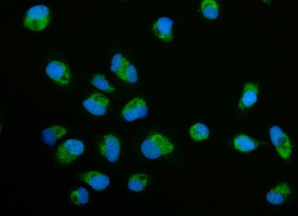
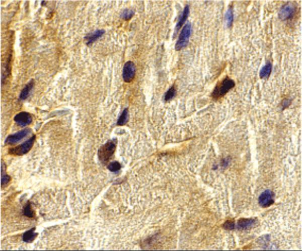


Comprehensive Human Protein Atlas IHC scoring per tissue (reliability: Approved — Medium consistency between antibody staining and RNA expression data.). Rows are taken directly from the HPA tissue chart — click any row's HPA link to view the source.
| Tissue | Cell type | Level | Evidence | Source |
|---|---|---|---|---|
| Adrenal gland | Glandular cells | High | Protein (IHC) | HPA → |
| Appendix | Glandular cells | High | Protein (IHC) | HPA → |
| Bronchus | Basal cells | High | Protein (IHC) | HPA → |
| Caudate | Neuronal cells | High | Protein (IHC) | HPA → |
| Cerebellum | Purkinje cells | High | Protein (IHC) | HPA → |
| Tissue | Cell type | Level | Evidence | Source |
|---|---|---|---|---|
| None in HPA: ECSIT is detected in all 45 scored tissues. Use a no-primary (secondary-only) and an isotype control instead. | ||||
Use the catalog antibody’s paraffin-section IHC conditions as the starting point, then assess staining by cell type and compartment.
ECSIT antibodies have IHC images from human paraffin sections and mouse heart cells (catalog image captions), plus IF images from human cells (catalog IF image captions).
A07966-3 has human paraffin-section IHC images and a PC-3 IF image (A07966-3 image captions); A07966 has a mouse heart IHC image (A07966 image caption). A07966-1 has a HeLa IF image and lists IF as a tested application (A07966-1 image caption and catalog applications).
Which to pick: For human tissue IHC, choose A07966-3: its own caption documents paraffin sections, EDTA retrieval at pH 8.0, and 2 μg/ml primary antibody; the fixative is unreported (A07966-3 IHC caption). For IF/ICC, A07966-3 lists both applications and has a PC-3 IF image, while A07966-1 lists IF and has a HeLa IF image (catalog applications and IF captions). For work involving human and mouse samples, consider A07966, which lists both species, although its IHC image shows mouse heart cells and reports no fixative (A07966 catalog reactivity and IHC caption).