This website uses cookies to ensure you get the best experience on our website.
- Table of Contents


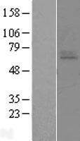
Facts about Endothelin-1 receptor.

Receptor for endothelin-1.
Mediates its action by association with G proteins that activate a phosphatidylinositol- calcium second messenger system.The rank order of binding affinities for ET-A is: ET1 > ET2 >> ET3. .
| Human | |
|---|---|
| Gene Name: | EDNRA |
| Uniprot: | P25101 |
| Entrez: | 1909 |

| Belongs to: |
|---|
| G-protein coupled receptor 1 family |

EDNRA; endothelin receptor type A; endothelin-1 receptor; endothelin-1-specific receptor; ET1-specific type; ETAR; ETRA; G protein-coupled receptor; hET-AR
Mass (kDA):
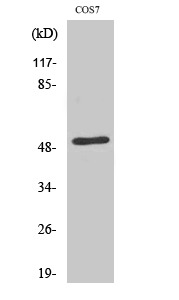
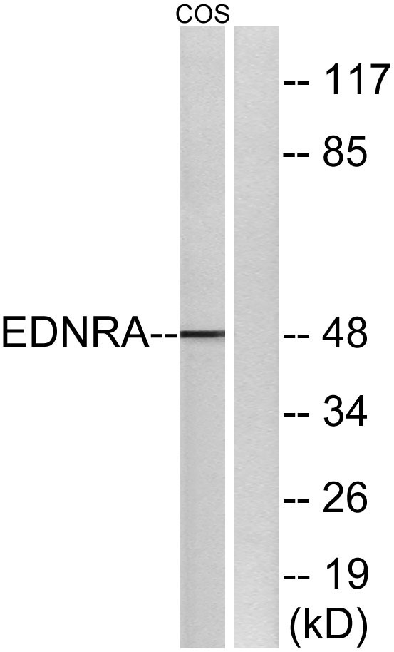
48.722 kDA

| Human | |
|---|---|
| Location: | 4q31.22-q31.23 |
| Sequence: | 4; NC_000004.12 (147480917..147544954) |
Isoform 1, isoform 3 and isoform 4 are expressed in a variety of tissues, with highest levels in the aorta and cerebellum, followed by lung, atrium and cerebral cortex, lower levels in the placenta, kidney, adrenal gland, duodenum, colon, ventricle and liver but no expression in umbilical vein endothelial cells. Within the placenta, isoform 1, isoform 2, isoform 3 and isoform 4 are expressed in the villi and stem villi vessels.
Cell membrane; Multi-pass membrane protein.


PMID: 1719979 by Adachi M., et al. Cloning and characterization of cDNA encoding human A-type endothelin receptor.
PMID: 1659806 by Cyr C., et al. Cloning and chromosomal localization of a human endothelin ETA receptor.
*More publications can be found for each product on its corresponding product page