EIF2AK2 / Interferon-induced, double-stranded RNA-activated protein kinase · IHC design guide

Design Immunohistochemistry for EIF2AK2

Plan chromogenic paraffin IHC for EIF2AK2 using its general cytoplasmic tissue pattern and high staining in bone marrow hematopoietic cells (HPA tissue IHC). Score nuclear signal separately because nuclear localization can increase in some cancers (UniProt).

Evidence assembled Oct 2026 · For research use; verify linked source records and product datasheet before use
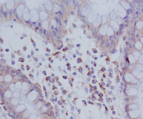
Immunohistochemistry protocol sheet for EIF2AK2 (IHC for EIF2AK2): expected localisation General cytoplasmic staining in tissue (HPA tissue IHC), antibody A01384-1, validated IHC image, and IHC protocol steps
Printable EIF2AK2 IHC protocol sheet — expected localisation General cytoplasmic staining in tissue (HPA tissue IHC), antibody A01384-1, controls and protocol steps. Open the full EIF2AK2 IHC guide →

EIF2AK2 Immunohistochemistry Experimental Design Guide

Expected localisation, validated protocols, controls and antibodies — the at-a-glance facts below, then the full design guide.

Must know before staining
Expected localisation General cytoplasmic staining in tissue (HPA tissue IHC)
Staining pattern Cytoplasmic staining in glandular and hematopoietic cells (HPA tissue IHC)
Antigen retrieval Citrate pH 6 HIER, heat-mediated (datasheet A01384-1)
Positive control ⓘ Adrenal gland+4 more · see all
Negative control ⓘ Adipose tissue
Important caveats
Reasons your staining may differ from the expected pattern.
Fixation Keep fixation consistent across sections (standard IHC practice; not target-specific)
Caveat Cancer samples may show increased nuclear staining (UniProt)
Regulation IFN induced expression (UniProt)
Isoform / epitope 2 isoforms; check epitope coverage; no transmembrane segment (UniProt)
Section 1

Recommended EIF2AK2 IHC & IF Protocols

The catalog antibody uses citrate pH 6 heat retrieval (datasheet A01384-1). One published pancreatic cancer IHC workflow provides additional parameters (PMC10561166).

Recommended immunohistochemistry (IHC-P) protocol parameters
SampleFormaldehyde-fixed, paraffin-embedded rat lung tissue (datasheet A01384-1)
FixationImage formalin-fixed; duration unreported (datasheet A01384-1); verify before use.
Sectioning4–5 µm sections on charged slides (standard)
DeparaffinisationXylene, graded ethanol series to water (standard)
Antigen retrievalHeat retrieval: Citrate pH 6 (datasheet A01384-1); 20 min, 95–100 °C (standard)
Peroxidase block3% H2O2, 10 min, room temperature (standard)
Blocking10% serum (datasheet A01384-1)
Primary antibodyRabbit anti-EIF2AK2, 2.5-5 μg/mL (datasheet A01384-1)
Primary incubationOvernight at 4 °C (standard)
DetectionHRP-polymer secondary, DAB chromogen 5–10 min (standard)
CounterstainHematoxylin, blue, dehydrate and mount (standard)
Expected resultEIF2AK2-positive staining in glandular cells of adrenal gland (HPA tissue IHC: High). HPA tissue profile: General cytoplasmic expression. No signal in the no-primary control.
💡Decision noteStart with citrate pH 6 heat retrieval (datasheet A01384-1); the published workflow heats sodium citrate at 95°C twice for 5 min (PMC10561166).
Section 2

What Is the Expected EIF2AK2 Staining Pattern?

EIF2AK2 staining in paraffin tissue sections should be predominantly cytoplasmic, including a possible perinuclear distribution (UniProt P19525 subcellular location; HPA: general cytoplasmic expression). High staining is reported in bone marrow hematopoietic cells and several glandular, respiratory epithelial, and neuronal cell populations (HPA tissue IHC). Nuclear localization can occur in specified disease settings (UniProt P19525 subcellular location). HPA rates its tissue IHC profile Approved, with medium consistency between staining and RNA expression (HPA tissue IHC reliability).

What am I looking at on my slide?
Cytoplasmic staining in bone marrow hematopoietic cells or breast glandular cells, with discernible cell boundaries (HPA: High in both populations).This fits the reported cell types and predominant compartment (HPA: general cytoplasmic expression; UniProt P19525 subcellular location). Compare intensity with neighboring cells on the same section; the HPA High category does not prescribe a chromogen score for every specimen.
Strong nuclear-only staining, with little cytoplasm, in an otherwise ordinary tissue section.This departs from the general cytoplasmic IHC profile (HPA tissue IHC). First assess counterstain, chromogen distribution, and controls. Nuclear EIF2AK2 can be genuine in the disease settings listed by UniProt, so nuclear signal alone does not establish an artefact (UniProt P19525 subcellular location).
Prominent staining of adipocytes while expected neighboring positive cells show little signal (HPA: adipocytes Not detected).Consider nonspecific antibody binding or endogenous detection activity, particularly if the same color appears in a no-primary control (general IHC practice). HPA's Not detected call describes its sampled adipocytes; it is a useful comparison, not proof that every adipocyte must be negative.
Haze across tissue and empty areas obscures cell boundaries.Diffuse signal that ignores tissue structure is difficult to score as EIF2AK2 (general IHC interpretation). Inspect a no-primary control and the location of precipitate; excess primary antibody, incomplete blocking, or detection background are general workflow possibilities, not documented EIF2AK2-specific effects.
No stain in bone marrow hematopoietic cells or breast glandular cells (HPA: High in both populations).Treat the run as inconclusive until a known-positive section stains as expected. Check that the catalog antibody is validated for IHC on paraffin sections, then review retrieval, antibody dilution, detection, and counterstain as general IHC workflow variables; no EIF2AK2-specific retrieval condition is supplied.
💡Expected EIF2AK2 appearanceA convincing positive shows cell-associated, predominantly cytoplasmic chromogen in an HPA High population such as bone marrow hematopoietic cells, while diffuse haze or staining in HPA Not detected adipocytes prompts a control check (HPA tissue IHC; general IHC interpretation).
How each factor affects the staining
Compartment and topologyEIF2AK2 is reported in cytoplasm, perinuclear cytoplasm, and nucleus, and has no transmembrane segment (UniProt P19525 subcellular location and topology). Interpret a rim near the nucleus in its cellular context; a membrane-only outline is unsupported by this record.
Tissue and cell contextHPA reports High staining in several distinct populations, Low staining in alveolar cells, cardiomyocytes, myocytes, smooth muscle cells, follicle cells, and fibroblasts, and Not detected staining in adipocytes (HPA tissue IHC). Choose comparisons by cell type; whole-organ averages can hide these differences.
Antibody evidence and molecular formsThree listed antibodies carry Approved IHC status; the supplied record does not give their epitopes (HPA antibody validation). UniProt lists 2 isoforms and phosphorylation sites, but these facts alone cannot predict which forms a particular IHC antibody detects (UniProt P19525 isoforms and modified residues).
Antigen retrievalUse the catalog antibody's IHC-P instructions as the starting point and optimize retrieval only as a general paraffin-IHC workflow step. Neither the supplied HPA staining data nor UniProt topology or modifications establishes an EIF2AK2-specific retrieval requirement or fixation sensitivity.
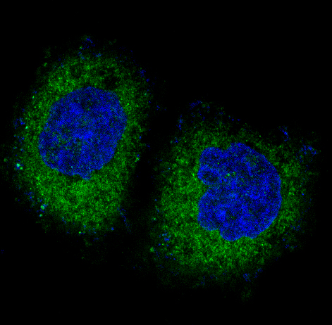
IF/ICC Q&A: where should signal appear?Predominantly in the cytosol: HPA's ICC-IF summary reports enhanced cytosolic localization (HPA subcellular ICC-IF). This supports a compartment comparison with IHC; the ICC-IF record does not specify an IF protocol here.
Why is my staining missing, weak or wrong?
SituationLikely causeNext action
Positive tissue is blank.The run may have failed, or the section and antibody may not match the intended IHC-P application; HPA reports High staining in bone marrow hematopoietic cells (HPA tissue IHC).Confirm an IHC-P-validated primary and its catalog instructions. Check a known-positive section and detection controls, then review retrieval, dilution, and chromogen development as general IHC steps.
Adipocytes show strong chromogen.This conflicts with HPA's Not detected adipocyte observation; nonspecific binding or endogenous detection activity is possible (HPA tissue IHC; general IHC practice).Compare a no-primary control and adjacent cell types. If the detection system uses an enzyme, assess its appropriate endogenous-activity block before interpreting the signal.
All compartments and empty spaces look stained.A uniform deposit does not match the reported general cytoplasmic pattern (HPA tissue IHC). Excess reagent or detection background is possible in routine IHC.Inspect the no-primary control, tissue edges, and blank slide areas; then adjust antibody concentration, washes, blocking, or chromogen development according to the detection system.
Nuclei dominate the stain.Nuclear EIF2AK2 is reported in certain cancers, acute leukemia, MDS, and advanced Creutzfeldt-Jakob disease neurocytes (UniProt P19525 subcellular location). The HPA tissue profile is generally cytoplasmic (HPA tissue IHC).Record the specimen context and whether cytoplasm is also stained. Compare controls before calling nuclear signal either specific or artifactual; do not apply the disease observation to an unrelated specimen.
A low-staining cell population is used as the sole positive control.HPA calls alveolar cells, cardiomyocytes, myocytes, smooth muscle cells, follicle cells, and fibroblasts Low, while other listed populations are High (HPA tissue IHC).Include a listed High population, such as bone marrow hematopoietic cells, for run assessment. Score each cell population separately rather than treating weak signal in a Low group as a failed assay.
Different IHC antibodies give different distributions.The listed antibodies have Approved IHC status, but no epitope mapping is supplied; EIF2AK2 has 2 isoforms (HPA antibody validation; UniProt P19525 isoforms). The source record does not identify the cause of disagreement.Compare staining in the same tissue and cell types, review each antibody's IHC-P documentation, and retain only patterns supported by appropriate controls. Do not attribute disagreement to an isoform or phosphorylation state without antibody-specific evidence.

Sample controls for EIF2AK2 IHC & IF

🧪Run bone marrow first: hematopoietic cells should stain (HPA: High in bone marrow hematopoietic cells). Use adipose tissue as the negative comparator, where adipocytes should lack specific staining (HPA: Not detected in adipocytes); HPA does not identify a negative cell type within bone marrow, so any internal negative cells should show no specific chromogen while retaining visible morphology.
Positive control tissue: Adrenal gland (Glandular cells, HPA High)
Negative control tissue: Adipose tissue (HPA Not detected)
ICC-IF cell lines (HPA subcellular resource): HPA ICC-IF images show EIF2AK2 in A-431, U-251MG, U2OS, SK-MEL-30, with annotated localisation: Cytosol (enhanced) (HPA subcellular).
Technical controls: Include no-primary (secondary-only) and host- and isotype-matched irrelevant IgG controls, plus EIF2AK2 knockout tissue if available or an immunizing-peptide block when appropriate (standard IHC practice). Quench endogenous peroxidase in bone marrow before HRP detection to reduce staining unrelated to the target (standard IHC practice; selected A01384-1 caption: HRP secondary).
⚠️Feasibility: No target-specific fixation window or fixation effect is reported in the supplied evidence; the selected A01384-1 caption describes formaldehyde fixation, although its structured fixative field says “not stated in the caption” (selected A01384-1 caption and fixative field). Heat retrieval in citrate buffer at pH 6 was used in that paraffin-section example, but a retrieval requirement is unreported (selected A01384-1 caption). Frozen-section feasibility is unreported; HPA documents cytosolic ICC-IF staining in cell lines for the separate IF/ICC application (HPA subcellular: cytosol, enhanced).

HPA tissue IHC evidence for EIF2AK2

Comprehensive Human Protein Atlas IHC scoring per tissue (reliability: Approved — Medium consistency between antibody staining and RNA expression data.). Rows are taken directly from the HPA tissue chart — click any row's HPA link to view the source.

Positive expression · recommended positive controls

TissueCell typeLevelEvidenceSource
Adrenal gland Glandular cells High Protein (IHC) HPA →
Bone marrow Hematopoietic cells High Protein (IHC) HPA →
Breast Glandular cells High Protein (IHC) HPA →
Bronchus Respiratory epithelial cells High Protein (IHC) HPA →
Caudate Neuronal cells High Protein (IHC) HPA →

Undetected expression · recommended negative controls

TissueCell typeLevelEvidenceSource
Adipose tissue Adipocytes Not detected Protein (IHC) HPA →
Section 3

Advanced EIF2AK2 IHC Tips

Use matched paraffin sections and cell-level controls to optimize EIF2AK2 staining while preserving interpretable cytoplasmic and nuclear patterns.

How should I optimize retrieval when EIF2AK2 staining is weak?
Start with heat-mediated citrate retrieval at pH 6 for paraffin sections (datasheet A01384-1). The selected rat-lung IHC used this buffer with 2.5 µg/ml primary antibody incubated overnight at 4 °C (caption A01384-1). Compare heating times on matched sections while holding cooling, antibody incubation, and chromogen development constant (standard IHC practice). If staining remains weak, test another retrieval condition only as a fallback alongside the citrate reference and a no-primary control (standard IHC practice). Accept a change when cellular signal improves without tissue damage or diffuse background (standard IHC practice).
Could fixation explain variable EIF2AK2 staining between paraffin blocks?
Target-specific fixation sensitivity for EIF2AK2 is unknown, so the supplied tissue patterns and protein features cannot establish an optimal fixation time (evidence scope: supplied record). Record fixation duration, cold ischemia, block age, and any decalcification for each specimen before comparing staining (standard IHC practice). If fixation is suspected, compare matched material fixed for 6 and 24 hours as an optimization experiment rather than an EIF2AK2 requirement (standard IHC practice). Keep citrate retrieval at pH 6 and detection settings constant across that comparison (datasheet A01384-1; standard IHC practice). Evaluate morphology and staining together, using the same control material in each run (standard IHC practice).
Should EIF2AK2 staining appear in cytoplasm, nucleus, or both?
Expect predominantly cytoplasmic staining in routine tissue IHC, consistent with the general HPA tissue profile (HPA: general cytoplasmic expression). Cytosolic localisation is also supported by cell imaging, while UniProt lists nuclear and perinuclear localisation (HPA: cytosol enhanced; UniProt P19525 localisation). Nuclear signal may therefore be plausible, particularly in the listed cancer and disease settings, but assess it separately from cytoplasmic signal (UniProt P19525 localisation). Compare staining with cell boundaries and a hematoxylin counterstain, and record the fraction of positive nuclei rather than merging compartments into one score (standard IHC practice). Confirm any unexpected pattern with controls and morphology (standard IHC practice).
How do isoforms and epitope location affect EIF2AK2 IHC interpretation?
EIF2AK2 has 2 reported isoforms, but the supplied antibody information does not map its recognized epitope (UniProt P19525 isoforms; evidence scope: supplied antibody information). Its RNA-binding domains span residues 9–77 and 100–167, and its kinase domain spans 267–538 (UniProt P19525 domains). Reported modifications include phosphorylation near residues 83–90, so an unmapped antibody cannot establish modification-specific staining (UniProt P19525 modified residues; evidence scope: supplied antibody information). Do not infer which isoform produces a stained cell from this IHC result alone (standard IHC interpretation). If isoform discrimination matters, obtain epitope mapping and validate it against isoform-specific reference material (standard IHC practice).
How can IF help assess EIF2AK2 localisation alongside a cell marker?
For multiplex IF, pair EIF2AK2 with a marker of the expected cell type, such as an epithelial marker when evaluating glandular cells (HPA: high staining in breast glandular cells; standard IF practice). Choose a far-red channel for the weaker signal when tissue autofluorescence affects shorter wavelengths, and inspect single-label controls for bleed-through (standard IF practice). EIF2AK2 has no transmembrane segment and is reported in the cytoplasm and nucleus, so permeabilisation should permit access to intracellular epitopes, including nuclei when nuclear signal is assessed (UniProt P19525 topology and localisation). Titrate permeabilisation against preserved morphology and background (standard IF practice). Interpret IF as a separate assay whose antibody performance requires its own controls (standard IF practice).
What should I check when EIF2AK2 chromogenic staining is diffuse?
First compare the stained slide with a no-primary control and inspect whether color follows cells, tissue edges, or damaged areas (standard IHC practice). Block endogenous peroxidase before HRP detection and shorten chromogen development if the control also develops color (standard IHC practice). The selected IHC caption used 10% serum blocking for 1 hour at room temperature and an HRP secondary at 1:250 (caption A01384-1). Titrate primary antibody around the reported 2.5 µg/ml while keeping retrieval at citrate pH 6 (caption A01384-1). Retain conditions that resolve cellular staining without diffuse deposits (standard IHC practice).
How should I quantify EIF2AK2 staining across specimens? ⚠ ANSWER MARKED FOR VERIFICATION
Define the tissue region and cell population before scoring, since HPA reports different staining levels among cell types and tissues (HPA: tissue IHC profile). For cellular IHC, record the percentage of positive cells and intensity on a 0–3 scale; an H-score then ranges from 0–300 (standard IHC practice). Score cytoplasmic and nuclear compartments separately because both are reported for EIF2AK2 (UniProt P19525 localisation). Normalize counts to the number of eligible cells, or report positive-cell density per mm² of viable tissue when cell counts are impractical (standard IHC practice). Apply identical thresholds, counterstain assessment, and exclusion rules across batches (standard IHC practice).
How can I distinguish true EIF2AK2 staining from artefact?
Favor staining that follows intact cells and the expected predominantly cytoplasmic pattern, while allowing separately assessed nuclear signal in relevant settings (HPA: general cytoplasmic expression; UniProt P19525 localisation). Check the cell population: HPA reports high staining in bone-marrow hematopoietic cells and low staining in lung alveolar cells (HPA: tissue IHC profile). Sharp staining confined to section edges, folds, or necrotic material warrants scrutiny as a preparation artefact (standard IHC practice). A no-primary control helps identify endogenous enzyme activity or detection-related deposits in chromogenic IHC (standard IHC practice). Do not interpret staining intensity alone as kinase activation; the protein has reported phosphorylation sites, but this antibody's modification specificity is unreported (UniProt P19525 modified residues; evidence scope: supplied antibody information).
Boster reagents

Best EIF2AK2 / Interferon-induced, double-stranded RNA-activated protein kinase IHC Antibodies

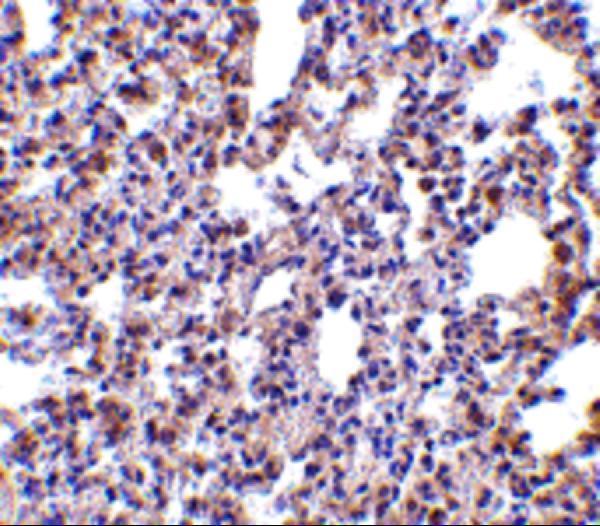
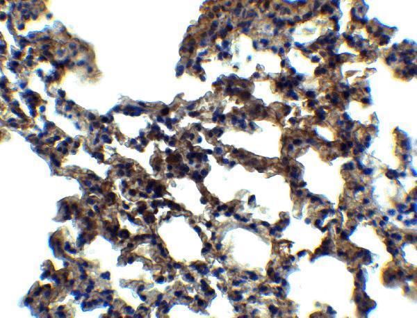
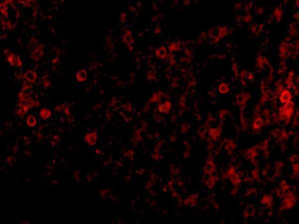
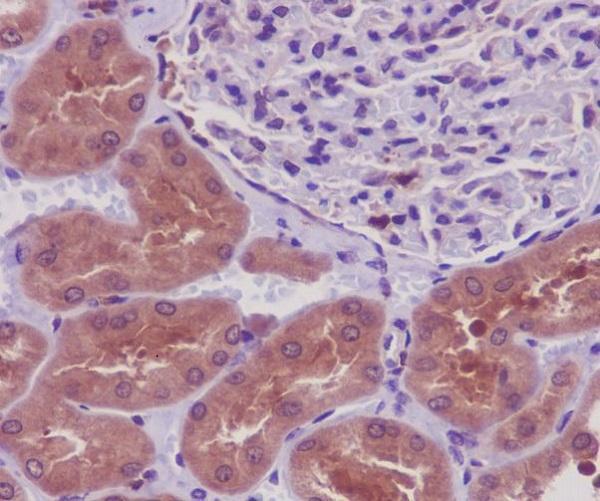
IHC figures cover rat and mouse lung, human kidney and colon; IF figures cover mouse lung and A549 cells (catalog image captions). Listed reactivity spans human, mouse and rat (catalog reactivity).

Real IHC data Immunohistochemistry Validation of PKR in Rat Lung Immunohistochemical analysis of paraffin-embedded rat lung tissue using anti-PKR antibody (A01384-1) at 2.5 μg/ml. Tissue was fixed with formaldehyde and blocked with 10% serum for 1 h at RT; antigen retrieval was by heat mediation with a citrate buffer (pH6). Samples were incubated with primary antibody overnight at 4˚C. A goat anti-rabbit IgG H&L (HRP) at 1/250 was used as secondary. Counter stained with Hematoxylin.
Anti-PKR EIF2AK2 Antibody
Cat # A01384-1
Real IHC data Immunohistochemical analysis of paraffin-embedded human kidney, using PKR Antibody.
Anti-PKR EIF2AK2 Rabbit Monoclonal Antibody
Cat # M01384
Real IHC data Immunohistochemical analysis of paraffin-embedded human colon, using Phospho-PKR (T446) Antibody.
Anti-Phospho-PKR (T446) EIF2AK2 Rabbit Monoclonal Antibody
Cat # P01384
Real IF data IF analysis of PKR/EIF2AK2 using anti-PKR/EIF2AK2 antibody (PB9803). PKR/EIF2AK2 was detected in an immunocytochemical section of A549 cells. Enzyme antigen retrieval was performed using IHC enzyme antigen retrieval reagent (AR0022) for 15 mins. The cells were blocked with 10% goat serum. And then incubated with 2 μg/mL rabbit anti-PKR/EIF2AK2 Antibody (PB9803) overnight at 4°C. DyLight488 Conjugated Goat Anti-Rabbit IgG (BA1127) was used as secondary antibody at 1:500 dilution and incubated for 30 minutes at 37°C. The section was counterstained with DAPI. Visualize using a fluorescence microscope and filter sets appropriate for the label used.
Anti-PKR/EIF2AK2 Antibody ®
Cat # PB9803

A01384-1 has IHC-P images in rat and mouse lung and IF images in mouse lung (A01384-1 image captions); M01384 has paraffin-section IHC in human kidney (M01384 IHC caption). P01384 has paraffin-section IHC in human colon (P01384 IHC caption); PB9803 has ICC/IF in A549 cells (PB9803 IF caption).

Which to pick: For tissue IHC, choose A01384-1 for formaldehyde-fixed, citrate-retrieved rat or mouse lung (A01384-1 IHC captions), M01384 for paraffin-section human kidney (M01384 IHC caption), or P01384 for phospho-PKR T446 staining in paraffin-section human colon (P01384 IHC caption); the fixative is unreported for M01384 and P01384 (M01384 and P01384 IHC captions). For IF/ICC, PB9803 has an A549-cell IF image (PB9803 IF caption), while the rabbit monoclonal M01384 lists ICC/IF and has a HeLa-cell IF image (catalog applications, clone and IF caption). For cross-species tissue IHC, A01384-1 lists human, mouse and rat reactivity, with IHC images specifically in mouse and rat (catalog reactivity; A01384-1 IHC captions).

Each figure is that product's own IHC / IF validation image from its datasheet.

References

  1. UniProt Consortium. UniProt entry P19525 (E2AK2_HUMAN, Interferon-induced, double-stranded RNA-activated protein kinase).
  2. Human Protein Atlas. EIF2AK2 tissue IHC expression (reliability: Approved).
  3. Human Protein Atlas. EIF2AK2 subcellular location (ICC-IF): Localized to the cytosol..
  4. Human Protein Atlas. EIF2AK2 antibody validation summary (3 antibodies).
  5. Eukaryotic translation initiation factor 2α kinase 2 in pancreatic cancer: An approach towards managing clinical prognosis and molecular immunological characterization. Oncology letters 2023 — PMC10561166.
  6. Mutation in Prkra results in cerebellar abnormality and reduced eIF2α phosphorylation in a model of DYT-PRKRA. Disease models & mechanisms 2024 — PMC11625895.
  7. A frameshift mutation in the murine Prkra gene causes dystonia and exhibits abnormal cerebellar development and reduced eIF2α phosphorylation. bioRxiv : the preprint server for biology 2024 — PMC11185611.
  8. Long non-coding RNA GRASLND enhances chondrogenesis via suppression of the interferon type II signaling pathway. eLife 2020 — PMC7202894.
  9. PubMed PMID:1695551 — UniProt-cited evidence.
  10. PubMed PMID:1373553 — UniProt-cited evidence.
  11. PubMed PMID:8921913 — UniProt-cited evidence.