EIF3D / Eukaryotic translation initiation factor 3 subunit D · IHC design guide

Design Immunohistochemistry for EIF3D

Plan chromogenic EIF3D IHC in paraffin sections using the general cytoplasmic tissue pattern (HPA tissue IHC). Tonsil non-germinal center cells show high staining, while staining and RNA expression have medium consistency (HPA tissue IHC).

Evidence assembled Oct 2026 · For research use; verify linked source records and product datasheet before use
Immunohistochemistry protocol sheet for EIF3D (IHC for EIF3D): expected localisation General cytoplasmic staining (HPA tissue IHC), antibody A30683, validated IHC image, and IHC protocol steps
Printable EIF3D IHC protocol sheet — expected localisation General cytoplasmic staining (HPA tissue IHC), antibody A30683, controls and protocol steps. Open the full EIF3D IHC guide →

EIF3D Immunohistochemistry Experimental Design Guide

Expected localisation, validated protocols, controls and antibodies — the at-a-glance facts below, then the full design guide.

Must know before staining
Expected localisation General cytoplasmic staining (HPA tissue IHC)
Staining pattern Cytoplasmic in tonsil non-germinal center cells (HPA tissue IHC)
Antigen retrieval Citrate pH 6.0 HIER, 95–98 °C, 20 min (rule: cytoplasmic / membrane antigen)
Positive control ⓘ Tonsil+4 more · see all
Negative control ⓘ Adipose tissue+2 more · see all
Important caveats
Reasons your staining may differ from the expected pattern.
Fixation Keep paraffin-section fixation consistent between samples. (standard IHC practice; not target-specific)
Caveat Staining and RNA expression have medium consistency (HPA tissue IHC)
Regulation Low tissue specificity (HPA tissue RNA)
Isoform / epitope 3 isoforms; epitope coverage needs checking (UniProt)
Section 1

Recommended EIF3D IHC & IF Protocols

The catalog antibody’s IHC-P protocol is followed by three published EIF3D protocols for gastric, ovarian, and renal tumor sections (PMC5661832; PMC5302064; PMC6945244).

Recommended immunohistochemistry (IHC-P) protocol parameters
SampleParaffin-embedded human breast carcinoma tissue; fixative not specified (datasheet A30683)
FixationImage fixative and duration unreported (datasheet A30683); verify before use.
Sectioning4–5 µm sections on charged slides (standard)
DeparaffinisationXylene, graded ethanol series to water (standard)
Antigen retrievalHeat-induced epitope retrieval in citrate buffer, pH 6.0, 20 min at 95–98 °C (standard rule: cytoplasmic / membrane antigen)
Peroxidase block3% H2O2, 10 min, room temperature (standard)
Blocking10% normal serum of the secondary host, 30 min, room temperature (standard)
Primary antibodyRabbit anti-EIF3D, 1:100 - 1:300 (datasheet A30683)
Primary incubationOvernight at 4 °C (standard)
DetectionHRP-polymer secondary, DAB chromogen 5–10 min (standard)
CounterstainHematoxylin, blue, dehydrate and mount (standard)
Expected resultEIF3D-positive staining in non-germinal center cells of tonsil (HPA tissue IHC: High). HPA tissue profile: General cytoplasmic expression. No signal in the no-primary control.
💡Decision noteStart with citrate pH 6.0 retrieval at 95–98 °C for 20 min (page antigen retrieval); compare the published conditions if staining needs optimization.
Section 2

What Is the Expected EIF3D Staining Pattern?

EIF3D should appear predominantly in the cytoplasm of stained cells (UniProt O15371: Cytoplasm; HPA tissue IHC: general cytoplasmic expression). Tonsil non-germinal center cells show High staining; several glandular and other cell populations show Medium staining (HPA tissue IHC). EIF3D has no transmembrane segment (UniProt O15371 topology). HPA rates its tissue staining Approved, with medium consistency against RNA data and external verification pending (HPA tissue IHC).

What am I looking at on my slide?
Cytoplasmic staining in tonsil non-germinal center cells, with weaker staining in some other tissues.This matches the reported High tonsil signal and Medium signal in adrenal, appendix and breast glandular cells (HPA tissue IHC). Compare the named cell populations rather than whole-section darkness; HPA reports general cytoplasmic expression (HPA tissue IHC).
Predominantly nuclear or sharply membranous staining, especially without cytoplasmic signal.Question target specificity: UniProt places EIF3D in the cytoplasm, and HPA reports cytoplasmic tissue staining (UniProt O15371: Cytoplasm; HPA tissue IHC). Review morphology and compare a no-primary control before interpreting the compartment as EIF3D (general IHC practice).
Strong staining in adipocytes, oral squamous epithelial cells or skeletal myocytes.Those populations were not detected in the supplied HPA tissue images (HPA tissue IHC). Consider cross-reactivity or endogenous chromogenic activity, particularly if a no-primary control also stains (general IHC practice). Treat the HPA observations as reference patterns, not universal absence claims.
Haze or precipitate across cells, stroma and empty slide areas.A widespread deposit that does not follow cellular boundaries is background rather than a convincing cytoplasmic pattern (general IHC practice; UniProt O15371: Cytoplasm). Check whether the no-primary control reproduces it, then inspect blocking, washing and chromogen development (general IHC practice).
No cytoplasmic signal in tonsil non-germinal center cells.The result conflicts with HPA's High staining in that population (HPA tissue IHC). First check that the expected cells are present and that detection controls worked; then review the antibody's validated IHC-P conditions (general IHC practice). A single blank slide cannot establish absent EIF3D expression.
💡Expected EIF3D appearanceA convincing positive is cytoplasmic staining, strongest in tonsil non-germinal center cells in the supplied tissue examples (UniProt O15371: Cytoplasm; HPA tissue IHC: High); dominant nuclear, membranous or cell-free deposit is suspect (general IHC practice).
How each factor affects the staining
Tissue and cell selectionUse tonsil non-germinal center cells as the clearest supplied positive reference (High, HPA tissue IHC). Adipocytes, oral squamous epithelial cells and skeletal myocytes were not detected in the supplied images (HPA tissue IHC); assess the named cells, not the tissue label alone.
Strength of the tissue evidenceHPA calls tissue staining Approved but reports medium consistency with RNA and pending external verification (HPA tissue IHC). Its examples guide pattern checks; they do not independently prove that every stained cell contains EIF3D.
Antibody validationHPA063330 and HPA066216 have IHC Approved status; HPA045882 has no IHC status in the supplied record (HPA antibody records). Validation status is antibody specific, so confirm which reagent produced the slide before comparing patterns.
Protein form and locationUniProt annotates 3 isoforms, a 1–548 chain, no signal peptide and no transmembrane segment (UniProt O15371). These annotations support a cytoplasmic expectation but do not predict isoform-specific staining or antibody epitope coverage.
IF/ICC Q&A: what should be visible?Q: Where should EIF3D signal localize? A: Mainly in the cytosol; HPA marks cytosol as supported and lists A-431, HEK293, U2OS and SK-MEL-30 ICC-IF images (HPA subcellular). Interpret IF/ICC using its own guide.
Why is my staining missing, weak or wrong?
SituationLikely causeNext action
Tonsil reference has no signal.A failed staining run or an unrecognized non-germinal center population is possible (general IHC practice); HPA reports High staining there (HPA tissue IHC).Verify morphology and run controls, then check the IHC-validated antibody's recommended dilution, retrieval and detection conditions (general IHC practice). Do not infer EIF3D absence until the run is valid.
Nuclei dominate the stain.The distribution conflicts with the cytoplasmic annotation and HPA tissue profile (UniProt O15371: Cytoplasm; HPA tissue IHC).Compare a no-primary control and inspect whether nuclear counterstain is being mistaken for chromogen; review antibody specificity if the nuclear chromogen persists (general IHC practice).
Adipocytes or myocytes stain strongly.These cells were not detected in the supplied HPA examples (HPA tissue IHC); cross-reactivity or background may explain the discrepancy (general IHC practice).Confirm the stained cell type, inspect a no-primary control and compare the same run with a known positive cell population (general IHC practice; HPA tissue IHC: tonsil non-germinal center cells High).
Staining covers tissue and blank slide areas.Diffuse deposit is consistent with nonspecific background or excess chromogen development (general IHC practice).Check no-primary and reagent controls; inspect washing, blocking and chromogen development before scoring cellular signal (general IHC practice).
Only a thin cell edge appears positive.An edge-only pattern does not match cytoplasmic EIF3D localization (UniProt O15371: Cytoplasm; HPA tissue IHC).Examine intact cells at higher magnification, compare the no-primary control and reassess specificity if cytoplasmic staining remains absent (general IHC practice).
Signal varies between supplied reference tissues.Variation can reflect the reported cell-level range: tonsil non-germinal center cells are High, while several glandular populations are Medium (HPA tissue IHC).Score the identified cell population and its cytoplasmic pattern within each tissue; do not require identical intensity across tissues (HPA tissue IHC; general IHC practice).

Sample controls for EIF3D IHC & IF

🧪Run tonsil first: non-germinal center cells should stain, with predominantly cytoplasmic signal (HPA: High in tonsil non-germinal center cells; UniProt O15371: Cytoplasm). Use adipose tissue as a negative because adipocytes are not detected (HPA: Not detected in adipocytes); on the tonsil slide, cells without specific staining should remain at background, but HPA does not identify a named internal negative cell type there (HPA: tonsil non-germinal center cells High).
Positive control tissue: Tonsil (Non-germinal center cells, HPA High)
Negative control tissue: Adipose tissue (HPA Not detected)
ICC-IF cell lines (HPA subcellular resource): HPA ICC-IF images show EIF3D in A-431, HEK293, U2OS, SK-MEL-30, with annotated localisation: Cytosol (supported) (HPA subcellular).
Technical controls: Run a no-primary (secondary-only) control and a host-species- and immunoglobulin-class-matched isotype control; assess specificity with an EIF3D knockout sample or peptide competition, as illustrated by the peptide-blocked section in the selected caption (caption: A30683). For chromogenic tonsil IHC, block endogenous peroxidase and check the no-primary slide for background from inflammatory cells (standard IHC practice).
⚠️Feasibility: A target-specific fixation window or fixation effect is unreported, and the selected A30683 paraffin-section caption does not state the fixative (caption: fixative not stated). Retrieval dependence is unreported, so optimize antigen retrieval empirically for paraffin IHC; the supplied evidence does not establish that frozen sections or IF/ICC are easier (standard IHC practice; HPA: ICC-IF images available). In tonsil, endogenous peroxidase in inflammatory cells can create misleading chromogenic signal (standard IHC practice).

HPA tissue IHC evidence for EIF3D

Comprehensive Human Protein Atlas IHC scoring per tissue (reliability: Approved — Medium consistency between antibody staining and RNA expression data. Pending external verification.). Rows are taken directly from the HPA tissue chart — click any row's HPA link to view the source.

Positive expression · recommended positive controls

TissueCell typeLevelEvidenceSource
Tonsil Non-germinal center cells High Protein (IHC) HPA →
Adrenal gland Glandular cells Medium Protein (IHC) HPA →
Appendix Glandular cells Medium Protein (IHC) HPA →
Bone marrow Hematopoietic cells Medium Protein (IHC) HPA →
Breast Glandular cells Medium Protein (IHC) HPA →

Undetected expression · recommended negative controls

TissueCell typeLevelEvidenceSource
Adipose tissue Adipocytes Not detected Protein (IHC) HPA →
Oral mucosa Squamous epithelial cells Not detected Protein (IHC) HPA →
Skeletal muscle Myocytes Not detected Protein (IHC) HPA →
Section 3

Advanced EIF3D IHC Tips

Troubleshoot EIF3D staining in paraffin sections by checking retrieval, cytoplasmic localisation, controls and scoring before interpreting biological differences.

What retrieval conditions should I try first for weak EIF3D staining?
Start with citrate buffer at pH 6.0, heated to 95–98 °C for 20 min (page retrieval setting). If cytoplasmic signal remains weak, compare a shorter and a longer heating interval on matched sections while holding antibody dilution and detection constant (standard IHC practice). Record tissue preservation alongside staining intensity, because excessive heating can damage morphology and make cell-level scoring unreliable (standard IHC practice). Only after that comparison should you test an alternative buffer as a fallback; a change in signal does not by itself establish specificity, so retain the peptide-blocked control shown for the catalog antibody (selected SKU A30683 caption).
How should I handle fixation when EIF3D staining varies between paraffin blocks?
EIF3D-specific sensitivity to fixation is unknown from the supplied evidence, and the catalog image states paraffin embedding without reporting its fixative (selected SKU A30683 caption). For new specimens, use one documented fixation process across comparison groups, then record fixative, duration, tissue thickness and processing dates (standard IHC practice). When blocks have different histories, stain matched sections in the same run and compare morphology before assigning biological meaning to intensity differences (standard IHC practice). Do not infer a preferred fixation time from cytoplasmic localisation, phosphorylation sites or tissue staining patterns; those observations do not measure fixation sensitivity (UniProt O15371; HPA tissue IHC).
Where should convincing EIF3D staining appear in a chromogenic section?
Prioritise interpretable cytoplasmic staining: EIF3D is annotated in the cytoplasm, and independent imaging places it mainly in the cytosol (UniProt O15371 subcellular; HPA subcellular). It has no annotated transmembrane segment or signal peptide, so an exclusively crisp membrane outline warrants investigation before scoring it as EIF3D (UniProt O15371 topology and processing). Compare the suspected signal with cytoplasmic staining in appropriate cells on the same section, and inspect nuclei, tissue edges and damaged areas separately (standard IHC practice). A nuclear-only pattern should prompt review of counterstain, detection background and antibody specificity rather than a new localisation claim (UniProt O15371 subcellular; standard IHC practice).
Can this IHC stain distinguish EIF3D isoforms or phosphorylation states?
Do not assign an EIF3D isoform from staining intensity alone: the record lists 3 isoforms, but the supplied catalog caption does not map the antibody epitope to them (UniProt O15371 isoforms; selected SKU A30683 caption). EIF3D also has annotated modified residues, including phosphoserines at positions 161, 528 and 529; no supplied IHC evidence establishes modification-specific recognition (UniProt O15371 modified residues). Check the antibody's documented immunogen or epitope against isoform sequences before claiming coverage, and use a separately validated modification-specific assay for phosphorylation claims (standard IHC practice). Peptide blocking supports binding to the competing peptide but cannot identify which isoform or modification produced tissue staining (selected SKU A30683 caption; standard IHC practice).
How can IF help resolve ambiguous EIF3D-positive cells seen by IHC?
Use IF as a complementary localisation check, multiplexing EIF3D with a validated marker for the cell population under study rather than assigning cell identity from EIF3D alone (standard IF practice). HPA reports supported cytosolic localisation and ICC/IF images in A-431, HEK293, U2OS and SK-MEL-30 cells, which provide context for the expected intracellular pattern (HPA subcellular). For tissue IF, choose fluorophores after inspecting an unstained section for autofluorescence, and favour a spectrally separated channel with low background (standard IF practice). Because the expected epitope is intracellular and EIF3D has no transmembrane segment, optimise gentle plasma-membrane permeabilisation and verify that it preserves morphology (UniProt O15371 topology; standard IF practice).
What should I check when EIF3D DAB staining is diffuse or widespread?
First inspect a no-primary control for detector background and include a peroxidase block before DAB development, both standard chromogenic IHC checks (standard IHC practice). Titrate the primary antibody and shorten chromogen development on matched sections if diffuse signal obscures cell boundaries (standard IHC practice). Broad cytoplasmic staining is plausible because HPA describes general cytoplasmic expression, but that observation does not validate every stained cell (HPA tissue IHC). The catalog image includes a synthesized-peptide blocking comparison in paraffin-embedded human breast carcinoma tissue; use it as supporting specificity evidence while assessing morphology, background controls and the expected cytoplasmic pattern together (selected SKU A30683 caption; UniProt O15371 subcellular).
How should I score EIF3D staining across tissue sections? ⚠ ANSWER MARKED FOR VERIFICATION
Define the cell population and region of interest before scoring, then record cytoplasmic intensity and the percentage of positive cells separately (standard IHC practice; UniProt O15371 subcellular). An H-score can combine intensity grades 0–3 with their cell percentages to give a 0–300 scale; report the grading rules and observer agreement (standard IHC practice). For mixed tissues, normalise counts to the number of eligible cells or report positive-cell density per mm² of viable tissue, rather than comparing raw positive-cell counts across unequal areas (standard IHC practice). Keep retrieval, staining batch, image exposure and positivity threshold consistent, and exclude necrotic or folded regions before comparing specimens (standard IHC practice).
How can I distinguish genuine EIF3D signal from staining artefact?
A credible pattern is cytoplasmic in morphologically intact cells, consistent with UniProt and HPA localisation evidence (UniProt O15371 subcellular; HPA subcellular). Compare cell populations cautiously: HPA reports high staining in tonsillar non-germinal-center cells and no detected staining in adipocytes, but its tissue IHC assessment is Approved with medium RNA-staining consistency and pending external verification (HPA tissue IHC). Signal restricted to edges, necrotic areas or an unexpected nuclear or membrane compartment should trigger review of section quality and control slides (standard IHC practice; UniProt O15371 topology). Residual endogenous peroxidase can mimic DAB signal, so check the no-primary control and peroxidase block before interpreting an isolated positive focus (standard IHC practice).
Boster reagents

Best EIF3D / Eukaryotic translation initiation factor 3 subunit D IHC Antibodies

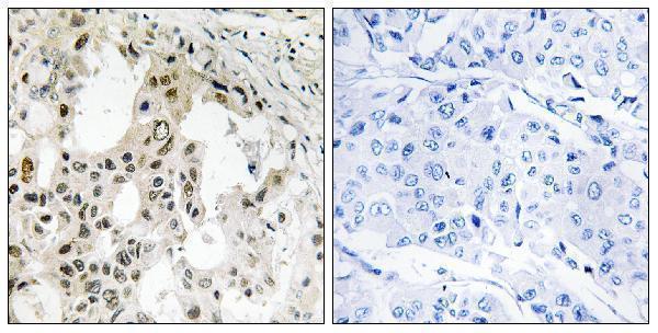
A30683 has IHC image evidence from paraffin-embedded human breast carcinoma tissue (A30683 image caption) and lists IF/ICC use with human and mouse reactivity (A30683 catalog).

Real IHC data Immunohistochemistry analysis of paraffin-embedded human breast carcinoma tissue, using EIF3D Antibody. The picture on the right is blocked with the synthesized peptide.
Anti-EIF3D Antibody
Cat # A30683

A30683 will render with an IHC image of paraffin-embedded human breast carcinoma tissue, including a peptide-blocked comparison (A30683 image caption). It lists IHC, IF and ICC applications and human and mouse reactivity (A30683 catalog).

Which to pick: Choose A30683 for paraffin-section IHC supported by the human breast carcinoma image; the fixative is unreported (A30683 image caption). For IF/ICC or human and mouse samples, A30683 is the rabbit polyclonal with those applications and species listed (A30683 catalog). For rat samples, A06212-2 lists rat reactivity, but its IHC dilution is specified for human tissue, and no IHC image is provided (A06212-2 catalog).

Each figure is that product's own IHC / IF validation image from its datasheet.

References

  1. UniProt Consortium. UniProt entry O15371 (EIF3D_HUMAN, Eukaryotic translation initiation factor 3 subunit D).
  2. Human Protein Atlas. EIF3D tissue IHC expression (reliability: Approved).
  3. Human Protein Atlas. EIF3D subcellular location (ICC-IF): Mainly localized to the cytosol..
  4. Human Protein Atlas. EIF3D antibody validation summary (3 antibodies).
  5. High expression of eIF3d is associated with poor prognosis in patients with gastric cancer. Cancer management and research 2017 — PMC5661832.
  6. Eukaryotic translation initiation factor 3 subunit D overexpression is associated with the occurrence and development of ovarian cancer. FEBS open bio 2016 — PMC5302064.
  7. Comprehensive analysis of 7-methylguanosine and immune microenvironment characteristics in clear cell renal cell carcinomas. Frontiers in genetics 2022 — PMC9393245.
  8. EIF3D promotes sunitinib resistance of renal cell carcinoma by interacting with GRP78 and inhibiting its degradation. EBioMedicine 2019 — PMC6945244.
  9. PubMed PMID:9341143 — UniProt-cited evidence.
  10. PubMed PMID:15461802 — UniProt-cited evidence.
  11. PubMed PMID:14702039 — UniProt-cited evidence.