ENO1 / Alpha-enolase · IHC design guide

Design Immunohistochemistry for ENO1

Plan chromogenic ENO1 IHC in paraffin sections using the 0.5–1 μg/mL range for A01250-1 (datasheet A01250-1). Use bronchial basal cells as a high-staining reference and assess cytoplasmic and nuclear staining (HPA tissue IHC).

Evidence assembled Oct 2026 · For research use; verify linked source records and product datasheet before use
Immunohistochemistry protocol sheet for ENO1 (IHC for ENO1): expected localisation Cytoplasmic and nuclear tissue staining (HPA tissue IHC), antibody A01250-1, validated IHC image, and IHC protocol steps
Printable ENO1 IHC protocol sheet — expected localisation Cytoplasmic and nuclear tissue staining (HPA tissue IHC), antibody A01250-1, controls and protocol steps. Open the full ENO1 IHC guide →

ENO1 Immunohistochemistry Experimental Design Guide

Expected localisation, validated protocols, controls and antibodies — the at-a-glance facts below, then the full design guide.

Must know before staining
Expected localisation Cytoplasmic and nuclear tissue staining (HPA tissue IHC)
Staining pattern Widespread cytoplasmic and nuclear staining (HPA tissue IHC)
Antigen retrieval EDTA pH 8.0 HIER, heat-mediated (datasheet A01250-1)
Positive control ⓘ Bronchus+4 more · see all
Negative control ⓘ None in HPA (detected in all 45 tissues); use no-primary + isotype controls
Important caveats
Reasons your staining may differ from the expected pattern.
Fixation Keep fixation consistent across paraffin sections (standard IHC practice; not target-specific)
Caveat Splice/transcript discrepancy may complicate interpretation (HPA tissue IHC)
Regulation Hypoxia tolerance; IHC effect unknown (UniProt)
Isoform / epitope 2 isoforms; no transmembrane segment; epitope scope unknown (UniProt)
Section 1

Recommended ENO1 IHC & IF Protocols

Compare the catalog antibody’s IHC-P protocol (datasheet A01250-1) with four published ENO1 tissue protocols (PMC12571633; PMC10931177; PMC7893569; PMC3233504).

Recommended immunohistochemistry (IHC-P) protocol parameters
SampleParaffin-embedded human liver cancer tissue; fixative not specified (datasheet A01250-1)
FixationImage fixative and duration unreported (datasheet A01250-1); verify before use.
Sectioning4–5 µm sections on charged slides (standard)
DeparaffinisationXylene, graded ethanol series to water (standard)
Antigen retrievalHeat retrieval: EDTA pH 8.0 (datasheet A01250-1); 20 min, 95–100 °C (standard)
Peroxidase block3% H2O2, 10 min, room temperature (standard)
Blocking10% goat serum (datasheet A01250-1)
Primary antibodyRabbit anti-ENO1, 0.5-1μg/ml (datasheet A01250-1)
Primary incubationOvernight at 4 °C (datasheet A01250-1)
DetectionStreptavidin-biotin complex (SABC), DAB chromogen (datasheet A01250-1)
CounterstainHematoxylin, blue, dehydrate and mount (standard)
Expected resultENO1-positive staining in basal cells of bronchus (HPA tissue IHC: High). HPA tissue profile: Ubiquitous cytoplasmic and nuclear expression. No signal in the no-primary control.
💡Decision noteStart with heat-mediated EDTA retrieval at pH 8.0 (datasheet A01250-1); published alternatives include EDTA pH 7.4 (PMC12571633) and citrate pH 6.0 (PMC3233504).
Section 2

What Is the Expected ENO1 Staining Pattern?

ENO1 should stain cytoplasm and may stain nuclei across many cell types; HPA describes a ubiquitous cytoplasmic and nuclear IHC pattern, with high staining in bronchial basal cells, fallopian tube non-ciliated cells and lung alveolar type I cells (HPA tissue IHC). UniProt also places ENO1 at the cell membrane and reports no transmembrane segment (UniProt P06733 topology). HPA rates the tissue pattern Supported, with medium staining–RNA consistency and a splice/transcript discrepancy (HPA tissue IHC).

What am I looking at on my slide?
Bronchial basal cells, fallopian tube non-ciliated cells or lung alveolar type I cells show strong cytoplasmic staining, with possible nuclear staining (HPA tissue IHC).This fits HPA's High staining calls and its broader cytoplasmic and nuclear profile (HPA tissue IHC). Score intensity and compartment in identified cells rather than treating every stained structure as equally informative (general IHC practice).
Staining is confined to an unexpected structure, with no discernible cytoplasmic signal in expected positive cells (HPA tissue IHC; UniProt P06733 subcellular location).Check morphology and detection background before assigning ENO1 localisation (general IHC practice). Nuclear or membrane staining alone is not automatically erroneous: both compartments have source support (HPA tissue IHC; UniProt P06733 subcellular location).
Fibroblasts dominate the signal while nearby expected positive cells are unstained (HPA tissue IHC).HPA lists soft-tissue fibroblasts as Low, so this reversal warrants a cross-reactivity or endogenous-detection check (HPA tissue IHC; general IHC practice). Low means weak expected staining, not an absolute negative control (HPA tissue IHC).
Chromogen forms a broad haze across tissue and spaces without a clear cellular pattern (general IHC practice).Diffuse background limits compartment and cell-level scoring; assess nonspecific detection, blocking and washing with appropriate reagent controls (general IHC practice). It cannot establish widespread ENO1 expression by itself.
A section containing bronchial basal cells has no interpretable signal in those cells (HPA tissue IHC).Those cells are an HPA High reference, so first check section integrity, antibody use and detection performance (HPA tissue IHC; general IHC practice). An absent signal alone cannot establish that the specimen lacks ENO1.
💡Expected ENO1 appearanceCall an IHC result positive when identifiable cells show cellular cytoplasmic staining, with nuclear staining permissible, and the named HPA High cell populations stain strongly (HPA tissue IHC); uniform haze or isolated deposits without cell boundaries are suspect background (general IHC practice).
How each factor affects the staining
Tissue and cell choiceBronchial basal cells, fallopian tube non-ciliated cells and lung alveolar type I cells are High references; cardiomyocytes, smooth muscle cells, skeletal myocytes and fibroblasts are Low, not confirmed negatives (HPA tissue IHC). Compare like cell populations when judging intensity (general IHC practice).
Compartment and topologyHPA reports cytoplasmic and nuclear tissue staining; UniProt lists cytoplasm, nucleus and cell membrane, with no transmembrane segment or signal peptide (HPA tissue IHC; UniProt P06733). These annotations do not predict a distinct membrane outline in every paraffin section.
Antibody evidenceCAB018614, CAB069394 and CAB080034 have Supported IHC status; HPA068284 and HPA068721 have no listed IHC status (HPA antibodies). Supported evidence helps set expectations but does not validate every section, detector or scoring threshold (general IHC practice).
Isoforms and epitope scopeUniProt lists alpha-enolase and MBP-1 isoforms, plus modified residues (UniProt P06733). The supplied record does not map an IHC antibody epitope or show which isoforms it detects, so a compartment pattern cannot identify an isoform.
IF/ICC Q: What pattern is reported?A: HPA describes mainly plasma-membrane and cytosol localisation in ICC-IF (HPA subcellular). That observation can inform compartment interpretation, but it is separate from HPA's tissue IHC staining profile and is not an IHC protocol (HPA tissue IHC; HPA subcellular).
Why is my staining missing, weak or wrong?
SituationLikely causeNext action
No stain in expected High cells (HPA tissue IHC).An uninformative section, failed detection or unsuitable assay conditions may explain absent signal (general IHC practice).Confirm the named cell type and section quality; run a known positive section and detection control, then review the antibody's IHC instructions, including retrieval if specified (general IHC practice). ENO1-specific retrieval sensitivity is unreported here.
Signal covers most tissue as a uniform brown haze (general IHC practice).Nonspecific antibody or detection background may obscure cellular staining (general IHC practice).Inspect a no-primary control, blocking, washing and detector settings; interpret only staining that resolves to cells and compartments (general IHC practice).
Chromogen appears in control sections without primary antibody (general IHC practice).Endogenous detection activity or a detector-related artifact is possible (general IHC practice).Apply the detector's appropriate endogenous-activity control or block and repeat the no-primary comparison before attributing signal to ENO1 (general IHC practice).
Fibroblasts stain strongly while bronchial basal cells are weak or blank (HPA tissue IHC).This reverses the supplied Low and High reference levels; cross-reactivity or a technical difference is possible (HPA tissue IHC; general IHC practice).Verify cell identity and compare an HPA High reference with controls under the same staining run; review antibody-specific IHC validation (HPA antibodies; general IHC practice).
A membrane edge or nuclear signal is dismissed as an artifact solely because it is not cytoplasmic (HPA tissue IHC; UniProt P06733).That rule overlooks reported nuclear and membrane localisation (HPA tissue IHC; UniProt P06733 subcellular location).Recheck cellular morphology and control staining; record compartments separately. Do not infer that membrane-associated ENO1 spans the membrane (UniProt P06733 topology; general IHC practice).
Low-staining muscle cells or fibroblasts are used as an absolute negative control (HPA tissue IHC).HPA labels these cells Low, while describing ENO1 tissue expression as ubiquitous (HPA tissue IHC).Use them only for relative intensity context; rely on reagent controls to assess detection background and a named High cell population to check positive performance (HPA tissue IHC; general IHC practice).

Sample controls for ENO1 IHC & IF

🧪Run bronchus first and look for staining in basal cells (HPA: High in bronchus basal cells). HPA lists no negative tissue and detects ENO1 in all 45 scored tissues, so use no-primary and isotype controls for the negative comparison; any unstained cells on the bronchus slide are a background reference, not a validated ENO1-negative population (HPA: no negative rows; ENO1 detected in all scored tissues).
Positive control tissue: Bronchus (Basal cells, HPA High)
Negative control tissue: None in HPA: ENO1 is detected in all 45 scored tissues. Use a no-primary (secondary-only) and an isotype control instead.
ICC-IF cell lines (HPA subcellular resource): HPA ICC-IF images show ENO1 in A-549, MCF-7, U2OS, HeLa, A-431, PC-3, with annotated localisation: Plasma membrane (enhanced), Cytosol (enhanced) (HPA subcellular).
Technical controls: Include a no-primary, secondary-only section, a rabbit control IgG matched as closely as possible to the primary antibody, and, if available, an ENO1 knockout specimen or validated peptide-block control (caption: rabbit anti-ENO1; standard IHC control practice). For bronchus sections, check endogenous peroxidase and biotin background when using the caption’s biotinylated secondary and SABC/DAB detection (caption: biotinylated goat anti-rabbit IgG and SABC/DAB; standard IHC practice).
⚠️Feasibility: No target-specific fixation window or fixation effect is reported, and the selected A01250-1 paraffin-section caption does not state a fixative (caption: fixative unreported). The reported IHC workflow uses heat retrieval in EDTA at pH 8.0, but the evidence does not establish whether retrieval is required under other conditions (caption: heat-mediated EDTA retrieval). HPA has ICC-IF images, while the supplied evidence does not establish that frozen sections or IF are easier; assess bronchus staining alongside endogenous peroxidase and biotin background controls (HPA: ICC-IF images; caption: SABC/DAB; standard IHC control practice).

HPA tissue IHC evidence for ENO1

Comprehensive Human Protein Atlas IHC scoring per tissue (reliability: Supported — Medium consistency between antibody staining and RNA expression data. Caution, Splice and/or transcript discrepancy exists.). Rows are taken directly from the HPA tissue chart — click any row's HPA link to view the source.

Positive expression · recommended positive controls

TissueCell typeLevelEvidenceSource
Bronchus Basal cells High Protein (IHC) HPA →
Fallopian tube Non-ciliated cells High Protein (IHC) HPA →
Lung Alveolar cells type I High Protein (IHC) HPA →
Adipose tissue Adipocytes Medium Protein (IHC) HPA →
Adrenal gland Glandular cells Medium Protein (IHC) HPA →

Undetected expression · recommended negative controls

TissueCell typeLevelEvidenceSource
None in HPA: ENO1 is detected in all 45 scored tissues. Use a no-primary (secondary-only) and an isotype control instead.
Section 3

Advanced ENO1 IHC Tips

Troubleshoot ENO1 staining in paraffin sections by checking retrieval, compartment patterns, controls and scoring before interpreting chromogenic signal.

Which antigen retrieval should I try first for ENO1 paraffin sections?
Start with heat-mediated retrieval in EDTA at pH 8.0 for the catalog antibody in paraffin sections (datasheet A01250-1). The selected tissue image used that retrieval, followed by 10% goat serum blocking and 1 µg/mL primary antibody overnight at 4°C (caption A01250-1). If staining is weak, compare a second retrieval condition on adjacent sections while holding antibody concentration and detection constant (standard IHC practice). Assess tissue preservation and background alongside signal, because stronger chromogen alone does not establish better antigen detection (standard IHC practice). Do not assume that a retrieval result from another antibody applies to this catalog antibody (standard IHC practice).
How should I troubleshoot fixation-related changes in ENO1 staining?
Target-specific sensitivity of ENO1 staining to fixation is unknown from the supplied evidence (caption A01250-1: fixative unreported). The selected image identifies paraffin-embedded tissue and EDTA retrieval, but does not state its fixative (caption A01250-1). Record the fixative, fixation duration and processing history for each case, and compare sections processed alike when investigating weak or variable signal (standard IHC practice). Use the documented pH 8.0 EDTA retrieval as the starting condition, then assess morphology, nuclear detail and background beside ENO1 staining (datasheet A01250-1; standard IHC practice). Do not attribute a staining difference to fixation without a controlled comparison (standard IHC practice).
What ENO1 staining compartments should I expect in IHC?
Expect widespread cytoplasmic staining with possible nuclear signal in tissue sections (HPA: ubiquitous cytoplasmic and nuclear expression). Membrane-associated signal is also biologically plausible: ENO1 can move to the plasma membrane, although it has no transmembrane segment (UniProt P06733: subcellular location and topology). HPA subcellular imaging emphasizes plasma membrane and cytosol, so assess these compartments separately when comparing IHC fields (HPA subcellular: enhanced membrane and cytosol; standard IHC practice). Confirm that apparent membrane rims follow intact cell borders rather than folds or section edges (standard IHC practice). Nuclear staining alone cannot identify which ENO1 isoform produced the signal (UniProt P06733: 2 isoforms; standard IHC interpretation).
Can this stain distinguish alpha-enolase from MBP-1 or modified ENO1?
Do not assign staining to one isoform without knowing which sequence the antibody recognizes (UniProt P06733: alpha-enolase and MBP-1; standard IHC interpretation). ENO1 has 2 listed isoforms and annotated modified residues, including acetylation and phosphorylation (UniProt P06733: isoforms and modified residues). An epitope overlapping an isoform-specific region or modified residue could change recognition, but the supplied caption does not map this antibody's epitope (UniProt P06733: isoforms and modified residues; caption A01250-1). Compare staining with an independently characterized antibody or an appropriate specificity control if isoform identity matters (standard IHC practice). Report the observed compartment and antibody identity without claiming isoform-specific detection (standard IHC interpretation).
How can I investigate ENO1 localisation with IF alongside IHC?
Use IF as a separate localisation experiment; the selected antibody caption documents chromogenic IHC in paraffin sections, not an IF procedure (caption A01250-1). Multiplex ENO1 with a marker for the expected cell type, then inspect overlap and individual channels before assigning signal to that population (standard IF practice). Choose fluorophores in channels with low tissue autofluorescence and include an unstained section to assess intrinsic fluorescence (standard IF practice). For cytosolic or nuclear epitopes, optimize permeabilisation; for a cell-surface epitope, compare staining without permeabilisation, since ENO1 lacks a transmembrane segment and its antibody epitope is unspecified (UniProt P06733: topology and localisation; standard IF practice).
How do I reduce diffuse or misleading ENO1 IHC background?
First compare the stained section with a no-primary control and inspect whether diffuse signal follows tissue structures or processing artefacts (standard IHC practice). The selected image used 10% goat serum before 1 µg/mL primary antibody overnight at 4°C; these are documented starting conditions for the catalog antibody (caption A01250-1). With its biotinylated secondary and avidin-biotin detection, check endogenous biotin interference, and include a peroxidase block before DAB development (caption A01250-1; standard IHC practice). Adjust blocking, washes, primary concentration and chromogen development one variable at a time (standard IHC practice). Remember that broad cytoplasmic ENO1 expression can be genuine, so diffuse staining alone is insufficient evidence of background (HPA: ubiquitous cytoplasmic expression).
How should I score ENO1 staining across tissue sections? ⚠ ANSWER MARKED FOR VERIFICATION
Predefine the cell population, compartment and regions to score before reviewing group labels (standard IHC practice). For cytoplasmic or nuclear ENO1, report the percentage of positive cells and an intensity-based H-score; score membrane signal separately when present (HPA: cytoplasmic and nuclear expression; UniProt P06733: membrane localisation; standard IHC practice). Normalize counts to eligible intact cells, or report positive-cell density per mm² of viable tissue when cell segmentation is impractical (standard IHC practice). Apply identical thresholds and chromogen exposure criteria across the comparison set (standard IHC practice). Record exclusions for folds, edges and necrotic regions so that tissue quality does not drive the apparent ENO1 difference (standard IHC practice).
How can I distinguish true ENO1 staining from artefact?
Judge signal against the expected broad cytoplasmic distribution, while allowing nuclear and membrane-associated staining where cell morphology supports it (HPA: ubiquitous cytoplasmic and nuclear expression; UniProt P06733: membrane localisation). Do not reject a cell solely because its intensity differs: HPA lists high staining in bronchial basal cells and low staining in cardiomyocytes (HPA tissue IHC: bronchus and heart muscle). Treat staining confined to cut edges, folds or necrotic material as suspect and inspect a no-primary control (standard IHC practice). In DAB IHC, assess endogenous peroxidase and biotin-related detection background before assigning isolated deposits to ENO1 (caption A01250-1: avidin-biotin/DAB detection; standard IHC practice).
Boster reagents

Best ENO1 / Alpha-enolase IHC Antibodies

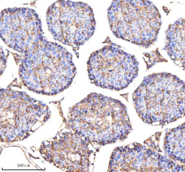
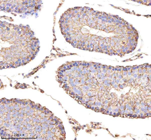
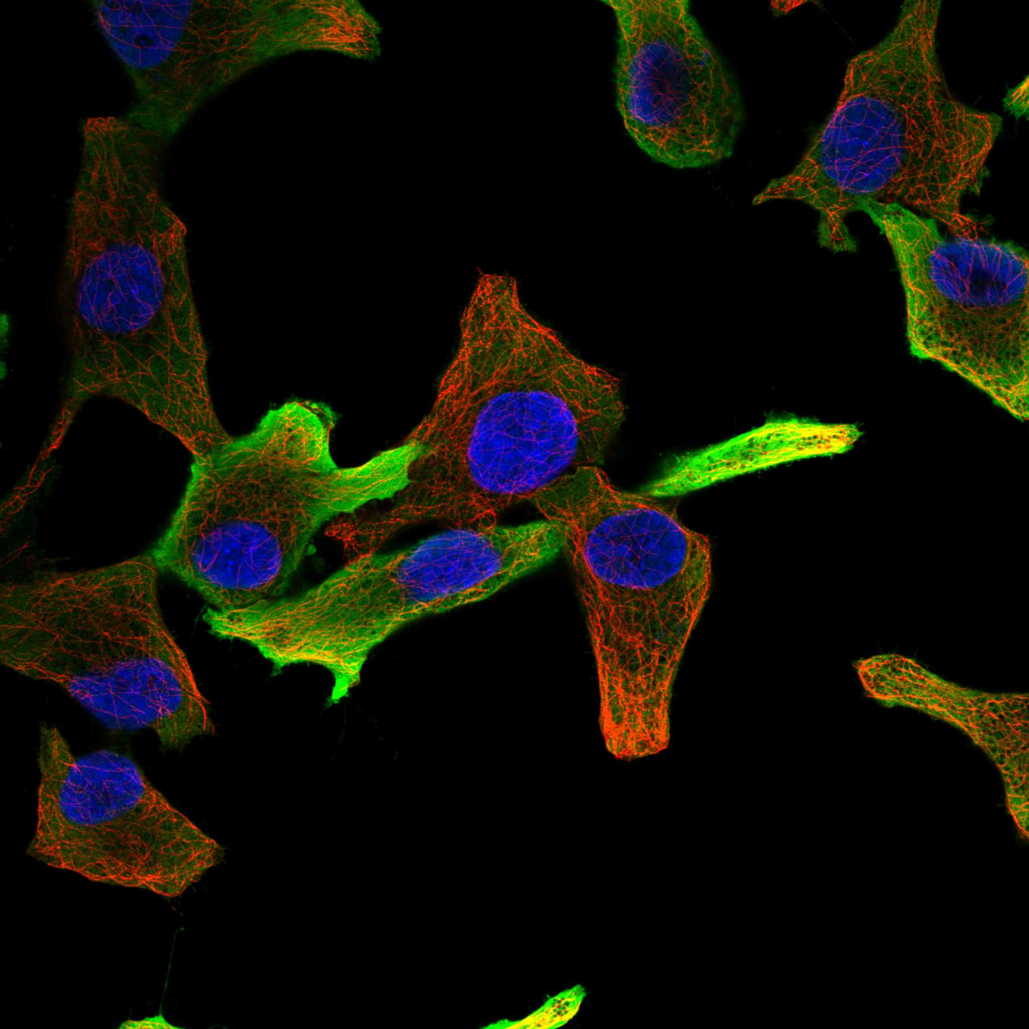
Anti-ENO1 IHC images cover paraffin sections of human liver cancer and cerebellum and mouse and rat testis; an IF image covers A431 cells (catalog image captions: A01250-1, A01250-3).

Real IHC data IHC analysis of ENO1 using anti-ENO1 antibody (A01250-1). ENO1 was detected in paraffin-embedded section of human liver cancer tissue. Heat mediated antigen retrieval was performed in EDTA buffer (pH8.0, epitope retrieval solution). The tissue section was blocked with 10% goat serum. The tissue section was then incubated with 1μg/ml rabbit anti-ENO1 Antibody (A01250-1) overnight at 4°C. Biotinylated goat anti-rabbit IgG was used as secondary antibody and incubated for 30 minutes at 37°C. The tissue section was developed using Strepavidin-Biotin-Complex (SABC) (Catalog # SA1022) with DAB as the chromogen.
Anti-ENO1 Antibody ®
Cat # A01250-1
Real IHC data IHC analysis of ENO1 using anti-ENO1 antibody (A01250-3). ENO1 was detected in a paraffin-embedded section of mouse testis tissue. Heat mediated antigen retrieval was performed in EDTA buffer (pH 8.0, epitope retrieval solution). The tissue section was blocked with 10% goat serum. The tissue section was then incubated with 2 μg/ml rabbit anti-ENO1 Antibody (A01250-3) overnight at 4°C. Peroxidase Conjugated Goat Anti-rabbit IgG was used as secondary antibody and incubated for 30 minutes at 37°C. The tissue section was developed using HRP Conjugated Rabbit IgG Super Vision Assay Kit (Catalog # SV0002) with DAB as the chromogen.
Anti-ENO1 Antibody ®
Cat # A01250-3
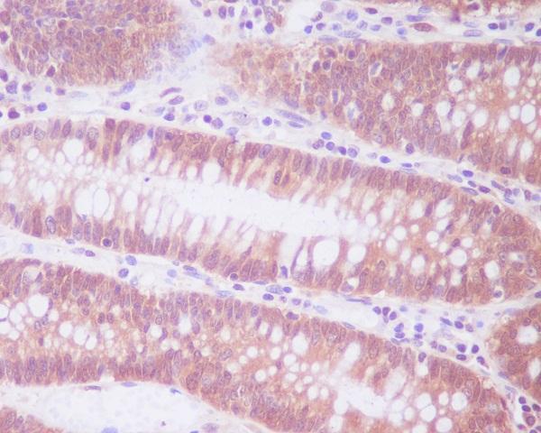
Real IHC data Immunohistochemical analysis of paraffin-embedded human colon, using ENO1 Antibody.
Anti-ENO1/Alpha Enolase Rabbit Monoclonal Antibody
Cat # M01250

A01250-1 lists IHC and IF/ICC, with IHC images from human liver cancer and mouse and rat testis and an IF image from A431 cells; A01250-3 lists IHC, with images from mouse and rat testis and human cerebellum (catalog applications and image captions). M01250 lists IF/ICC and has a paraffin human colon IHC image, although IHC is absent from its application list and no IF image is supplied (catalog applications and image captions).

Which to pick: For tissue IHC, choose A01250-1 at 0.5–1 μg/ml or A01250-3 at 2–5 μg/ml, matching the sample to each SKU’s own paraffin IHC captions; the captions do not report the fixative (catalog dilutions and IHC captions). For IF/ICC, A01250-1 has an A431 IF image at 2 μg/ml, while monoclonal M01250 (clone AEA-5) lists IF/ICC at 1:50 without an IF image (catalog IF caption, applications, clone field and dilutions). For human, mouse and rat tissue IHC, both A01250-1 and A01250-3 have species matched IHC captions; A01250-1 also lists monkey reactivity, without a monkey IHC caption (catalog reactivity and IHC captions).

Each figure is that product's own IHC / IF validation image from its datasheet.

References

  1. UniProt Consortium. UniProt entry P06733 (ENOA_HUMAN, Alpha-enolase).
  2. Human Protein Atlas. ENO1 tissue IHC expression (reliability: Supported).
  3. Human Protein Atlas. ENO1 subcellular location (ICC-IF): Mainly localized to the plasma membrane and cytosol..
  4. Human Protein Atlas. ENO1 antibody validation summary (5 antibodies).
  5. Immunohistochemical analysis of Enolase-1 sublocalization in benign and malignant breast tumors: potential implications for tumor progression and prognosis. Frontiers in oncology 2025 — PMC12571633.
  6. High Expression of ENO1 and Low Levels of Circulating Anti-ENO1 Autoantibodies in Patients with Myelodysplastic Neoplasms and Acute Myeloid Leukaemia. Cancers 2024 — PMC10931177.
  7. Impact of Tissue Enolase 1 Protein Overexpression in Esophageal Cancer Progression. International journal of medical sciences 2021 — PMC7893569.
  8. Arsenic, cadmium and neuron specific enolase (ENO2, γ-enolase) expression in breast cancer. Cancer cell international 2011 — PMC3233504.
  9. PubMed PMID:3529090 — UniProt-cited evidence.
  10. PubMed PMID:2373081 — UniProt-cited evidence.
  11. PubMed PMID:2005901 — UniProt-cited evidence.