This website uses cookies to ensure you get the best experience on our website.


- Table of Contents
Plan chromogenic ENTPD2 IHC-P with the catalog antibody at 1:50–1:100 (datasheet: A07914-1). Compare cytoplasmic tissue staining with membrane and ER expectations, and consider cerebellar granular-layer cells as a positive control (HPA tissue IHC; UniProt).

Expected localisation, validated protocols, controls and antibodies — the at-a-glance facts below, then the full design guide.
| Expected localisation | Cytoplasmic staining in tissue; membrane and ER localisation expected from topology (HPA tissue IHC; UniProt) | |
| Staining pattern | Cytoplasmic staining across several tissue types (HPA tissue IHC) | |
| Antigen retrieval | Citrate pH 6.0 HIER, 95–98 °C, 20 min (rule: cytoplasmic / membrane antigen) | |
| Positive control | Adrenal gland+4 more · see all | |
| Negative control | Adipose tissue+2 more · see all |
| Fixation | Formalin-fixed paraffin sections are documented (selected-SKU IHC image A07914-1); Matched tissue-IHC evidence does not establish the fixation claim. Validate the specimen-specific method before use. (selected-SKU IHC image A07914-1) | |
| Caveat | Medium staining–RNA consistency limits tissue prediction (HPA tissue IHC) | |
| Regulation | No expression regulator reported (UniProt) | |
| Isoform / epitope | 3 isoforms; check whether the N-terminal epitope spans cytoplasmic or extracellular sequence (UniProt; datasheet: A07914-1) |
The catalog antibody’s IHC-P protocol is provided separately. The published protocols below describe ENTPD2 staining in human hepatocellular carcinoma and gastric tissue (PMC7438573; PMC13066996).
| Sample | FFPE human brain tissue (datasheet A07914-1) |
| Fixation | Image formalin-fixed; duration unreported (datasheet A07914-1); verify before use. |
| Sectioning | 4–5 µm sections on charged slides (standard) |
| Deparaffinisation | Xylene, graded ethanol series to water (standard) |
| Antigen retrieval | Heat-induced epitope retrieval in citrate buffer, pH 6.0, 20 min at 95–98 °C (standard rule: cytoplasmic / membrane antigen) |
| Peroxidase block | 3% H2O2, 10 min, room temperature (standard) |
| Blocking | 10% normal serum of the secondary host, 30 min, room temperature (standard) |
| Primary antibody | Rabbit anti-ENTPD2, 1:50-1:100 (datasheet A07914-1) |
| Primary incubation | Overnight at 4 °C (standard) |
| Detection | HRP-polymer secondary, DAB chromogen 5–10 min (standard) |
| Counterstain | Hematoxylin, blue, dehydrate and mount (standard) |
| Expected result | ENTPD2-positive staining in glandular cells of adrenal gland (HPA tissue IHC: High). HPA tissue profile: Cytoplasmic expression in several different tissue types. No signal in the no-primary control. |
ENTPD2 is annotated at the cell and endoplasmic reticulum membranes, with two transmembrane segments flanking an extracellular domain (UniProt Q9Y5L3 topology). In tissue IHC, expect staining in selected epithelial and glandular cells and brain structures, often appearing cytoplasmic (HPA tissue IHC: high staining in listed sites; cytoplasmic profile). HPA rates the tissue IHC evidence Approved, with medium consistency between staining and RNA data (HPA tissue IHC: reliability).
| Distinct staining in colon or duodenal glandular cells, with a cytoplasmic or membrane-associated appearance. | This fits the observed high glandular-cell staining and cytoplasmic tissue profile (HPA tissue IHC: colon and duodenum, High; cytoplasmic profile). Membrane and endoplasmic reticulum localisation offer biological context, but a chromogenic section may not resolve either membrane precisely (UniProt Q9Y5L3 subcellular location; standard IHC interpretation). |
| Strong nuclear-only staining, without a convincing cell boundary or cytoplasmic component. | Treat this as a compartment mismatch requiring investigation: the supplied locations are cell membrane and endoplasmic reticulum membrane, while HPA describes cytoplasmic tissue staining (UniProt Q9Y5L3 subcellular location; HPA tissue IHC: profile). Check the control slide and antibody specificity before calling nuclei positive (standard IHC practice). |
| Apparent positivity concentrated in adipocytes, marrow hematopoietic cells, or liver cholangiocytes. | These are reported as not detected in the specified cell populations (HPA tissue IHC: adipose tissue, bone marrow and liver). Unexpected chromogen may reflect cross-reactivity or endogenous detection activity; assess morphology and detection controls before assigning ENTPD2 expression (standard IHC practice). |
| A uniform haze extends across tissue, stroma, and empty areas rather than following cell morphology. | This is background rather than a persuasive cell-specific pattern (standard IHC interpretation). Compare a matched control and inspect blocking, washes, and chromogen development; the HPA profile describes cytoplasmic expression in several tissue types, not a uniform section-wide signal (HPA tissue IHC: profile; standard IHC practice). |
| No staining appears in colon glandular cells on the same run. | Colon glandular cells are a supplied high-staining reference, so an absent signal makes the run inconclusive (HPA tissue IHC: colon, High; standard IHC practice). Confirm tissue preservation and control performance, then review the antibody and detection workflow before interpreting other unstained tissue as negative (standard IHC practice). |
| Compartment and topology (UniProt Q9Y5L3 topology; HPA tissue IHC: profile) | ENTPD2 has two transmembrane segments and an extracellular region spanning residues 29–462 (UniProt Q9Y5L3 topology). HPA describes cytoplasmic staining in tissue (HPA tissue IHC: profile). Interpret apparent cytoplasmic signal alongside the endoplasmic reticulum membrane annotation; neither source establishes which structures a given chromogenic stain resolves (UniProt Q9Y5L3 subcellular location; standard IHC interpretation). |
| Reference tissue selection (HPA tissue IHC: listed cell staining) | Colon and duodenal glandular cells are reported High; adipocytes, marrow hematopoietic cells, and liver cholangiocytes are Not detected (HPA tissue IHC: listed cell staining). Use these as expected-pattern comparators, while judging the named cell populations rather than treating an entire organ as uniformly positive or negative (standard IHC interpretation). |
| Evidence limits and discordance (HPA tissue IHC: reliability; UniProt Q9Y5L3 tissue specificity) | HPA calls tissue IHC Approved with medium consistency against RNA and reports high bronchial respiratory epithelial staining (HPA tissue IHC: reliability; bronchus, High). UniProt cites no expression in adult lung from a tissue-specificity study (UniProt Q9Y5L3 tissue specificity). Record the sampled cell type and method when results differ; these sources do not resolve that discrepancy. |
| IF/ICC Q&A: Is the IHC pattern validated for fluorescence? (HPA subcellular; HPA antibodies) | HPA summarizes the location as Membrane but supplies no ICC-IF image cell lines or main location, and lists no ICC status for antibody HPA017676 (HPA subcellular; HPA antibodies). The tissue IHC pattern can guide an IF comparison, but it does not establish an IF staining result or an IF protocol (HPA tissue IHC: profile; HPA subcellular). |
| Antigen retrieval and assay conditions (standard IHC practice) | For paraffin IHC, assess retrieval and antibody conditions with a positive tissue control and the chosen antibody's IHC-P instructions (standard IHC practice). The supplied sources give no ENTPD2-specific retrieval setting, dilution, or fixation sensitivity; a change in signal after optimization alone cannot establish a target-specific fixation effect (source scope; standard IHC interpretation). |
| Situation | Likely cause | Next action |
|---|---|---|
| Colon glandular cells are blank in an otherwise readable section (HPA tissue IHC: colon, High). | The positive control has failed or assay sensitivity is insufficient; the slide alone cannot identify which step failed (standard IHC interpretation). | Check section quality, antibody identity, retrieval, detection reagents, and a same-run positive control; repeat before scoring other tissues negative (standard IHC practice). |
| Staining is nuclear-only across several tissues (standard IHC observation). | The compartment conflicts with annotated membrane locations and the cytoplasmic tissue profile (UniProt Q9Y5L3 subcellular location; HPA tissue IHC: profile). | Review morphology and controls, then reassess antibody specificity and detection conditions before accepting the pattern (standard IHC practice). |
| Brown signal is widespread, including stroma or cell-free areas (standard IHC observation). | Background may arise from nonspecific reagent binding, endogenous peroxidase activity, or excessive chromogen development (standard chromogenic IHC practice). | Inspect matched detection controls; review blocking, washes, and development time, then compare the corrected slide with the expected cell pattern (standard chromogenic IHC practice; HPA tissue IHC: profile). |
| Signal appears in adipocytes, marrow hematopoietic cells, or cholangiocytes (HPA tissue IHC: Not detected in these cells). | The cell-specific result conflicts with the supplied HPA observations; cross-reactivity or endogenous detection activity is possible (HPA tissue IHC: listed negatives; standard IHC interpretation). | Confirm cell identity on the counterstain, compare the negative control, and require a convincing morphology-matched pattern before reporting positivity (standard IHC practice). |
| A lung section stains although UniProt reports no adult lung expression (UniProt Q9Y5L3 tissue specificity). | HPA reports High staining specifically in bronchial respiratory epithelial cells, so sampling and evidence type may account for different descriptions (HPA tissue IHC: bronchus, High; UniProt Q9Y5L3 tissue specificity). | Document the stained cell population and compare it with bronchial epithelium; describe the source discrepancy without forcing a tissue-wide positive or negative call (HPA tissue IHC: bronchus, High; standard IHC interpretation). |
| A fluorescence experiment shows a pattern unlike tissue IHC (HPA subcellular; HPA tissue IHC). | The supplied HPA record has no ICC-IF images or ICC validation status for HPA017676, so an IF pattern is not established here (HPA subcellular; HPA antibodies). | Evaluate fluorescence controls and localisation within the separate IF/ICC workflow; do not treat the tissue IHC result as IF validation (standard IF practice; HPA antibodies). |



Comprehensive Human Protein Atlas IHC scoring per tissue (reliability: Approved — Medium consistency between antibody staining and RNA expression data.). Rows are taken directly from the HPA tissue chart — click any row's HPA link to view the source.
| Tissue | Cell type | Level | Evidence | Source |
|---|---|---|---|---|
| Adrenal gland | Glandular cells | High | Protein (IHC) | HPA → |
| Bronchus | Respiratory epithelial cells | High | Protein (IHC) | HPA → |
| Cerebellum | Cells in granular layer | High | Protein (IHC) | HPA → |
| Cerebral cortex | Neuropil | High | Protein (IHC) | HPA → |
| Colon | Glandular cells | High | Protein (IHC) | HPA → |
Troubleshoot ENTPD2 chromogenic IHC in paraffin sections using matched controls, compartment-aware interpretation, and the retrieval conditions specified for this page.
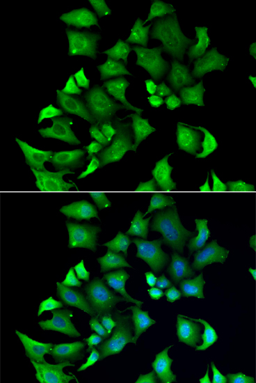
Human-reactive anti-ENTPD2 antibodies have IHC data from formalin-fixed, paraffin-embedded human brain and IF data from A549 cells (catalog reactivity; IHC and IF image captions).
A07914-1 is listed for human IHC-P and shown with DAB staining of formalin-fixed, paraffin-embedded human brain (catalog applications/reactivity; IHC image caption). A07914 is listed for human IF/ICC and shown by IF in A549 cells with DAPI counterstain (catalog applications/reactivity; IF image caption).
Which to pick: Choose A07914-1 for human paraffin-section IHC at 1:50–1:100; it is polyclonal and its IHC image uses formalin-fixed, paraffin-embedded human brain (catalog applications/dilution/clonality; IHC image caption). Choose A07914 for IF/ICC at 1:50–1:100, supported by an IF image of A549 cells (catalog applications/dilution; IF image caption). Neither SKU has reported reactivity beyond human, so neither has catalog support for cross-species use (catalog reactivity).