This website uses cookies to ensure you get the best experience on our website.


- Table of Contents
Plan EPHA1 paraffin-section IHC around the cytoplasmic and membranous staining reported in most tissues (HPA tissue IHC). Use glandular cells with medium staining as a reference and account for epitope location when interpreting results (HPA tissue IHC; UniProt).

Expected localisation, validated protocols, controls and antibodies — the at-a-glance facts below, then the full design guide.
| Expected localisation | Membranous and cytoplasmic in most tissues (HPA tissue IHC) | |
| Staining pattern | Most tissues show cytoplasmic and membranous staining (HPA tissue IHC) | |
| Antigen retrieval | Citrate pH 6.0 HIER, 95–98 °C, 20 min (rule: cytoplasmic / membrane antigen) | |
| Positive control | Adrenal gland+4 more · see all | |
| Negative control | Smooth muscle |
| Fixation | Matched tissue-IHC evidence does not establish the fixation claim. Validate the specimen-specific method before use. (selected-SKU IHC image PA1573) | |
| Caveat | Phospho-Tyr605 staining depends on site phosphorylation (UniProt; catalog A03024Y605-1) | |
| Regulation | Overexpressed in several carcinomas (UniProt) | |
| Isoform / epitope | 3 isoforms; map epitopes to extracellular or cytoplasmic regions (UniProt) |
Compare the catalog antibody’s IHC-P protocol with three published EPHA1 IHC methods (PMC7133419; PMC4234387; PMC8700455).
| Sample | Tissue sections; selected-image fixative not specified (standard IHC workflow) |
| Fixation | Image fixative and duration unreported (datasheet PA1573); verify before use. |
| Sectioning | 4–5 µm sections on charged slides (standard) |
| Deparaffinisation | Xylene, graded ethanol series to water (standard) |
| Antigen retrieval | Heat-induced epitope retrieval in citrate buffer, pH 6.0, 20 min at 95–98 °C (standard rule: cytoplasmic / membrane antigen) |
| Peroxidase block | 3% H2O2, 10 min, room temperature (standard) |
| Blocking | 10% normal serum of the secondary host, 30 min, room temperature (standard) |
| Primary antibody | Rabbit anti-EPHA1, 0.5-1μg/ml (datasheet PA1573) |
| Primary incubation | Overnight at 4 °C (standard) |
| Detection | HRP-polymer secondary, DAB chromogen 5–10 min (standard) |
| Counterstain | Hematoxylin, blue, dehydrate and mount (standard) |
| Expected result | EPHA1-positive staining in glandular cells of adrenal gland (HPA tissue IHC: Medium). HPA tissue profile: Cytoplasmic and membranous expression in most tissues, including parathyroid gland. No signal in the no-primary control. |
EPHA1 is a single-pass cell-membrane receptor with an extracellular region at residues 26–547 and a cytoplasmic region at 569–976 (UniProt P21709 topology). In paraffin-section IHC, expect membranous staining with possible cytoplasmic staining in epithelial or glandular cells; HPA reports this pattern across most tissues (HPA tissue IHC). Treat the pattern as provisional: HPA rates the tissue IHC reliability Uncertain (HPA tissue IHC).
| Membrane-associated staining in glandular or respiratory epithelial cells, with some cytoplasmic signal. | This fits the reported cytoplasmic and membranous tissue pattern (HPA tissue IHC) and EPHA1 cell-membrane location (UniProt P21709). Compare cells within the same section: HPA reports Medium staining in adrenal, appendix, breast, and cervix glandular cells and bronchial respiratory epithelial cells (HPA tissue IHC). |
| Strong nuclear-only staining, with little signal at cell borders or in cytoplasm. | Nuclear-only staining does not fit the reported membrane location (UniProt P21709; HPA subcellular summary) or the tissue IHC pattern (HPA tissue IHC). Check morphology and staining controls before assigning this signal to EPHA1; the antibody's tissue IHC reliability is Uncertain (HPA tissue IHC). |
| Staining is strongest in a cell population expected to be unstained, while the expected cells are faint. | For example, HPA reports smooth muscle cells as Not detected and bronchial respiratory epithelial cells as Medium (HPA tissue IHC). A reversed pattern raises concern for cross-reactivity or endogenous chromogenic detection activity; verify the cell identities and examine a no-primary control. |
| Uniform color covers tissue and spaces between cells, obscuring cell borders. | Diffuse background cannot establish EPHA1 localization. In chromogenic IHC, inspect a no-primary control for detection-related signal, then review blocking, antibody concentration, washes, and imaging exposure as general workflow checks. HPA's Uncertain reliability makes a clear cellular pattern especially useful (HPA tissue IHC). |
| No staining appears in a comparison section selected for reported EPHA1 staining. | HPA reports Medium staining in several glandular and epithelial cell populations, but its tissue IHC reliability is Uncertain (HPA tissue IHC). Confirm that the sampled section contains the specified cells, then check the primary antibody and detection run. One negative section alone cannot distinguish absent antigen from assay failure. |
| Compartment and unknown antibody epitope | EPHA1 spans the membrane, with extracellular residues 26–547 and cytoplasmic residues 569–976 (UniProt P21709 topology). The supplied antibody record gives no epitope, so topology cannot predict which domain CAB026144 recognizes or specify an EPHA1-specific antigen-retrieval condition. |
| Tissue and cell selection | HPA reports Medium staining in listed glandular, respiratory epithelial, and selected neural cells; smooth muscle cells are Not detected (HPA tissue IHC). Score the named cell population rather than treating every cell in a tissue as equally informative. These are reported patterns, not guaranteed controls (HPA reliability: Uncertain). |
| Antibody validation | CAB026144 is a mouse monoclonal antibody with IHC status Uncertain and no ICC status supplied (HPA antibodies). The tissue profile also has Uncertain reliability (HPA tissue IHC). Interpret weak or unexpected staining against tissue morphology and controls; no independently confirmed IHC pattern is supplied here. |
| IF/ICC expectation? | HPA summarizes EPHA1 as Membrane, but supplies no main ICC-IF location or cell lines with ICC-IF images (HPA subcellular). Membrane localization is a reasonable expectation (UniProt P21709; HPA subcellular), but this record cannot validate an IF/ICC staining pattern or supply an IF/ICC protocol. |
| Situation | Likely cause | Next action |
|---|---|---|
| Reported Medium-staining cells are blank in paraffin-section IHC. | The section may lack the named cells, or the antibody and chromogenic detection run may have failed; the reported level is provisional (HPA tissue IHC: Medium; reliability Uncertain). | Confirm cell identity and tissue preservation on the counterstained section. Run a documented positive assay control, then review the primary-antibody, retrieval, and detection steps as general IHC workflow checks; no EPHA1-specific retrieval condition is supplied. |
| Only nuclei stain. | Nuclear-only staining conflicts with EPHA1's reported membrane location and tissue IHC pattern (UniProt P21709; HPA tissue IHC). Its source remains unresolved from appearance alone. | Check the no-primary control and review nuclear counterstain separately. Reassess whether membrane or cytoplasmic staining is present in the HPA-reported cells before interpreting the nuclear signal as EPHA1 (HPA tissue IHC). |
| Smooth muscle cells stain more strongly than adjacent reported positive cells. | HPA lists smooth muscle cells as Not detected (HPA tissue IHC). Cross-reactivity or endogenous chromogenic activity is possible; staining alone does not identify which. | Verify cell boundaries and identities, inspect a no-primary control, and compare with a section containing HPA-reported Medium cells (HPA tissue IHC). If the unexpected pattern persists, treat the antibody result as unresolved. |
| Diffuse brown signal obscures cell borders. | Background may arise during general chromogenic IHC detection or from excessive nonspecific staining; it does not demonstrate EPHA1's membrane location (UniProt P21709). | Inspect no-primary and reagent controls. Review blocking, primary-antibody concentration, wash stringency, and chromogen development as general IHC steps; retain only cell-associated staining for interpretation. |
| Cytoplasmic staining appears without a sharp membrane rim. | HPA reports cytoplasmic as well as membranous tissue expression (HPA tissue IHC), so cytoplasmic signal alone is not proof of an artefact. The supplied record does not establish the antibody epitope or a processing explanation. | Check whether staining follows the HPA-reported cell distribution and whether background controls are clean (HPA tissue IHC). Record the compartment honestly; avoid calling a weak or diffuse cytoplasmic result confirmed membrane staining. |
| An IF/ICC image shows an unexpected distribution. | The supplied HPA ICC-IF record has a Membrane summary but no imaged cell lines or main location, and CAB026144 has no ICC status supplied (HPA subcellular; HPA antibodies). | Treat the IF/ICC result as unvalidated by this IHC evidence. Assess it with controls and the dedicated IF/ICC guide; do not transfer an IHC-P interpretation or protocol to IF/ICC. |


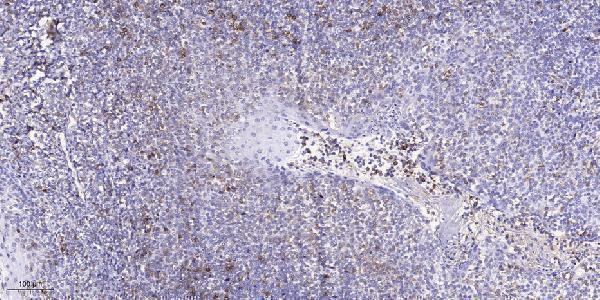


Comprehensive Human Protein Atlas IHC scoring per tissue (reliability: Uncertain — External characterization data supports antibody staining.). Rows are taken directly from the HPA tissue chart — click any row's HPA link to view the source.
| Tissue | Cell type | Level | Evidence | Source |
|---|---|---|---|---|
| Adrenal gland | Glandular cells | Medium | Protein (IHC) | HPA → |
| Appendix | Glandular cells | Medium | Protein (IHC) | HPA → |
| Breast | Glandular cells | Medium | Protein (IHC) | HPA → |
| Bronchus | Respiratory epithelial cells | Medium | Protein (IHC) | HPA → |
| Caudate | Glial cells | Medium | Protein (IHC) | HPA → |
| Tissue | Cell type | Level | Evidence | Source |
|---|---|---|---|---|
| Smooth muscle | Smooth muscle cells | Not detected | Protein (IHC) | HPA → |
Troubleshoot EPHA1 staining in paraffin section chromogenic IHC using its membrane topology (UniProt P21709) and tissue staining evidence rated Uncertain (HPA tissue IHC).
Two anti-EPHA1 antibodies have IHC figures from rat brain, human colon cancer, and human tonsil (catalog: IHC image captions); catalog reactivity covers human, mouse, and rat (catalog: reactivity lists).
PA1573 is listed for IHC and shows rat brain and human colon cancer tissue IHC(P) (catalog: PA1573 applications and IHC captions). A03024Y605-1 is listed for IHC and IF, with an IHC figure from a paraffin-embedded human tonsil section (catalog: A03024Y605-1 applications and IHC caption).
Which to pick: For tissue IHC targeting EPHA1, choose PA1573; its captions show rat brain and human colon cancer IHC(P), and its listed reactivity includes human, mouse, and rat (catalog: PA1573 IHC captions and reactivity). For phospho-Tyr605 IHC or IF, choose A03024Y605-1; it is listed as polyclonal, reacts with human and mouse, and has a paraffin-section IHC figure from human tonsil (catalog: A03024Y605-1 title, dilution_raw, reactivity, applications, and IHC caption). The figure captions support paraffin-section use but do not report a fixative; ICC is not separately listed (catalog: IHC captions and applications).