This website uses cookies to ensure you get the best experience on our website.
- Table of Contents


1 Citations
Facts about Ephrin type-B receptor 2.

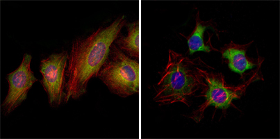
Involved in the guidance of commissural axons, that form a significant interhemispheric connection between the 2 temporal lobes of the cerebral cortex. Also involved in guidance of contralateral inner ear efferent growth cones at the midline and of retinal ganglion cell axons to the optic disk.
| Human | |
|---|---|
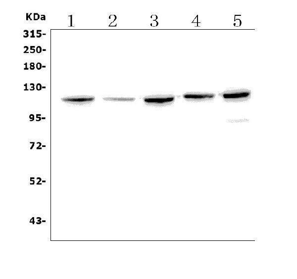
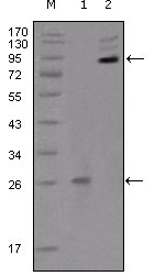
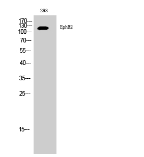
| Gene Name: | EPHB2 |
| Uniprot: | P29323 |
| Entrez: | 2048 |

| Belongs to: |
|---|
| protein kinase superfamily |

CAPB; Cek5; Drt; DRTEphB2; EC 2.7.10; EC 2.7.10.1; EK5; elk-related tyrosine kinase; EPH receptor B2; eph tyrosine kinase 3; EphB2; EPH-like kinase 5; ephrin type-B receptor 2; EPHT3MGC87492; EPTH3; Erk; ERKHek5; Hek5; Nuk; PCBC; protein-tyrosine kinase HEK5; Qek2; Renal carcinoma antigen NY-REN-47; Sek3; Tyro5; Tyrosine-protein kinase receptor EPH-3; Tyrosine-protein kinase TYRO5
Mass (kDA):
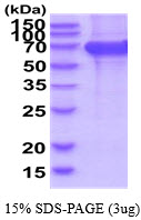
117.493 kDA

| Human | |
|---|---|
| Location: | 1p36.12 |
| Sequence: | 1; NC_000001.11 (22710770..22921500) |
Brain, heart, lung, kidney, placenta, pancreas, liver and skeletal muscle. Preferentially expressed in fetal brain.
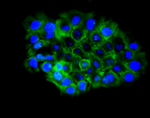
Cell membrane; Single-pass type I membrane protein. Cell projection, axon. Cell projection, dendrite.


PMID: 8033077 by Kiyokawa E., et al. Overexpression of ERK, an EPH family receptor protein tyrosine kinase, in various human tumors.
PMID: 8589679 by Ikegaki N., et al. Molecular characterization and chromosomal localization of DRT (EPHT3): a developmentally regulated human protein-tyrosine kinase gene of the EPH family.