This website uses cookies to ensure you get the best experience on our website.


- Table of Contents
Plan ERLIN2 paraffin-section IHC around cytoplasmic tissue staining (HPA tissue IHC) and ER membrane localisation (UniProt). Use breast glandular cells as a high-staining reference and cardiomyocytes as a not-detected reference when evaluating staining (HPA tissue IHC).

Expected localisation, validated protocols, controls and antibodies — the at-a-glance facts below, then the full design guide.
| Expected localisation | Cytoplasmic tissue staining (HPA tissue IHC) | |
| Staining pattern | Ubiquitous cytoplasmic staining (HPA tissue IHC) | |
| Antigen retrieval | EDTA pH 8.0 HIER, heat-mediated (datasheet M07042-1) | |
| Positive control | Breast+4 more · see all | |
| Negative control | Heart muscle |
| Fixation | Keep fixation consistent across compared paraffin sections. (selected-SKU IHC image M07042-1) | |
| Caveat | Staining and RNA show medium consistency (HPA tissue IHC) | |
| Regulation | No expression regulator specified (UniProt) | |
| Isoform / epitope | 3 isoforms; antibody epitope is unspecified (UniProt) |
The catalog antibody’s IHC-P protocol is accompanied by published ERLIN2 workflows for lung adenocarcinoma and breast tissue microarrays (PMC7793841; PMC10818095).
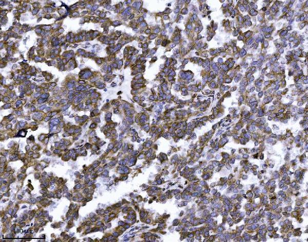
| Sample | Paraffin-embedded human ovarian serous carcinoma tissue; fixative not specified (datasheet M07042-1) |
| Fixation | Image fixative and duration unreported (datasheet M07042-1); verify before use. |
| Sectioning | 4–5 µm sections on charged slides (standard) |
| Deparaffinisation | Xylene, graded ethanol series to water (standard) |
| Antigen retrieval | Heat retrieval: EDTA pH 8.0 (datasheet M07042-1); 20 min, 95–100 °C (standard) |
| Peroxidase block | 3% H2O2, 10 min, room temperature (standard) |
| Blocking | 10% goat serum (datasheet M07042-1) |
| Primary antibody | Mouse monoclonal (clone 3H9A2) anti-ERLIN2, 2 μg/ml (datasheet M07042-1) |
| Primary incubation | Overnight at 4 °C (datasheet M07042-1) |
| Detection | HRP-conjugated secondary, DAB chromogen (datasheet M07042-1) |
| Counterstain | Hematoxylin, blue, dehydrate and mount (standard) |
| Expected result | ERLIN2-positive staining in glandular cells of breast (HPA tissue IHC: High). HPA tissue profile: Ubiquitous cytoplasmic expression. No signal in the no-primary control. |
ERLIN2 is an endoplasmic reticulum (ER) membrane protein with residues 25–339 facing the ER lumen (UniProt O94905 topology). In paraffin sections, expect cytoplasmic staining across many cell types: HPA describes ubiquitous cytoplasmic expression, with high staining in breast glandular cells and bronchial respiratory epithelial cells (HPA tissue IHC). HPA rates its tissue IHC profile Supported, with medium consistency between antibody staining and RNA expression (HPA tissue IHC).
| Cytoplasmic staining in breast glandular cells or bronchial respiratory epithelial cells. | This matches the reported high staining in both cell types (HPA tissue IHC). An ER-associated distribution is biologically consistent with ERLIN2's membrane location (UniProt O94905); chromogenic IHC alone cannot prove that each stained structure is ER. |
| Predominantly nuclear or sharply plasma-membrane staining, with little cytoplasmic signal. | That compartment conflicts with ERLIN2's ER membrane location (UniProt O94905) and HPA's cytoplasmic tissue profile (HPA tissue IHC). Check the negative control and antibody specificity before scoring it as ERLIN2. |
| Strong cardiomyocyte staining, especially when expected positive cells also stain. | HPA reports ERLIN2 as not detected in cardiomyocytes (HPA tissue IHC). Consider cross-reactivity or endogenous chromogen activity; compare matched controls and do not treat one unexpected cell type as proof of a new expression pattern. |
| Diffuse stain covers connective tissue, extracellular space, and cells alike. | A broad haze lacks the cell-associated cytoplasmic pattern reported by HPA (HPA tissue IHC). Assess background with a primary-antibody omission control, then review blocking, washes, and detection conditions as general IHC checks. |
| No visible stain in breast glandular cells or bronchial respiratory epithelial cells. | Both are reported high-staining cell populations (HPA tissue IHC), so their absence raises a technical concern. Confirm tissue identity and control performance, then review retrieval, antibody dilution, and detection; a single negative section cannot establish ERLIN2 absence. |
| Which compartment should guide scoring? | Score cell-associated cytoplasmic signal: ERLIN2 resides in the ER membrane (UniProt O94905), while HPA describes ubiquitous cytoplasmic tissue staining (HPA tissue IHC). Routine chromogenic IHC may not resolve the ER network. |
| How should tissue choice affect interpretation? | Breast glandular and bronchial respiratory epithelial cells are reported High; cardiomyocytes are Not detected (HPA tissue IHC). Use the former as expected-positive examples and interpret the latter at cell-type level, not as a whole-heart negative. |
| What does the antibody validation establish? | HPA lists IHC as Supported for HPA002025 and CAB014894, and rates the tissue profile Supported with medium staining–RNA consistency (HPA tissue IHC). These ratings support use of the reported pattern but do not verify every signal in a new section. |
| Does sequence information predict an IHC failure? | ERLIN2 has one transmembrane segment at residues 4–24, three isoforms, and a glycosylation site at residue 106 (UniProt O94905). The supplied sources give no antibody epitope or target-specific fixation-sensitivity result, so these features do not justify a retrieval or staining prediction. |
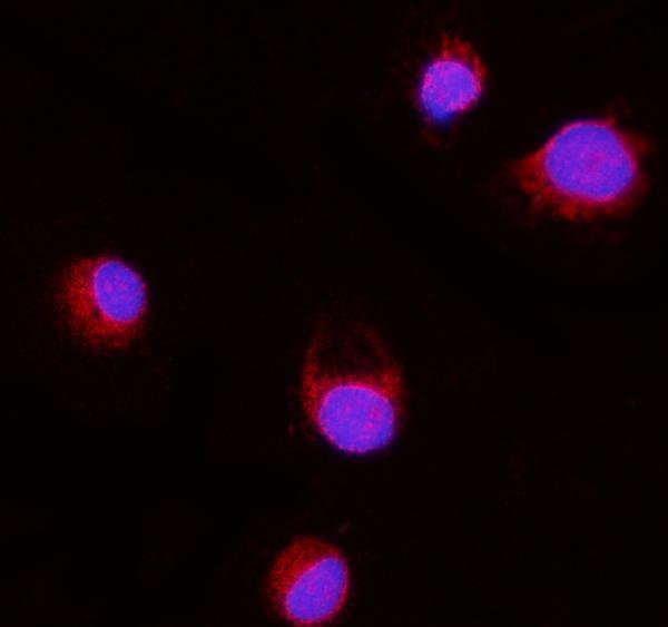
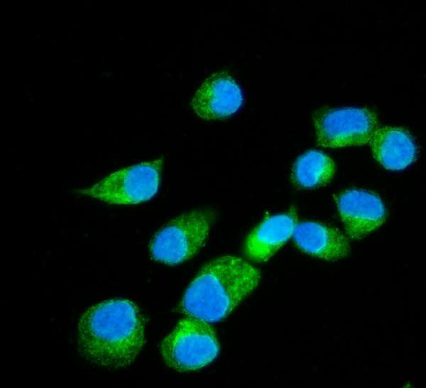
| IF/ICC: what localization is expected? | Mainly ER localization is reported and supported in HPA ICC-IF (HPA subcellular). This answers the localization question only; IF/ICC conditions belong in its separate guide. |
| Situation | Likely cause | Next action |
|---|---|---|
| Expected-positive tissue has no signal. | A failed staining step is possible when high-staining cells are absent from the result (HPA tissue IHC). | Check section identity and assay controls. As general IHC practice, review retrieval, primary dilution, detection reagents, and counterstain before interpreting a biological negative. |
| Signal is mostly nuclear. | Nuclear dominance does not fit the ER membrane assignment (UniProt O94905) or HPA's cytoplasmic profile (HPA tissue IHC). | Compare the expected-positive cells and omission control; review antibody specificity and scoring before accepting the nuclear signal. |
| Cardiomyocytes appear strongly positive. | HPA reports cardiomyocytes as Not detected (HPA tissue IHC); unexpected signal may reflect nonspecific binding or endogenous detection activity. | Inspect cell identity and matched controls. If the control also colors cardiomyocytes, troubleshoot the detection system before assigning ERLIN2 positivity. |
| The whole section has diffuse brown background. | Uniform staining obscures the cell-associated cytoplasmic pattern reported for ERLIN2 (HPA tissue IHC). | Use an omission control, then review blocking, wash stringency, and chromogen development as general chromogenic IHC checks. |
| Only low-staining tissue looks negative. | HPA lists squamous epithelial cells of oral mucosa and vagina as Low, while selected glandular and respiratory epithelial cells are High (HPA tissue IHC). | Run an expected high-staining cell population alongside it; score the named cell type rather than declaring a whole tissue negative. |
| Staining is present but its intracellular pattern is unclear. | The ER membrane assignment (UniProt O94905) and cytoplasmic IHC profile (HPA tissue IHC) set the expectation, but chromogenic resolution may limit compartment assignment. | Record the observable cytoplasmic distribution and control results. For finer localization, consult the separate IF/ICC guide; HPA reports supported ER localization by ICC-IF (HPA subcellular). |
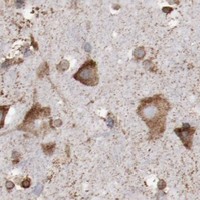
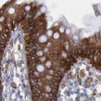
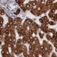

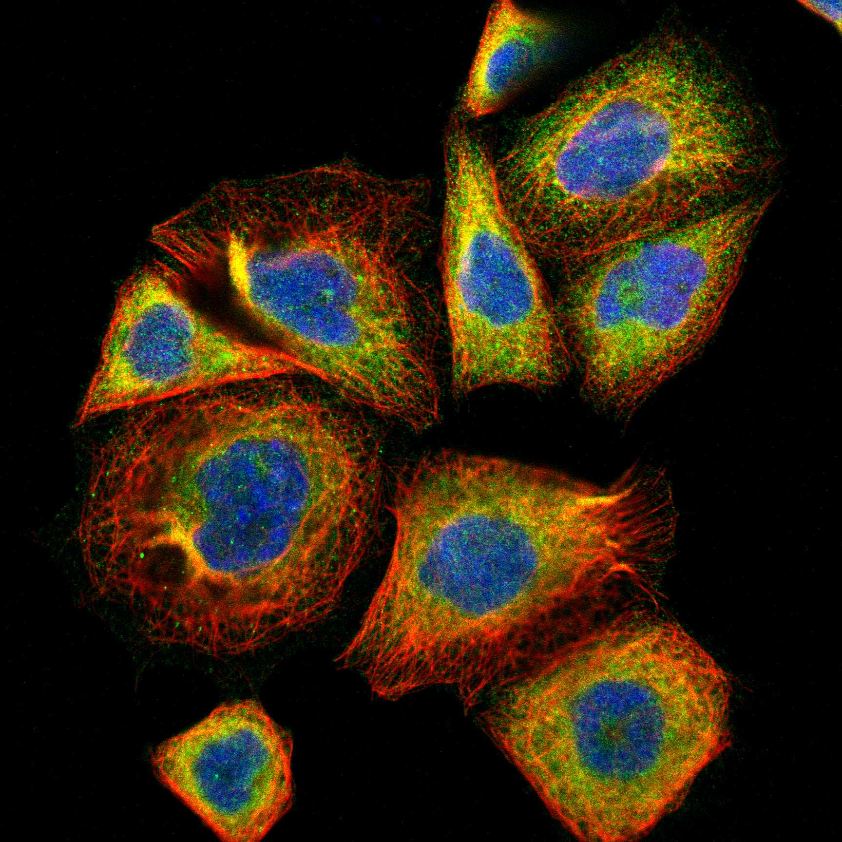
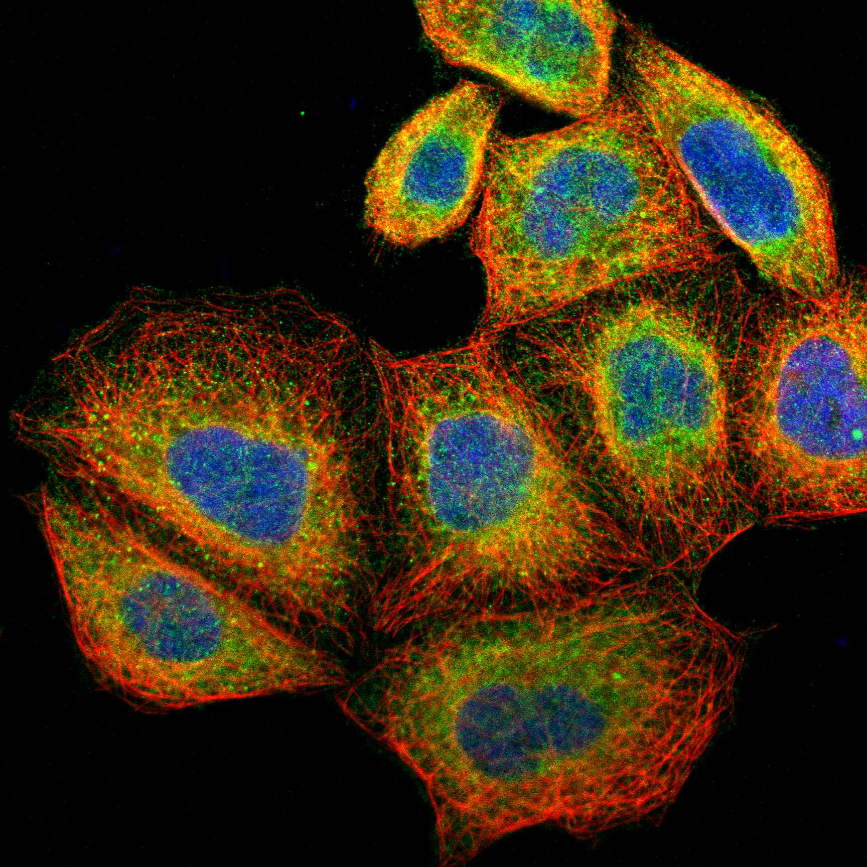
Comprehensive Human Protein Atlas IHC scoring per tissue (reliability: Supported — Medium consistency between antibody staining and RNA expression data.). Rows are taken directly from the HPA tissue chart — click any row's HPA link to view the source.
| Tissue | Cell type | Level | Evidence | Source |
|---|---|---|---|---|
| Breast | Glandular cells | High | Protein (IHC) | HPA → |
| Bronchus | Respiratory epithelial cells | High | Protein (IHC) | HPA → |
| Caudate | Glial cells | High | Protein (IHC) | HPA → |
| Cerebellum | Cells in granular layer | High | Protein (IHC) | HPA → |
| Cerebral cortex | Glial cells | High | Protein (IHC) | HPA → |
| Tissue | Cell type | Level | Evidence | Source |
|---|---|---|---|---|
| Heart muscle | Cardiomyocytes | Not detected | Protein (IHC) | HPA → |
Troubleshoot ERLIN2 chromogenic IHC by checking retrieval, processing, cellular localisation and controls before comparing staining across sections.
ERLIN2 antibody cards show human paraffin-section IHC and human cell IF images (catalog captions); one IF/ICC antibody also lists mouse and rat reactivity (catalog: A07042-2).
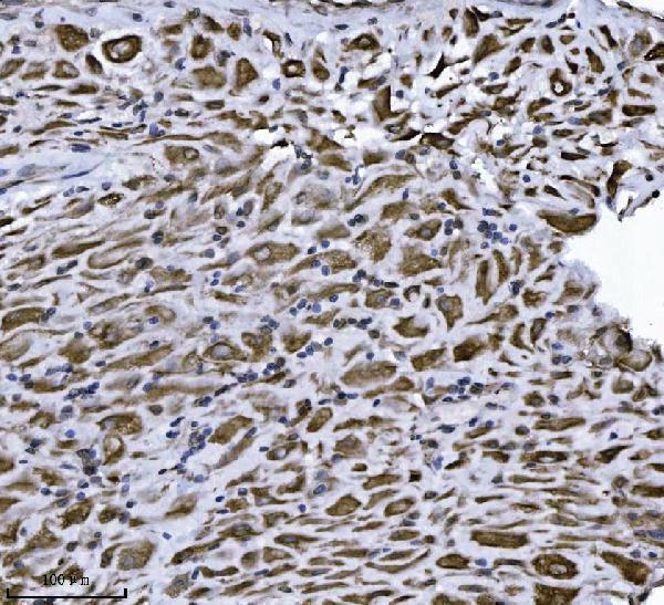
M07042-1 shows IHC staining in human paraffin-embedded ovarian serous carcinoma, placenta, squamous cell lung carcinoma and thyroiditis sections (M07042-1 IHC captions). A07042-2 shows IF staining in human T-47D cells (A07042-2 IF caption).
Which to pick: Choose M07042-1 for human paraffin-section IHC: its own captions show staining after EDTA pH 8.0 retrieval at 2 μg/ml (M07042-1 IHC captions); the fixative is unreported (M07042-1 IHC captions). Choose A07042-2 for human IF/ICC: its own caption shows T-47D cell staining at 5 μg/ml (A07042-2 IF caption). For mouse or rat work, A07042-2 lists both species as reactive, but its pictured IF example is human only (A07042-2 catalog reactivity and IF caption).