EVA1A / Protein eva-1 homolog A · IHC design guide

Design Immunohistochemistry for EVA1A

Use hepatocyte and renal tubule cytoplasmic staining to plan and assess EVA1A chromogenic IHC in paraffin sections (HPA tissue IHC). Include controls when interpreting signal because HPA reports presumed off-target binding and low agreement between staining and RNA expression (HPA tissue IHC).

Evidence assembled Oct 2026 · For research use; verify linked source records and product datasheet before use
Immunohistochemistry protocol sheet for EVA1A (IHC for EVA1A): expected localisation Cytoplasmic in tissue (HPA tissue IHC); ER and lysosome membranes (UniProt), antibody A11580-1, validated IHC image, and IHC protocol steps
Printable EVA1A IHC protocol sheet — expected localisation Cytoplasmic in tissue (HPA tissue IHC); ER and lysosome membranes (UniProt), antibody A11580-1, controls and protocol steps. Open the full EVA1A IHC guide →

EVA1A Immunohistochemistry Experimental Design Guide

Expected localisation, validated protocols, controls and antibodies — the at-a-glance facts below, then the full design guide.

Must know before staining
Expected localisation Cytoplasmic in tissue (HPA tissue IHC); ER and lysosome membranes (UniProt)
Staining pattern Cytoplasmic staining in hepatocytes and renal tubule cells (HPA tissue IHC)
Antigen retrieval Citrate pH 6 HIER, heat-mediated (datasheet A11580-1)
Positive control ⓘ Liver+2 more · see all
Negative control ⓘ Adipose tissue+4 more · see all
Important caveats
Reasons your staining may differ from the expected pattern.
Fixation Keep fixation consistent across paraffin sections. (standard IHC practice; not target-specific)
Caveat Presumed off-target binding can confound staining (HPA tissue IHC)
Regulation Liver-enriched RNA expression (HPA RNA)
Isoform / epitope No annotated isoforms; epitope sidedness is unspecified (UniProt)
Section 1

Recommended EVA1A IHC & IF Protocols

The catalog antibody uses citrate pH 6 retrieval (datasheet A11580-1). The 3 published protocols below report EVA1A staining in paraffin sections (PMC9588234; PMC5945723; PMC10961441).

Recommended immunohistochemistry (IHC-P) protocol parameters
SampleParaffin-embedded human liver cancer tissue; fixative not specified (datasheet A11580-1)
FixationImage fixative and duration unreported (datasheet A11580-1); verify before use.
Sectioning4–5 µm sections on charged slides (standard)
DeparaffinisationXylene, graded ethanol series to water (standard)
Antigen retrievalHeat retrieval: Citrate pH 6, 20 min (datasheet A11580-1)
Peroxidase block3% H2O2, 10 min, room temperature (standard)
Blocking10% goat serum (datasheet A11580-1)
Primary antibodyRabbit anti-EVA1A, 1μg/ml (datasheet A11580-1)
Primary incubationOvernight at 4 °C (datasheet A11580-1)
DetectionStreptavidin-biotin complex (SABC), DAB chromogen (datasheet A11580-1)
CounterstainHematoxylin, blue, dehydrate and mount (standard)
Expected resultEVA1A-positive staining in hepatocytes of liver (HPA tissue IHC: High). HPA tissue profile: Cytoplasmic expression in several tissues, including hepatocytes and renal tubules kidney. No signal in the no-primary control.
💡Decision noteStart with heat-mediated citrate pH 6 retrieval (datasheet A11580-1; PMC9588234). The other published heat-retrieval protocol does not specify a buffer (PMC5945723).
Section 2

What Is the Expected EVA1A Staining Pattern?

EVA1A is an endoplasmic reticulum and lysosome membrane protein with one transmembrane segment at residues 35–55 (UniProt Q9H8M9). In paraffin IHC, expect cytoplasmic staining in hepatocytes and renal tubular cells (HPA tissue IHC). HPA rates its tissue IHC profile Approved, while reporting low agreement with RNA data, disregarded presumed off-target staining, and pending external verification (HPA tissue IHC).

What am I looking at on my slide?
Strong cytoplasmic staining in hepatocytes; moderate staining in renal tubular cells.This matches the reported liver High and kidney Medium cell-specific patterns (HPA tissue IHC). Judge the stained cells and compartment together; staining intensity alone does not establish specificity, given HPA’s stated reliability caveats (HPA tissue IHC).
Predominantly nuclear staining in hepatocytes or renal tubular cells.A nuclear-dominant pattern conflicts with the reported cytoplasmic tissue pattern (HPA tissue IHC) and ER/lysosome membrane localisation (UniProt Q9H8M9). Treat it as suspect; compare with the negative control and reassess detection conditions (general IHC practice).
Strong staining in adipocytes or appendix glandular cells.These cell types are reported Not detected (HPA tissue IHC). Consider cross-reactivity or endogenous detection activity before assigning the signal to EVA1A (general IHC practice), especially because HPA reports presumed off-target binding in its tissue assessment (HPA tissue IHC).
Diffuse chromogen across tissue and background, obscuring cell boundaries.This cannot support a cell-specific EVA1A call (general IHC practice). Check the negative control, blocking, antibody concentration, washes, and chromogen development as general IHC variables; HPA’s reported cytoplasmic staining does not validate a uniform background pattern (HPA tissue IHC).
No staining in hepatocytes on a run that includes liver.The absence conflicts with reported High hepatocyte staining (HPA tissue IHC), but does not by itself prove loss of EVA1A. Check tissue preservation, retrieval, primary-antibody step, and detection with appropriate run controls (general IHC practice). No EVA1A-specific fixation sensitivity is reported in the supplied sources.
💡Expected EVA1A appearanceCall a positive IHC result when hepatocytes show strong cytoplasmic staining, with renal tubular cells providing a moderate comparison (HPA tissue IHC); dominant nuclear signal or strong staining in reported negative cell types is suspect (UniProt Q9H8M9; HPA tissue IHC).
How each factor affects the staining
Membrane topology and compartmentEVA1A has one transmembrane segment at residues 35–55 and localises to ER and lysosome membranes (UniProt Q9H8M9). HPA describes the paraffin tissue pattern as cytoplasmic (HPA tissue IHC); the record does not require a visibly outlined organelle membrane in chromogenic IHC.
Choice of tissue comparisonHepatocytes and Leydig cells are reported High; renal tubular cells are Medium (HPA tissue IHC). Adipocytes and appendix glandular cells are reported Not detected (HPA tissue IHC). Use these as pattern comparisons, while retaining HPA’s stated concerns about RNA agreement and off-target staining (HPA tissue IHC).
Strength of antibody evidenceAntibody HPA008055 is rated Approved for IHC and Supported for ICC (HPA antibodies). HPA’s tissue profile remains pending external verification and notes presumed off-target binding that was disregarded (HPA tissue IHC). Interpret a matching stain as supportive evidence, alongside controls and morphology (general IHC practice).
IF/ICC Q&A: what localisation is expected?HPA reports mainly vesicles, with additional plasma-membrane localisation in ICC-IF; vesicles are Supported and plasma membrane is Approved (HPA subcellular). These ICC-IF findings help interpret fluorescence images but do not require visible plasma-membrane staining in paraffin chromogenic IHC (HPA subcellular; HPA tissue IHC).
Why is my staining missing, weak or wrong?
SituationLikely causeNext action
Liver hepatocytes are blank.A failed run or weak detection is possible; hepatocytes are reported High (HPA tissue IHC).Confirm tissue and run controls, then review retrieval, antibody application, and detection steps (general IHC practice). Do not infer an EVA1A-specific fixation effect; none is reported in the supplied sources.
Kidney signal is weaker than liver.This can fit the reported Medium renal tubular and High hepatocyte levels (HPA tissue IHC).Score tubular cells against liver and the run controls, accounting for cell identity and background (general IHC practice). Do not require equal chromogen intensity across the two tissues.
Staining is mainly nuclear.That compartment conflicts with cytoplasmic tissue staining (HPA tissue IHC) and ER/lysosome membrane localisation (UniProt Q9H8M9).Check the negative control and detection background; review morphology before scoring nuclei as positive (general IHC practice).
Adipocytes stain strongly.Adipocytes are reported Not detected (HPA tissue IHC); cross-reactivity or endogenous detection activity is possible (general IHC practice).Compare the primary-omission and other appropriate detection controls, then review blocking and detection chemistry (general IHC practice). HPA’s off-target caveat warrants caution (HPA tissue IHC).
The whole section looks brown.Diffuse background can obscure the reported cell-specific cytoplasmic pattern (HPA tissue IHC; general IHC practice).Review negative controls, washing, antibody concentration, and chromogen development before interpreting the section (general IHC practice).
IF/ICC appears punctate or peripheral.Vesicles are the main reported ICC-IF location; plasma membrane is an additional location (HPA subcellular).Interpret that fluorescence localisation using the IF/ICC guide and its controls (general IF practice). For this paraffin IHC result, score the reported tissue cells and cytoplasmic pattern (HPA tissue IHC).

Sample controls for EVA1A IHC & IF

🧪Run liver first: hepatocytes should stain (HPA: High in hepatocytes); use adipose tissue as the negative tissue, assessing adipocytes (HPA: Not detected in adipocytes). On the liver slide, nonhepatocyte cells should be assessed for background staining, but their EVA1A-negative status is unverified by the supplied HPA rows.
Positive control tissue: Liver (Hepatocytes, HPA High)
Negative control tissue: Adipose tissue (HPA Not detected)
ICC-IF cell lines (HPA subcellular resource): HPA ICC-IF images show EVA1A in A-431, U-251MG, U2OS, with annotated localisation: Vesicles (supported) (HPA subcellular).
Technical controls: Include a no-primary secondary-only control, a host- and clonality-matched rabbit IgG isotype control, and EVA1A-knockout tissue or a validated peptide-block control (standard IHC practice; caption: rabbit primary antibody). For liver sections, quench endogenous peroxidase and check for endogenous biotin before using the caption’s biotin-based DAB detection (standard IHC practice; caption: biotinylated secondary, SABC and DAB).
⚠️Feasibility: A target-specific fixation window or fixation effect is unreported, and the selected SKU A11580-1 caption does not state the fixative (caption: fixative not stated). The reported paraffin-section staining used heat retrieval in citrate buffer at pH 6 for 20 minutes; retrieval dependence and whether frozen sections or IF are easier remain unestablished (caption: retrieval conditions; supplied evidence). For IF/ICC, assess vesicular staining separately from the tissue IHC result (HPA: vesicles supported in ICC-IF); in liver IHC, endogenous biotin can complicate the reported biotin-based detection (standard IHC practice; caption: SABC detection).

HPA tissue IHC evidence for EVA1A

Comprehensive Human Protein Atlas IHC scoring per tissue (reliability: Approved — Low consistency between antibody staining and RNA expression data. Presumed off target binding observed and disregarded. Pending external verification.). Rows are taken directly from the HPA tissue chart — click any row's HPA link to view the source.

Positive expression · recommended positive controls

TissueCell typeLevelEvidenceSource
Liver Hepatocytes High Protein (IHC) HPA →
Testis Leydig cells High Protein (IHC) HPA →
Kidney Cells in tubules Medium Protein (IHC) HPA →

Undetected expression · recommended negative controls

TissueCell typeLevelEvidenceSource
Adipose tissue Adipocytes Not detected Protein (IHC) HPA →
Appendix Glandular cells Not detected Protein (IHC) HPA →
Bone marrow Hematopoietic cells Not detected Protein (IHC) HPA →
Breast Adipocytes Not detected Protein (IHC) HPA →
Cerebellum Cells in granular layer Not detected Protein (IHC) HPA →
Section 3

Advanced EVA1A IHC Tips

Use compartment and cell type controls to interpret EVA1A staining in paraffin sections; treat published tissue patterns as reference points requiring local validation.

Which retrieval condition should I start with for EVA1A in paraffin sections?
Start with heat mediated retrieval in citrate buffer at pH 6 for 20 minutes (datasheet A11580-1). The same paraffin section example used 1 µg/mL catalog antibody overnight at 4°C, providing a matched starting condition for assessing retrieval (datasheet A11580-1). Compare a retrieved section with an otherwise matched section without retrieval, keeping section thickness, detection and exposure to DAB consistent so any gain is interpretable (standard IHC practice). If staining remains weak, optimise heating time on adjacent sections before trying another retrieval buffer, and watch for damaged morphology or increased background (standard IHC practice).
How should I troubleshoot fixation when EVA1A staining is weak or uneven?
Target specific fixation sensitivity is unknown because the selected paraffin section caption does not state a fixative (datasheet A11580-1). Record the fixative, fixation duration and processing history for each specimen, then compare sections processed alike before attributing differences to EVA1A expression (standard IHC practice). Inspect morphology and staining across the section, since poorly preserved or unevenly processed areas can confound chromogenic interpretation (standard IHC practice). If archival blocks vary, optimise retrieval and antibody conditions on a consistently processed control block first, then assess whether the same settings produce comparable signal in the other blocks (standard IHC practice).
Where should convincing EVA1A chromogenic staining appear?
Interpret EVA1A as a membrane associated protein of the endoplasmic reticulum and lysosome, with a transmembrane segment at residues 35–55 (UniProt Q9H8M9 topology and subcellular annotation). HPA describes cytoplasmic tissue staining, including hepatocytes and renal tubule cells, while its cellular imaging places EVA1A mainly in vesicles (HPA tissue IHC; HPA subcellular). In paraffin sections, assess intracellular granular or cytoplasmic staining against tissue architecture rather than expecting every membrane to resolve individually (standard IHC practice). Compare the same compartment in neighbouring positive and negative cells, and investigate dominant nuclear staining before assigning it to EVA1A (UniProt Q9H8M9 subcellular annotation; standard IHC practice).
Could epitope position or modification explain inconsistent EVA1A staining?
The record lists a single 152 amino acid EVA1A chain and no annotated isoforms, so an isoform specific staining explanation is unsupported here (UniProt Q9H8M9 processing and isoforms). Its transmembrane segment spans residues 35–55, and annotated phosphorylation includes residues 106 and 114 (UniProt Q9H8M9 topology and modified residues). The catalog evidence supplied here does not identify the antibody epitope, so its location relative to the membrane or those modifications cannot be assigned (datasheet A11580-1; UniProt Q9H8M9). When staining differs between samples, check retrieval and tissue preservation first, then seek epitope mapping or independent antibody validation before proposing a modification dependent explanation (standard IHC practice).
How can IF help assess the cell type and compartment behind an IHC signal?
Use IF as a separate cross check of an ambiguous chromogenic pattern, rather than transferring the paraffin IHC incubation conditions directly (datasheet A11580-1; standard IHC/IF practice). Multiplex EVA1A with a marker for the expected cell type, such as hepatocytes when examining liver, and compare signals within identified cells (HPA tissue IHC: high in hepatocytes; standard IF practice). Choose spectrally separated fluorophores and inspect unstained tissue for autofluorescence before interpreting overlap (standard IF practice). Because the epitope side of the EVA1A membrane is unspecified, establish whether permeabilisation is needed empirically and assess effects on membrane and vesicular patterns (UniProt Q9H8M9 topology; HPA subcellular; standard IF practice).
How do I distinguish EVA1A staining from chromogenic background?
The selected paraffin example used 10% goat serum blocking, a biotinylated secondary and DAB detection, giving a defined workflow to evaluate locally (datasheet A11580-1). Run a section without primary antibody to reveal secondary reagent, endogenous enzyme or chromogen related signal under the same detection conditions (standard IHC practice). Include an appropriate peroxidase block and assess whether endogenous biotin contributes background when using a biotin based detection system (standard IHC practice). Compare suspect staining with tissue morphology and a consistently processed positive control; diffuse staining in every compartment needs further optimisation before being scored as EVA1A (UniProt Q9H8M9 subcellular annotation; standard IHC practice).
What is a defensible way to quantify EVA1A IHC across specimens? ⚠ ANSWER MARKED FOR VERIFICATION
Define the cell population and intracellular compartment before scoring, since HPA reports high staining in hepatocytes but medium staining in kidney tubule cells (HPA tissue IHC). For a fixed cell population, report the percentage of positive cells and an intensity based H score with the scoring thresholds stated in advance (standard IHC practice). If cell abundance varies, report positive cells per mm² of viable, annotated tissue and normalise comparisons to the same eligible cell population or analysed area (standard IHC practice). Hold retrieval, detection, imaging and threshold settings constant across the comparison, and exclude folds, edges and necrotic regions by a prespecified rule (standard IHC practice).
When should an EVA1A positive IHC result be questioned?
Question staining confined to nuclei or other unexpected compartments, because the record places EVA1A at endoplasmic reticulum and lysosome membranes and HPA reports mainly vesicular cellular localisation (UniProt Q9H8M9 subcellular annotation; HPA subcellular). Check cell identity: HPA reports high signal in hepatocytes, medium signal in kidney tubules and no detected signal in several listed cell populations (HPA tissue IHC). Treat staining limited to section edges, necrotic regions or structures positive in the no primary control as possible processing or detection artefact (standard IHC practice). HPA flags low consistency with RNA data and presumed off target binding, so corroborate an unexpected pattern with controls or independent evidence before assigning biological meaning (HPA tissue IHC reliability description).
Boster reagents

Best EVA1A / Protein eva-1 homolog A IHC Antibodies

A11580-1 has real IHC images from human paraffin sections (catalog IHC captions); its listed reactivity covers human, mouse and rat (catalog reactivity). No IF image is supplied (catalog IF images).

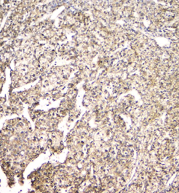
Real IHC data IHC analysis of EVA1A using anti-EVA1A antibody (A11580-1). EVA1A was detected in paraffin-embedded section of human liver cancer tissue. Heat mediated antigen retrieval was performed in citrate buffer (pH6, epitope retrieval solution) for 20 mins. The tissue section was blocked with 10% goat serum. The tissue section was then incubated with 1μg/ml rabbit anti-EVA1A Antibody (A11580-1) overnight at 4°C. Biotinylated goat anti-rabbit IgG was used as secondary antibody and incubated for 30 minutes at 37°C. The tissue section was developed using Strepavidin-Biotin-Complex (SABC)(Catalog # SA1022) with DAB as the chromogen.
Anti-TMEM166/EVA1A Antibody ®
Cat # A11580-1

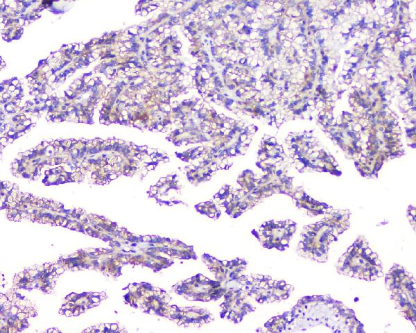
A11580-1 will render with an IHC figure from a human liver cancer paraffin section; its other IHC caption shows a human renal cancer paraffin section (catalog IHC captions). The antibody lists IHC, IHC-F and ICC applications and human, mouse and rat reactivity (catalog applications; catalog reactivity).

Which to pick: Choose A11580-1 for tissue IHC: its own captions document paraffin sections with citrate pH 6 retrieval, while the fixative is unreported (catalog IHC captions). For IF/ICC, A11580-1 lists IHC-F and ICC, but supplies no IF image or IF dilution (catalog applications; catalog IF images; catalog dilutions). It is also the listed cross-species option because this rabbit polyclonal antibody lists human, mouse and rat reactivity; the supplied IHC images show human tissue only (catalog host; catalog dilution_raw; catalog reactivity; catalog IHC captions).

Each figure is that product's own IHC / IF validation image from its datasheet.

References

  1. UniProt Consortium. UniProt entry Q9H8M9 (EVA1A_HUMAN, Protein eva-1 homolog A).
  2. Human Protein Atlas. EVA1A tissue IHC expression (reliability: Approved).
  3. Human Protein Atlas. EVA1A subcellular location (ICC-IF): Mainly localized to vesicles. In addition localized to the plasma membrane..
  4. Human Protein Atlas. EVA1A antibody validation summary (1 antibodies).
  5. EVA1A responds to endoplasmic reticulum stress to regulate renal fibrosis by promoting TGF-β signaling pathway. Journal of advanced research 2026 — PMC13453889.
  6. Down-regulation of EVA1A by miR-103a-3p promotes hepatocellular carcinoma cells proliferation and migration. Cellular & molecular biology letters 2022 — PMC9588234.
  7. MicroRNA-125b reverses oxaliplatin resistance in hepatocellular carcinoma by negatively regulating EVA1A mediated autophagy. Cell death & disease 2018 — PMC5945723.
  8. EVA1A, a novel and promising prognostic biomarker in colorectal cancer. Frontiers in oncology 2024 — PMC10961441.
  9. PubMed PMID:14702039 — UniProt-cited evidence.
  10. PubMed PMID:15498874 — UniProt-cited evidence.
  11. PubMed PMID:15815621 — UniProt-cited evidence.