This website uses cookies to ensure you get the best experience on our website.
- Table of Contents
17 Citations 4 Q&As
13 Citations
33 Citations
8 Citations
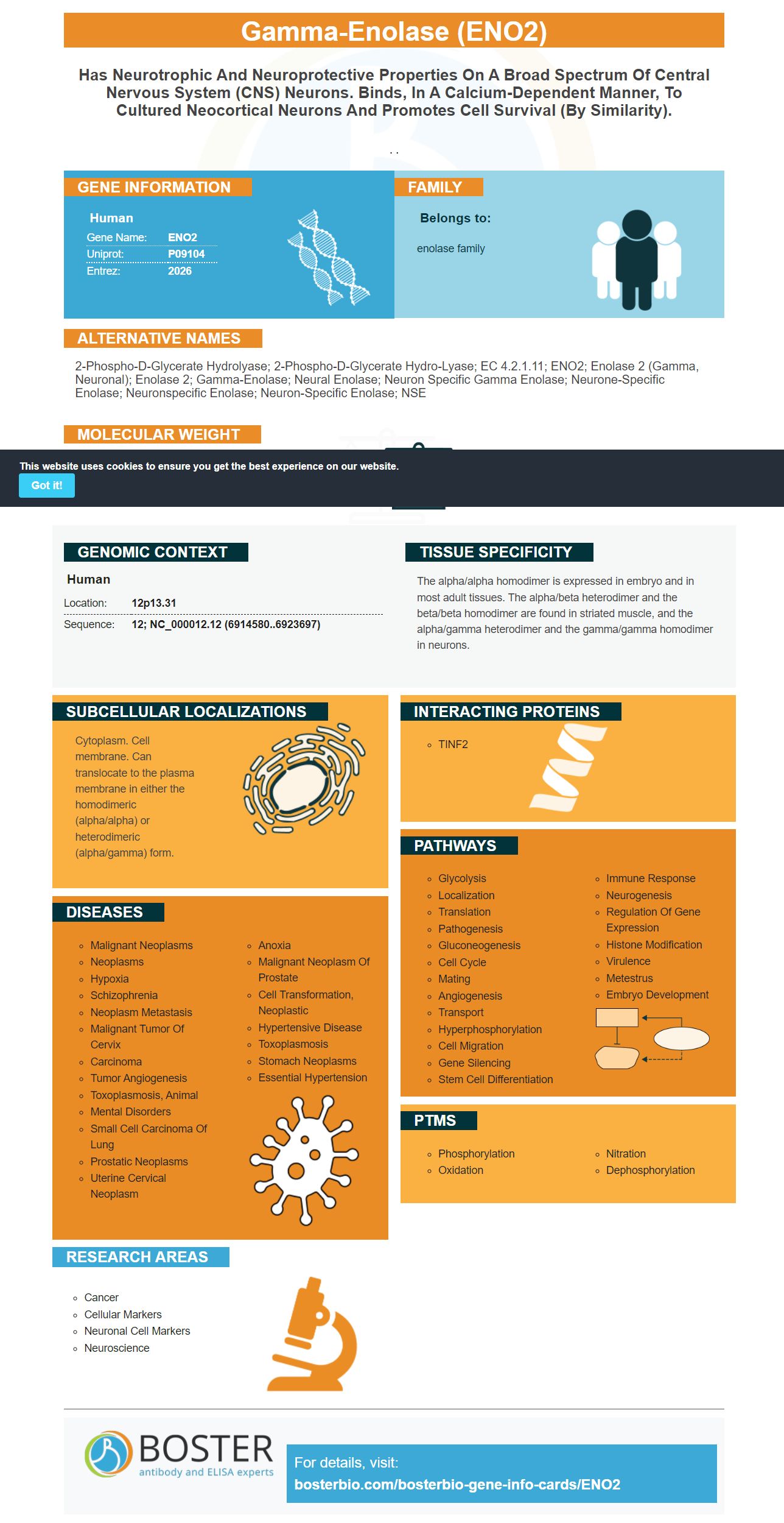
Facts about Gamma-enolase.
. .
Human | |
---|---|
Gene Name: | ENO2 |
Uniprot: | P09104 |
Entrez: | 2026 |
Belongs to: |
---|
enolase family |
2-phospho-D-glycerate hydrolyase; 2-phospho-D-glycerate hydro-lyase; EC 4.2.1.11; ENO2; enolase 2 (gamma, neuronal); Enolase 2; gamma-Enolase; Neural enolase; neuron specific gamma enolase; neurone-specific enolase; Neuronspecific Enolase; Neuron-specific enolase; NSE
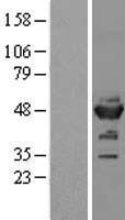
Mass (kDA):
47.269 kDA
Human | |
---|---|
Location: | 12p13.31 |
Sequence: | 12; NC_000012.12 (6914580..6923697) |
The alpha/alpha homodimer is expressed in embryo and in most adult tissues. The alpha/beta heterodimer and the beta/beta homodimer are found in striated muscle, and the alpha/gamma heterodimer and the gamma/gamma homodimer in neurons.
Cytoplasm. Cell membrane. Can translocate to the plasma membrane in either the homodimeric (alpha/alpha) or heterodimeric (alpha/gamma) form.
Researchers can submit results to the Boster Bio: The Best Uses for the ENO2 Marker in many ways when using it. Boster scientists can benefit from credit for their results. This credit is available to researchers from all over the world. Here are the top applications of the ENO2 Marker:
Each step in an ELISA experiment requires a decision. These choices range from preparation of the sample to the choice of blocking buffer. Utilizing an optimization tool from Boster Bio will help you answer these questions and boost the efficiency of your experiment. It is important to ensure that antibodies are concentrated. If you are unsure of how to optimize your ELISA then you can look up these optimization tips and guidelines.
One of the limitations of using a PCR-based marker such as ENO1 is that it isn't sensitive enough for detection of ENO1-delete cancers. This is a common mistake which should be avoided whenever possible. Therefore, it is imperative to choose an instrument that is more sensitive, a bioassay with greater range of samples, or to use an ELISA kit that includes ENO1 and ENO2 markers.
To increase the sensitivity of ENO2 markers it is possible to replace them with cell lines that express ENO1 in totality. The Gli56 cell line is homozygous ENO1 deletion cell line, which is selectively sensitive to POMHEX. This cell line has been shown to expand in pyruvate-free DMEM.
The Limit of Blank (LoB), is the measurement of the lowest concentrations of an analytical substance within the sample. In other words, it is the lowest concentration at which the test results that are positive would be considered the limit of detection. In a typical assay the LoB is determined by measuring the number of replicates of the analyte and then formulating the standard deviation and mean of the results.
When analysing your results When interpreting your results, the Limit of Blank (LoD), is vital. Another way to reduce the matrix effect is to use concentrations that are known. The analytical response of low concentration samples can be compared to the response of the blank sample to determine the concentration of the analyte. Boster Bio ELISAs are suitable for urine samples where the proteins are in their original state. Additionally, Boster Bio has mastered the technique of coating ELISA plates to lessen the impact of this factor.
You should inspect the product and packaging as soon as it arrives. Boster Biological Technology should be informed immediately if you notice any damage or shortage. If you received a defective product, you can return it to receive a full refund, or a replacement. Boster Biotech will give you a refund or exchange for the product, depending on which is the most appropriate for your purposes. Boster Bio replacements are guaranteed for a minimum of 12 months.
The study assessed ENO2 gene expression in 60 duplicates of normal tissue samples from 10 healthy women. The results showed that 28.5 copies/uL were measured that was not significantly different from levels found in the positive control. Additionally, there was no significant difference between the negative and positive controls in the amount of ENO2. ENO2 could not detect the presence of PNET cells within the ovarian tissues.
The biological half-life of ENO2 in cerebrospinal fluid determines the limit of detection. In diseases that lead to rapid degeneration of nerve cells, increased levels of neuron-specific enolase have been observed. The detection limit for the ENO2 marker must therefore be determined by a strict standard. When making estimates of the LOD of chromatographic techniques there are certain guidelines.
The limits of detection of GFAP were determined using plasma samples taken from healthy individuals. Plasma samples were spiked with a low level of endogenous GFAP to activate the recombinant GFAP calibration. Three samples were spiked with a concentration of fifty to one hundred and twenty-one percent of ULOQ. The serial dilutions that resulted were lower than the LLOD. The resultant samples were assayed to determine the percent recovery.
A GFAP immunoassay can be useful in clinical trials and research. A robust blood GFAP assay can catalyze cost-effective translational studies, improve the reliability of the blood samples, and help researchers choose the best treatment options for patients. As a biomarker for a variety of neurological disorders, circulating GFAP is promising for early diagnosis and treatment.
Simoa measures the plasma GFAP concentrations in plasma. The PTM signature indicates that GFAP is insufficiently controlled in the presence of severe AxD. The biosensor is sensitive enough to detect these changes in plasma. It also detects one molecules. The detection limits of GFAP with the ENO2 marker are within the range of clinical studies. A low-sensitivity ELISA test can detect only one molecule.
The GFET biosensor exhibits a good, real-time response to samples of patients and PBS. A GFAP concentration of 0.56 pg/mL increased the current intensity of the sensor in comparison to the control plasma sample. The GFAP biosensor is able to detect patient samples in just 200 seconds (three minutes) after injection. The biosensor can also provide an analytical resolution that ranges from 10-1 pg/mL.
The study is a continuation of earlier findings on the GFAP gene. It included more than 1,400 patients from 18 trauma centers. It ran for four years. It is now approved by the FDA to be used as a measure to determine whether patients need an CT scan to detect an injury to the brain that is mildly traumatized. The protein has been studied over the years and was used along with S100B to determine whether patients require CT scans.
PMID: 3208766 by McAleese S.M., et al. Complete amino acid sequence of the neurone-specific gamma isozyme of enolase (NSE) from human brain and comparison with the non-neuronal alpha form (NNE).
PMID: 3385803 by van Obberghen E., et al. Human gamma enolase: isolation of a cDNA clone and expression in normal and tumor tissues of human origin.
*Showing only the more recent 20. More publications can be found for each product on its corresponding product page