This website uses cookies to ensure you get the best experience on our website.


- Table of Contents
Plan FADS2 chromogenic IHC on paraffin sections using liver hepatocytes as a high-staining reference (HPA tissue IHC). This guide covers the catalog antibody’s 1:25 IHC-P dilution (datasheet), expected cytoplasmic staining (HPA tissue IHC), and interpretation in light of low staining–RNA consistency (HPA tissue IHC).

Expected localisation, validated protocols, controls and antibodies — the at-a-glance facts below, then the full design guide.
| Expected localisation | Cytoplasmic staining (HPA tissue IHC); ER membrane location (UniProt) | |
| Staining pattern | Cytoplasmic staining in most tissues (HPA tissue IHC) | |
| Antigen retrieval | Citrate pH 6.0 HIER, 95–98 °C, 20 min (rule: cytoplasmic / membrane antigen) | |
| Positive control | Adrenal gland+4 more · see all | |
| Negative control | Adipose tissue+4 more · see all |
| Fixation | Keep fixation consistent across sections. (standard IHC practice; not target-specific) | |
| Caveat | Staining has low consistency with RNA expression (HPA tissue IHC) | |
| Regulation | Lower in breast tumors than normal tissue (UniProt) | |
| Isoform / epitope | Four isoforms; epitope coverage needs validation (UniProt) |
Compare the catalog antibody’s IHC-P protocol with published FADS2 IHC methods for osteosarcoma (PMC12214843) and esophageal biopsies (PMC9099135).
| Sample | Paraffin-embedded tissue sections; fixative not specified (datasheet A01220-1; sample unspecified) |
| Fixation | Image fixative and duration unreported (datasheet A01220-1); verify before use. |
| Sectioning | 4–5 µm sections on charged slides (standard) |
| Deparaffinisation | Xylene, graded ethanol series to water (standard) |
| Antigen retrieval | Heat-induced epitope retrieval in citrate buffer, pH 6.0, 20 min at 95–98 °C (standard rule: cytoplasmic / membrane antigen) |
| Peroxidase block | 3% H2O2, 10 min, room temperature (standard) |
| Blocking | 10% normal serum of the secondary host, 30 min, room temperature (standard) |
| Primary antibody | Rabbit anti-FADS2, 1:25 (datasheet A01220-1) |
| Primary incubation | Overnight at 4 °C (standard) |
| Detection | HRP-polymer secondary, DAB chromogen 5–10 min (standard) |
| Counterstain | Hematoxylin, blue, dehydrate and mount (standard) |
| Expected result | FADS2-positive staining in glandular cells of adrenal gland (HPA tissue IHC: High). HPA tissue profile: Cytoplasmic expression in most tissues. No signal in the no-primary control. |
FADS2 is an endoplasmic reticulum membrane protein with four transmembrane segments, so expect predominantly cytoplasmic staining, potentially concentrated around the nucleus (UniProt O95864 topology; HPA subcellular). HPA reports cytoplasmic staining in most tissues, including high staining in hepatocytes and adrenal glandular cells (HPA tissue IHC). Treat intensity comparisons cautiously: HPA rates its tissue IHC profile Approved but reports low consistency between antibody staining and RNA expression (HPA tissue IHC).
| Cytoplasmic staining in hepatocytes or adrenal glandular cells, with little nuclear signal. | This matches HPA's High staining in those cell types and its broad cytoplasmic profile (HPA tissue IHC). An ER-associated pattern is consistent with FADS2 topology (UniProt O95864); chromogenic IHC alone need not resolve individual ER membranes. |
| Strong signal confined to nuclei, with little cytoplasmic staining. | A nuclear-only pattern does not fit the reported ER membrane location (UniProt O95864; HPA subcellular). Review the counterstain and controls before calling it FADS2; HPA also reports nuclear membrane localization in ICC-IF, which is distinct from diffuse nuclear staining (HPA subcellular). |
| Prominent staining in adipocytes or lymph node germinal center cells. | HPA reports FADS2 as Not detected in these respective cell types (HPA tissue IHC). Check for antibody cross-reactivity or endogenous chromogenic activity before interpreting the signal; an HPA negative is a reference observation, not proof that every specimen must be negative. |
| Similar diffuse color across cells and surrounding tissue. | Uniform background obscures the cell-specific cytoplasmic pattern reported by HPA (HPA tissue IHC). Compare a control without primary antibody; nonspecific reagent binding or incomplete blocking can produce background in chromogenic IHC (standard IHC practice). |
| No staining in hepatocytes on an otherwise readable section. | Hepatocytes are reported High by HPA, making them a useful positive reference (HPA tissue IHC). A blank result warrants a check of the staining run and tissue quality before concluding FADS2 is absent; HPA's low RNA–staining consistency also limits certainty from a single specimen (HPA tissue IHC). |
| Compartment and epitope orientation (UniProt O95864 topology). | FADS2 has four membrane spans and cytoplasmic and lumenal segments (UniProt O95864). Epitope position could affect access in a given assay (standard IHC practice), but no antibody epitope or target-specific retrieval response is supplied; do not infer one from topology. |
| Tissue and cell selection (HPA tissue IHC). | HPA reports High staining in several glandular populations and hepatocytes, Low staining in salivary and prostate glandular cells, and Not detected staining in adipocytes (HPA tissue IHC). Score the named cell population rather than treating the whole section as uniformly positive or negative. |
| Strength of tissue-pattern evidence (HPA tissue IHC; HPA antibodies). | HPA labels the tissue profile Approved while noting low antibody-staining versus RNA consistency (HPA tissue IHC). HPA006741 has Approved IHC status; the supplied HPA record does not give HPA079284 an IHC status (HPA antibodies). Use the tissue pattern as a reference, with controls. |
| Isoforms and processing (UniProt O95864). | UniProt lists four isoforms and a full-length 1–444 chain, without an annotated signal peptide, propeptide, or glycosylation site (UniProt O95864). These annotations do not establish which isoforms an antibody detects or predict its paraffin-section sensitivity; epitope coverage remains unspecified. |
| Situation | Likely cause | Next action |
|---|---|---|
| Positive-reference liver has no hepatocyte signal (HPA tissue IHC). | The result conflicts with HPA's High hepatocyte staining, but the record does not identify a target-specific cause (HPA tissue IHC). | Check section integrity, primary-antibody addition, detection reagents, and a run control; review the antibody's IHC-P instructions for retrieval and dilution (standard IHC practice). Avoid assigning a FADS2 fixation effect without evidence. |
| Staining is mostly nuclear rather than cytoplasmic. | Diffuse nuclear staining is discordant with ER membrane FADS2 and HPA's cytoplasmic tissue profile (UniProt O95864; HPA tissue IHC). | Compare nuclear counterstain and a control without primary antibody; assess whether signal is truly intranuclear or outlines the nuclear membrane (standard IHC practice; HPA subcellular). |
| Adipocytes or lymph node germinal center cells stain strongly. | Those cells are listed as Not detected in HPA tissue IHC; cross-reactivity or endogenous detection activity is possible (HPA tissue IHC; standard IHC practice). | Compare a control without primary antibody and the expected positive cell population in the same run; evaluate background before scoring the unexpected cells (standard IHC practice). |
| Brown precipitate appears broadly across the section. | Diffuse deposit can arise from nonspecific binding or residual endogenous chromogenic activity (standard IHC practice). It does not resemble HPA's cell-associated cytoplasmic profile (HPA tissue IHC). | Inspect the control without primary antibody, blocking, washes, and the chromogenic detection step; interpret only signal distinguishable from background (standard IHC practice). |
| Only faint staining appears in salivary or prostate glandular cells. | HPA records Low staining in these populations, so weak signal can match the reference (HPA tissue IHC). Its Approved profile also carries a low RNA–staining consistency warning (HPA tissue IHC). | Compare cells within the section and a High reference tissue, such as liver, processed in the same run; score intensity in the specified cells (HPA tissue IHC; standard IHC practice). |
| Q: Does ICC-IF show the same localization as tissue IHC? | HPA reports the ER as the main approved ICC-IF location, with additional approved nuclear and plasma membrane locations (HPA subcellular). Tissue IHC is described mainly as cytoplasmic (HPA tissue IHC). | A: Use the ER-centered ICC-IF pattern as a localization cross-check, while interpreting paraffin-section chromogenic staining by its tissue and cell pattern; the ICC-IF record does not establish an IHC-P protocol (HPA subcellular; HPA tissue IHC). |


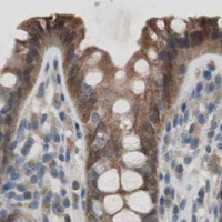
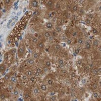
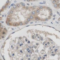


Comprehensive Human Protein Atlas IHC scoring per tissue (reliability: Approved — Low consistency between antibody staining and RNA expression data.). Rows are taken directly from the HPA tissue chart — click any row's HPA link to view the source.
| Tissue | Cell type | Level | Evidence | Source |
|---|---|---|---|---|
| Adrenal gland | Glandular cells | High | Protein (IHC) | HPA → |
| Appendix | Glandular cells | High | Protein (IHC) | HPA → |
| Cerebellum | Purkinje cells | High | Protein (IHC) | HPA → |
| Duodenum | Glandular cells | High | Protein (IHC) | HPA → |
| Epididymis | Glandular cells | High | Protein (IHC) | HPA → |
| Tissue | Cell type | Level | Evidence | Source |
|---|---|---|---|---|
| Adipose tissue | Adipocytes | Not detected | Protein (IHC) | HPA → |
| Lymph node | Germinal center cells | Not detected | Protein (IHC) | HPA → |
| Ovary | Ovarian stroma cells | Not detected | Protein (IHC) | HPA → |
| Parathyroid gland | Glandular cells | Not detected | Protein (IHC) | HPA → |
| Soft tissue | Fibroblasts | Not detected | Protein (IHC) | HPA → |
Troubleshoot FADS2 chromogenic IHC in paraffin sections using compartment, cell type, and control patterns; IF/ICC considerations are covered separately.
The catalog includes one human-reactive anti-FADS2 antibody with a DAB IHC image from a paraffin-embedded brain section (catalog: A01220-1; image caption). No IF image is supplied (catalog: A01220-1).
A01220-1 will render with its human brain paraffin-section IHC image, showing 1:25 primary dilution and DAB detection (image caption). Its listed applications include IHC-P, flow cytometry and WB; reactivity is listed as human (catalog: A01220-1).
Which to pick: Choose A01220-1 for human paraffin-section IHC: it is a rabbit polyclonal antibody listed for IHC-P, with a brain-section IHC image (catalog: A01220-1; image caption). The caption does not report the fixative (image caption). There is no IF/ICC application or image, or nonhuman reactivity, in this catalog entry, so it does not establish an IF/ICC or cross-species choice (catalog: A01220-1).