This website uses cookies to ensure you get the best experience on our website.


- Table of Contents
Plan FAM120A IHC-P around the general cytoplasmic tissue pattern (HPA tissue IHC). The guide identifies high-staining colon glandular cells and adipocytes with no detected staining as comparison populations (HPA tissue IHC).

Expected localisation, validated protocols, controls and antibodies — the at-a-glance facts below, then the full design guide.
| Expected localisation | General cytoplasmic staining in tissue sections (HPA tissue IHC) | |
| Staining pattern | Cytoplasmic staining across multiple tissue cell types (HPA tissue IHC) | |
| Antigen retrieval | Citrate pH 6 HIER, heat-mediated (datasheet M09208) | |
| Positive control | Appendix+4 more · see all | |
| Negative control | Adipose tissue |
| Fixation | Keep fixation consistent across sections (standard IHC practice; not target-specific); Matched tissue-IHC evidence does not establish the fixation claim. Validate the specimen-specific method before use. (selected-SKU IHC image M09208) | |
| Caveat | Medium staining–RNA consistency; verify specificity (HPA tissue IHC) | |
| Regulation | Widely expressed; regulation unspecified (UniProt) | |
| Isoform / epitope | 5 isoforms; epitope coverage is unknown (UniProt) |
Compare the catalog antibody’s IHC-P protocol (datasheet M09208) with published bladder tissue microarray (PMC13602547) and patient and mouse tissue protocols (PMC10987584).
| Sample | Formaldehyde-fixed, paraffin-embedded tissue sections (datasheet M09208; sample unspecified) |
| Fixation | Image formalin-fixed; duration unreported (datasheet M09208); verify before use. |
| Sectioning | 4–5 µm sections on charged slides (standard) |
| Deparaffinisation | Xylene, graded ethanol series to water (standard) |
| Antigen retrieval | Heat retrieval: Citrate pH 6 (datasheet M09208); 20 min, 95–100 °C (standard) |
| Peroxidase block | 3% H2O2, 10 min, room temperature (standard) |
| Blocking | 10% normal serum of the secondary host, 30 min, room temperature (standard) |
| Primary antibody | Mouse monoclonal (clone 1501CT803.19.85.5) anti-FAM120A, 1:25 (datasheet M09208) |
| Primary incubation | 1 hours at 37°C (datasheet M09208) |
| Detection | HRP-polymer secondary, DAB chromogen 5–10 min (standard) |
| Counterstain | Hematoxylin, blue, dehydrate and mount (standard) |
| Expected result | FAM120A-positive staining in glandular cells of appendix (HPA tissue IHC: High). HPA tissue profile: General cytoplasmic expression. No signal in the no-primary control. |
FAM120A should appear mainly in the cytoplasm of many cell types, including glandular epithelium, respiratory epithelium, neurons and glia (HPA: general cytoplasmic expression; HPA: High in these cells). Its cytosolic and cell membrane annotations, with no transmembrane segment, support a predominantly nonnuclear pattern (UniProt Q9NZB2: localisation and topology). HPA rates tissue IHC Approved, with medium agreement between staining and RNA data and external verification pending (HPA: tissue IHC reliability).
| Cytoplasmic chromogen in glandular cells of appendix, breast, cervix or colon; adjacent cells vary in intensity. | This fits the reported distribution: HPA scores those glandular populations High and describes general cytoplasmic expression (HPA: tissue IHC). Judge staining in the named cells, rather than requiring uniform colour across the section. Approved IHC reliability still carries a medium consistency and pending verification caveat (HPA: tissue IHC reliability). |
| Strong, predominantly nuclear staining with little cytoplasmic signal. | Treat this as a compartment mismatch: the expected tissue IHC pattern is cytoplasmic, and HPA ICC-IF places FAM120A in the cytosol (HPA: tissue IHC; HPA: subcellular). Review controls and detection conditions before assigning nuclear staining to FAM120A (general IHC practice). |
| Adipocytes stain strongly while the expected glandular or neural cells do not. | HPA reports FAM120A as Not detected in adipocytes, while several glandular and neural populations score High (HPA: tissue IHC). That reversal raises concern for antibody cross-reactivity or endogenous detection activity; it does not prove either cause (general IHC interpretation). |
| Chromogen spreads through stroma, lumina or much of the section without a cellular cytoplasmic pattern. | Diffuse colour is difficult to score as target staining because HPA describes a cellular, general cytoplasmic pattern (HPA: tissue IHC). Check reagent-only background, endogenous detection activity and nonspecific antibody binding with appropriate controls; these are general IHC checks, not established FAM120A-specific effects (general IHC practice). |
| No convincing signal in the expected cell population of a known-positive section. | An absent signal conflicts with HPA's High calls for the specified cells, but a single negative section cannot establish absent protein (HPA: tissue IHC). Confirm that the tissue contains those cells, then review antibody application, retrieval and detection controls (general IHC practice). |
| Cell population and tissue context | HPA reports High staining in glandular cells of appendix, breast, cervix and colon, respiratory epithelium of bronchus, and selected neurons and glia; adipocytes are Not detected (HPA: tissue IHC). Compare the named cells rather than whole-section colour. These calls describe observed staining, not a universal positive or negative guarantee (HPA: tissue IHC reliability). |
| Expected compartment and stimulus context | HPA tissue IHC describes general cytoplasmic expression, and HPA ICC-IF identifies the cytosol as the main location (HPA: tissue IHC; HPA: subcellular). UniProt also annotates cell membrane localisation and cytosol-to-plasma-membrane movement after UV irradiation (UniProt Q9NZB2: subcellular location). Routine tissue IHC has no supplied UV exposure context, so membrane enrichment is not required. |
| Antibody evidence | HPA019734 and HPA055800 each have Approved IHC status and Enhanced ICC status (HPA: antibody validation). Those labels support use of the reported patterns while retaining the tissue IHC caveat of medium staining–RNA consistency and pending external verification (HPA: tissue IHC reliability). ICC validation does not specify a paraffin-section dilution or retrieval condition. |
| Isoforms and epitope information | UniProt lists 5 FAM120A isoforms, A, B, D, E and F (UniProt Q9NZB2: isoforms). The supplied record does not locate the IHC antibody epitope or establish which isoforms it detects. Interpret a distribution mismatch against the HPA tissue pattern, without assigning it to a particular isoform. |
| Situation | Likely cause | Next action |
|---|---|---|
| Expected glandular cells show no stain. | The slide may lack the relevant cell population, or the IHC workflow may have failed; neither is established by an unstained section (HPA: High in specified glandular cells; general IHC practice). | Confirm cell identity on the counterstain, compare a documented positive tissue, and check the antibody, retrieval and chromogenic detection steps using their supplied instructions (general IHC practice). |
| Adipocytes or other unexpected cells dominate the stain. | Strong adipocyte staining conflicts with the HPA Not detected call and may reflect nonspecific binding or endogenous detection activity (HPA: adipocytes; general IHC interpretation). | Run a no-primary control and review blocking and detection conditions; require cytoplasmic staining in an HPA-positive cell population before calling the result supportive (general IHC practice; HPA: tissue IHC). |
| Background obscures cellular borders throughout the section. | Diffuse chromogen can arise from background binding or endogenous enzyme activity in chromogenic IHC (general IHC practice). | Use a no-primary control to locate detection background, then optimise blocking, antibody concentration and wash steps according to the assay instructions (general IHC practice). Rescore only discernible cellular cytoplasm (HPA: tissue IHC pattern). |
| Signal appears mainly nuclear or exclusively membranous. | A nuclear-dominant pattern disagrees with HPA's cytoplasmic observations; UniProt notes membrane translocation after UV, but no UV context is supplied for this section (HPA: tissue IHC; UniProt Q9NZB2: localisation). | Check the positive control and cellular counterstain, and repeat with the IHC-validated antibody if needed (general IHC practice). Record membrane enrichment as an observation without assuming UV-related translocation. |
| IF/ICC check: where should FAM120A fluorescence appear? | HPA reports a cytosolic main location in ICC-IF and Enhanced ICC validation for HPA019734 and HPA055800 (HPA: subcellular; HPA: antibody validation). These are cell-image observations, not an IHC-P protocol. | Expect predominantly cytosolic fluorescence and use appropriate fluorescence controls to assess background (HPA: subcellular; general IF practice). Interpret the paraffin-section chromogenic result against HPA tissue IHC separately. |


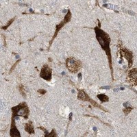


Comprehensive Human Protein Atlas IHC scoring per tissue (reliability: Approved — Medium consistency between antibody staining and RNA expression data. Pending external verification.). Rows are taken directly from the HPA tissue chart — click any row's HPA link to view the source.
| Tissue | Cell type | Level | Evidence | Source |
|---|---|---|---|---|
| Appendix | Glandular cells | High | Protein (IHC) | HPA → |
| Breast | Glandular cells | High | Protein (IHC) | HPA → |
| Bronchus | Respiratory epithelial cells | High | Protein (IHC) | HPA → |
| Caudate | Glial cells | High | Protein (IHC) | HPA → |
| Cerebellum | Purkinje cells | High | Protein (IHC) | HPA → |
| Tissue | Cell type | Level | Evidence | Source |
|---|---|---|---|---|
| Adipose tissue | Adipocytes | Not detected | Protein (IHC) | HPA → |
Troubleshoot FAM120A staining in paraffin sections by checking retrieval, cell type, subcellular pattern, and controls together.
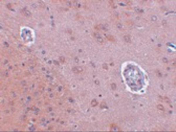
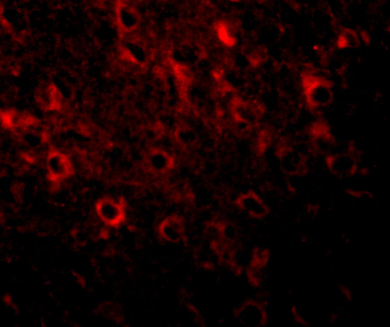
Anti-FAM120A antibodies have IHC images from human and rat brain tissue and IF images from human HeLa and rat brain cells (catalog image captions). Listed reactivity spans human, mouse, and rat (catalog reactivity).
M09208 has an IHC-P image of formaldehyde-fixed, paraffin-embedded human brain and an IF image of HeLa cells (M09208 image captions). A09208 has an IHC image of rat brain tissue and an IF image of rat brain cells; its IHC caption does not report the fixative (A09208 image captions).
Which to pick: For human paraffin-section IHC, choose the mouse monoclonal M09208: its IHC caption documents that preparation, and IHC-P is listed as an application (M09208 catalog entry and IHC image caption). For IF/ICC, choose M09208 for human HeLa cells or A09208 for rat brain cells, matching their respective IF images (M09208 and A09208 IF image captions). For work across species, consider A09208, a rabbit antibody with human, mouse, and rat listed as reactive; its IHC image shows rat brain tissue, but the caption does not report fixation or paraffin processing (A09208 catalog entry and IHC image caption).