FANCD2 / Fanconi anemia group D2 protein · IHC design guide

Design Immunohistochemistry for FANCD2

This FANCD2 chromogenic IHC guide centers on nuclear staining in glandular epithelia and subsets of testicular and lymphoid cells (HPA tissue IHC). It gives a catalog antibody starting range of 1:100–1:300 (datasheet A00563) and highlights cell-cycle-dependent nuclear foci when interpreting staining (UniProt).

Evidence assembled Oct 2026 · For research use; verify linked source records and product datasheet before use
Immunohistochemistry protocol sheet for FANCD2 (IHC for FANCD2): expected localisation Nuclear in most glandular epithelia (HPA tissue IHC), antibody A00563, validated IHC image, and IHC protocol steps
Printable FANCD2 IHC protocol sheet — expected localisation Nuclear in most glandular epithelia (HPA tissue IHC), antibody A00563, controls and protocol steps. Open the full FANCD2 IHC guide →

FANCD2 Immunohistochemistry Experimental Design Guide

Expected localisation, validated protocols, controls and antibodies — the at-a-glance facts below, then the full design guide.

Must know before staining
Expected localisation Nuclear in most glandular epithelia (HPA tissue IHC)
Staining pattern Nuclear glandular cells; subsets in testis and germinal centers (HPA tissue IHC)
Antigen retrieval Tris-EDTA pH 8.0 HIER, heat-mediated (datasheet A00563)
Positive control ⓘ Testis+4 more · see all
Negative control ⓘ Adipose tissue+4 more · see all
Important caveats
Reasons your staining may differ from the expected pattern.
Fixation Keep fixation consistent across paraffin sections. (standard IHC practice; not target-specific)
Caveat Mitotic redistribution may reduce nuclear-only scoring (UniProt)
Regulation S phase or DNA damage promotes nuclear foci (UniProt)
Isoform / epitope 4 isoforms; epitope coverage is unspecified (UniProt)
Section 1

Recommended FANCD2 IHC & IF Protocols

Compare the catalog antibody’s IHC-P protocol (datasheet A00563) with four published FANCD2 chromogenic IHC workflows (PMC10210052; PMC2096638; PMC10425492; PMC6403512).

Recommended immunohistochemistry (IHC-P) protocol parameters
SampleParaffin-embedded Human lung cancer tissue; fixative not specified (datasheet A00563)
FixationImage fixative and duration unreported (datasheet A00563); verify before use.
Sectioning4–5 µm sections on charged slides (standard)
DeparaffinisationXylene, graded ethanol series to water (standard)
Antigen retrievalHeat retrieval: Tris-EDTA pH 8.0 (datasheet A00563); 20 min, 95–100 °C (standard)
Peroxidase block3% H2O2, 10 min, room temperature (standard)
Blocking10% normal serum of the secondary host, 30 min, room temperature (standard)
Primary antibodyRabbit anti-FANCD2, 1:100-1:300 (datasheet A00563)
Primary incubationOvernight at 4 °C (standard)
DetectionHRP-polymer secondary, DAB chromogen 5–10 min (standard)
CounterstainHematoxylin, blue, dehydrate and mount (standard)
Expected resultFANCD2-positive staining in pachytene spermatocytes of testis (HPA tissue IHC: High). HPA tissue profile: Nuclear expression in most glandular epithelia, subset of cells in testis and in germinal centers of lymphoid tissues. No signal in the no-primary control.
💡Decision noteStart with heat-mediated Tris-EDTA pH 8.0 retrieval for the catalog antibody (datasheet A00563); follow the stated retrieval conditions when reproducing a published protocol.
Section 2

What Is the Expected FANCD2 Staining Pattern?

FANCD2 should stain predominantly in nuclei, especially in pachytene spermatocytes of testis and glandular epithelial cells of colon, duodenum and rectum (HPA tissue IHC). Nuclear foci may occur during S phase or after genotoxic stress (UniProt Q9BXW9). HPA rates its tissue IHC profile Approved, with medium consistency between antibody staining and RNA expression; FANCD2 has no transmembrane segment (HPA tissue IHC; UniProt Q9BXW9).

What am I looking at on my slide?
High nuclear staining in pachytene spermatocytes; medium nuclear staining in colon or duodenal glandular cells.This matches the reported positive cell types and levels (HPA tissue IHC). Score the relevant cells and their nuclear compartment, since the HPA profile describes expression in a subset of testis cells rather than every cell (HPA tissue IHC).
Prominent cytoplasmic staining with little or no nuclear signal in an expected positive cell population.Question this paraffin-section IHC pattern: FANCD2 is predominantly nuclear (UniProt Q9BXW9; HPA tissue IHC). UniProt describes transient cytoplasmic diffusion at mitotic onset, so an isolated cell outside the usual nuclear pattern is less decisive than widespread cytoplasmic staining (UniProt Q9BXW9).
Strong staining in adipocytes of adipose tissue or another cell population reported as undetected.HPA reports FANCD2 as not detected in adipose-tissue adipocytes (HPA tissue IHC). Check whether the signal follows that cell type and whether a no-primary control also develops color; cross-reactivity or endogenous detection activity are possible explanations (general IHC practice).
Color spreads across the section without a clear nuclear boundary or cell-type pattern.This is difficult to score as FANCD2 because the expected tissue pattern is nuclear and cell selective (HPA tissue IHC). Broad background can arise from nonspecific binding or detection chemistry; compare a no-primary control before interpreting weak positive cells (general IHC practice).
No nuclear staining in pachytene spermatocytes of a testis section.This conflicts with the high signal reported for that cell population (HPA tissue IHC). First confirm that pachytene spermatocytes are present and identifiable; then review the catalog antibody's IHC-P conditions and the detection controls (general IHC practice).
💡Expected FANCD2 appearanceCall a result positive when the appropriate cells show predominantly nuclear signal—strongest in pachytene spermatocytes and potentially moderate in listed glandular epithelia; broad cytoplasmic or cell-independent color is suspect (HPA tissue IHC; UniProt Q9BXW9; general IHC practice).
How each factor affects the staining
Tissue and cell selectionTestis pachytene spermatocytes provide a reported high reference; colon, duodenum and rectum glandular cells are medium, while adipose-tissue adipocytes are not detected (HPA tissue IHC). A negative field is meaningful only after its cell types are identified (general IHC practice).
Cell-cycle contextFANCD2 concentrates in nuclear foci during S phase and after genotoxic stress; it can leave chromosomes and diffuse into cytoplasm at mitotic onset (UniProt Q9BXW9). These are localization contexts, not promised frequencies or staining intensities in routine tissue IHC (UniProt Q9BXW9).
IHC antibody validationHPA marks CAB016117 IHC as Approved and HPA063742 IHC as unlisted; Approved tissue staining has medium consistency with RNA expression (HPA antibodies; HPA tissue IHC). Antibody-specific IHC validation matters when comparing slide patterns (HPA antibodies).
Detection interpretationA no-primary control can reveal background from the chromogenic detection system, while a known-positive tissue checks the staining run (general IHC practice). These controls help interpret unexpected color; they do not establish antibody specificity by themselves (general IHC practice).
IF/ICC Q: Should nuclear puncta appear in fluorescence images?Yes, nuclear bodies and nucleoplasm are supported main locations in HPA ICC-IF, and UniProt describes stress- and S-phase-associated nuclear foci (HPA subcellular; UniProt Q9BXW9). Nucleoli are an additional supported location; cytosol is uncertain in HPA ICC-IF (HPA subcellular).
Why is my staining missing, weak or wrong?
SituationLikely causeNext action
Testis control lacks staining in identifiable pachytene spermatocytes.Those cells are reported high by HPA, so the observation may reflect a failed staining run or a mismatch between antibody and method (HPA tissue IHC; general IHC practice).Verify cell identity and section quality, then check the catalog antibody's IHC-P instructions and run-level positive and no-primary controls (general IHC practice).
Colon glands stain weakly compared with testis.A difference can be expected: HPA reports medium colon glandular staining and high pachytene-spermatocyte staining (HPA tissue IHC).Score each tissue against its own reported cell type and level; use the same detection and counterstain assessment across compared sections (HPA tissue IHC; general IHC practice).
Color appears mainly in cytoplasm throughout positive tissue.A broad cytoplasmic pattern conflicts with the predominant nuclear tissue profile, although UniProt notes transient cytoplasmic diffusion at mitotic onset (HPA tissue IHC; UniProt Q9BXW9).Compare nuclear boundaries against the counterstain and examine a no-primary control; treat widespread cytoplasmic color cautiously when nuclear staining is absent (general IHC practice).
Adipocytes show convincing-looking chromogenic signal.Adipose-tissue adipocytes are reported as not detected; nonspecific binding or endogenous detection activity could explain unexpected color (HPA tissue IHC; general IHC practice).Check the no-primary control in the same tissue and compare localization with a positive tissue's nuclear pattern before scoring adipocytes as positive (HPA tissue IHC; general IHC practice).
Most tissue compartments develop diffuse brown background.The expected HPA pattern is nuclear and cell selective; diffuse color can arise from detection background or nonspecific binding (HPA tissue IHC; general IHC practice).Inspect the no-primary control, blocking and wash steps, then assess whether identifiable nuclei retain a cell-type-specific signal above background (general IHC practice).
Discrete nuclear dots differ between otherwise comparable positive cells.FANCD2 can form nuclear foci during S phase and following genotoxic stress; variable dots alone do not establish a staining failure (UniProt Q9BXW9).Record diffuse nuclear signal and puncta separately, with tissue and cell identity; avoid assigning a DNA-damage state from IHC appearance alone (UniProt Q9BXW9; general IHC practice).

Sample controls for FANCD2 IHC & IF

🧪Run testis first and look for staining in pachytene spermatocytes (HPA: High in pachytene spermatocytes). Use adipose tissue adipocytes as the negative tissue (HPA: Not detected in adipocytes); on the testis slide, compare neighboring non-pachytene cells, but do not assume they are FANCD2-negative from the supplied HPA row (HPA: High in pachytene spermatocytes).
Positive control tissue: Testis (Pachytene spermatocytes, HPA High)
Negative control tissue: Adipose tissue (HPA Not detected)
ICC-IF cell lines (HPA subcellular resource): HPA ICC-IF images show FANCD2 in U-251MG, A-549, SiHa, U2OS, with annotated localisation: Nucleoplasm (supported), Nuclear bodies (supported) (HPA subcellular).
Technical controls: Include a no-primary, secondary-only control and a nonimmune control matched to the primary antibody’s host and isotype, using host-matched nonimmune serum if the primary is polyclonal (standard IHC practice); use a FANCD2 knockout sample or an immunogen-peptide block as a biological specificity control (caption: peptide-preabsorbed negative control). For chromogenic testis IHC, quench endogenous peroxidase and check background in the no-primary control (standard IHC practice).
⚠️Feasibility: A target-specific fixation window or fixation effect is unreported, and the selected A00563 paraffin-section caption does not state the fixative (caption: fixative unreported). The caption reports high-pressure, high-temperature Tris-EDTA retrieval at pH 8.0, but provides no retrieval comparison that establishes a dependency (caption: Tris-EDTA retrieval, pH 8.0). The supplied evidence does not establish whether frozen sections or IF are easier; in testis, score the specified pachytene spermatocytes rather than treating all cells as equivalent, and assess nuclear signal with cell-cycle context (HPA: High in pachytene spermatocytes; UniProt Q9BXW9: nuclear foci during S phase and genotoxic stress).

HPA tissue IHC evidence for FANCD2

Comprehensive Human Protein Atlas IHC scoring per tissue (reliability: Approved — Medium consistency between antibody staining and RNA expression data.). Rows are taken directly from the HPA tissue chart — click any row's HPA link to view the source.

Positive expression · recommended positive controls

TissueCell typeLevelEvidenceSource
Testis Pachytene spermatocytes High Protein (IHC) HPA →
Colon Glandular cells Medium Protein (IHC) HPA →
Duodenum Glandular cells Medium Protein (IHC) HPA →
Esophagus Squamous epithelial cells Medium Protein (IHC) HPA →
Nasopharynx Ciliated cells (cell body) Medium Protein (IHC) HPA →

Undetected expression · recommended negative controls

TissueCell typeLevelEvidenceSource
Adipose tissue Adipocytes Not detected Protein (IHC) HPA →
Adrenal gland Glandular cells Not detected Protein (IHC) HPA →
Breast Adipocytes Not detected Protein (IHC) HPA →
Bronchus Basal cells Not detected Protein (IHC) HPA →
Caudate Glial cells Not detected Protein (IHC) HPA →
Section 3

Advanced FANCD2 IHC Tips

Troubleshoot FANCD2 staining in paraffin sections by checking retrieval, nuclear localisation and cell specific controls before comparing chromogenic scores.

How should I retrieve FANCD2 when nuclear staining is weak?
Use heat-mediated Tris-EDTA, pH 8.0, antigen retrieval for FANCD2 paraffin-section IHC (datasheet A00563). The selected antibody image used high-pressure, high-temperature retrieval in this buffer, followed by antibody at 1:100 overnight at 4 °C (caption A00563). If nuclei remain weak, compare a fresh retrieval run with a shorter or longer heating interval while keeping buffer composition and detection conditions fixed (standard IHC practice). Check a concurrently processed section containing expected positive cells, such as testicular pachytene spermatocytes, before increasing primary antibody concentration (HPA: High in pachytene spermatocytes; standard IHC practice). Excessive heating can damage section morphology, so judge signal alongside intact nuclear detail (standard IHC practice).
Could fixation explain inconsistent FANCD2 staining between paraffin blocks?
FANCD2-specific fixation sensitivity is unknown from the supplied evidence; the selected paraffin-section image does not report its fixative (caption A00563). Record each block’s fixative and fixation duration, then compare blocks using the same Tris-EDTA, pH 8.0, retrieval and staining run (datasheet A00563; standard IHC practice). Variable fixation can alter antigen accessibility and morphology in paraffin IHC, but that general effect does not establish a FANCD2-specific fixation requirement (standard IHC practice). Include a concurrently processed positive control and assess nuclear detail before attributing a weaker result to lower FANCD2 abundance (HPA: nuclear expression in glandular epithelia; standard IHC practice). Do not infer fixation tolerance from tissue expression patterns or protein modifications (HPA tissue IHC; UniProt Q9BXW9).
Is diffuse cytoplasmic FANCD2 staining plausible in chromogenic IHC?
Prioritise nuclear staining when assessing FANCD2 in paraffin sections: its main localisation is nuclear, with foci during S phase and after genotoxic stress (UniProt Q9BXW9). HPA reports supported nucleoplasmic and nuclear-body localisation, while its cytosolic assignment is uncertain (HPA subcellular). Compare chromogen with a nuclear counterstain and score only cells whose compartment can be resolved without overlapping neighbours (standard IHC practice). Diffuse cytoplasmic signal without convincing nuclei warrants review of primary antibody concentration, blocking and detection controls before biological interpretation (UniProt Q9BXW9; standard IHC practice). Mitotic localisation can differ because FANCD2 leaves chromosomes at mitotic onset and returns to nuclei after division (UniProt Q9BXW9).
Can this IHC stain distinguish FANCD2 isoforms or modified protein?
Do not assign an isoform from chromogenic intensity alone: the record lists 4 FANCD2 isoforms, but supplies no mapped epitope for this antibody (UniProt Q9BXW9; caption A00563). FANCD2 has annotated phosphorylation sites and participates in ubiquitin-dependent interactions, so modification state is biologically relevant without proving that this antibody distinguishes those states (UniProt Q9BXW9). Request an epitope map or isoform-reactivity evidence before claiming selective detection, and document the antibody identity alongside each staining run (standard IHC practice). The selected image includes an immunogen-peptide preabsorption control, which supports evaluation of that image’s staining but does not establish isoform or modification specificity (caption A00563). Score identifiable nuclear staining separately from nonspecific diffuse signal (UniProt Q9BXW9; standard IHC practice).
How should I assess FANCD2 by IF alongside this IHC guide?
Treat IF/ICC as a separate validation exercise: the selected antibody evidence here describes paraffin-section chromogenic IHC, with no supplied IF/ICC protocol for this antibody (caption A00563). For multiplex IF, pair FANCD2 with a validated marker identifying the expected cell population, such as pachytene spermatocytes, and compare signals within the same nuclei (HPA: High in pachytene spermatocytes; standard IF practice). Choose a fluorophore channel with low tissue autofluorescence and include single-colour and no-primary controls to identify bleed-through and background (standard IF practice). FANCD2 is nuclear and has no transmembrane segment, so permeabilisation must permit access through cellular and nuclear barriers; optimise it against preserved morphology (UniProt Q9BXW9; standard IF practice). Evaluate foci separately from diffuse nuclear signal (UniProt Q9BXW9).
What should I change when FANCD2 IHC shows widespread background?
First determine whether the colour lies in nuclei, cytoplasm, extracellular material or damaged tissue, using the counterstain and an adjacent section for orientation (standard IHC practice). FANCD2 is predominantly nuclear, so widespread nonnuclear chromogen is a reason to check specificity before scoring (UniProt Q9BXW9; HPA subcellular). Titrate the primary antibody around the image’s 1:100 overnight condition, and compare matched no-primary and detection-only controls (caption A00563; standard IHC practice). For peroxidase detection, verify the endogenous-peroxidase block and assess whether secondary reagent or chromogen development is driving background (standard IHC practice). The selected image’s peptide-preabsorbed control can help evaluate antibody-dependent staining, but it cannot replace tissue and detection controls across your run (caption A00563; standard IHC practice).
How can I quantify FANCD2 across paraffin sections fairly? ⚠ ANSWER MARKED FOR VERIFICATION
Define the cell compartment and region before scoring, then record the percentage of nuclei above a fixed threshold or a nuclear H-score using consistent intensity bins (UniProt Q9BXW9; standard IHC practice). If assessing focal staining, report positive nuclei per mm² of evaluable tissue and specify how individual nuclear foci were counted (UniProt Q9BXW9; standard IHC practice). Normalise positive counts to all eligible nuclei in the same annotated cell population, rather than to the whole section when cell composition varies (standard IHC practice). Use identical retrieval, detection, counterstaining and image-analysis settings for compared sections, with a shared control on each staining run (datasheet A00563; standard IHC practice). Report nuclear intensity and focal pattern separately because FANCD2 can concentrate in foci during S phase or genotoxic stress (UniProt Q9BXW9).
When is a FANCD2-positive IHC result credible?
A credible result places chromogen in identifiable nuclei of relevant cells and remains interpretable beside a clean negative control (UniProt Q9BXW9; standard IHC practice). Testicular pachytene spermatocytes show high HPA staining, whereas adipocytes in adipose tissue were not detected, providing context for cell-specific expectations rather than universal positive and negative controls (HPA tissue IHC). Treat signal confined to cut edges, necrotic areas or extracellular debris as suspect, and exclude those regions from scoring (standard IHC practice). Check no-primary and peroxidase-block controls when colour appears in the wrong compartment or follows endogenous enzyme activity (standard IHC practice). Interpret focal nuclear staining in light of cell cycle and damage state, while avoiding claims that chromogenic positivity alone proves active DNA repair (UniProt Q9BXW9).
Boster reagents

Best FANCD2 / Fanconi anemia group D2 protein IHC Antibodies

Catalog anti-FANCD2 antibodies have IHC images from human paraffin sections and an IF/ICC image from human cells; all four list Human, Mouse and Rat reactivity (catalog applications, image captions and reactivity).

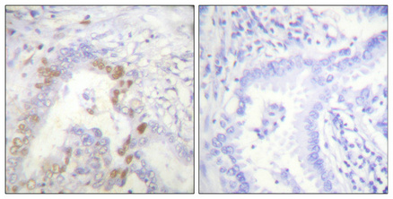
Real IHC data Immunohistochemical analysis of paraffin-embedded Human lung cancer. Antibody was diluted at 1:100 (4° overnight). High-pressure and temperature Tris-EDTA, pH8.0 was used for antigen retrieval. Negetive contrl (right) obtaned from antibody was pre-absorbed by immunogen peptide.
Anti-FANCD2 Antibody
Cat # A00563
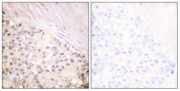
Real IHC data Immunohistochemistry (IHC) analyzes of FANCD2 (P216) pAb in paraffin-embedded human breast carcinoma tissue at 1:100.
Anti-FANCD2 (P216) Antibody
Cat # A00563-1
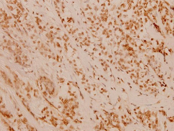
Real IHC data Immunohistochemical analysis of paraffin-embedded human spleen, using FANCD2 Antibody.
Anti-FANCD2 Rabbit Monoclonal Antibody
Cat # M00563
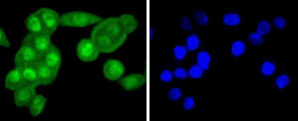
Real IF data ICC staining FANCD2 in SW480 cells (green). The nuclear counter stain is DAPI (blue). Cells were fixed in paraformaldehyde, permeabilised with 0.25% Triton X100/PBS.
Anti-FANCD2 Antibody
Cat # A00563-2

A00563 shows IHC in human lung cancer and breast carcinoma paraffin sections; A00563-1 shows IHC in human breast carcinoma paraffin sections (A00563 and A00563-1 image captions). M00563 shows IHC in human spleen paraffin sections, while A00563-2 shows IF/ICC in SW480 cells fixed with paraformaldehyde (M00563 and A00563-2 image captions).

Which to pick: For tissue IHC, choose A00563 for its peptide-absorption controls, A00563-1 for its breast carcinoma example, or monoclonal M00563 for its spleen example; their paraffin-section captions do not report the fixative (A00563, A00563-1 and M00563 image captions; catalog clone field). For IF/ICC, choose A00563-2 for its SW480 cell image and listed IF/ICC applications (A00563-2 image caption and catalog applications). All four list Human, Mouse and Rat reactivity, but the supplied images document human samples only (catalog reactivity and image captions).

Each figure is that product's own IHC / IF validation image from its datasheet.

References

  1. UniProt Consortium. UniProt entry Q9BXW9 (FACD2_HUMAN, Fanconi anemia group D2 protein).
  2. Human Protein Atlas. FANCD2 tissue IHC expression (reliability: Approved).
  3. Human Protein Atlas. FANCD2 subcellular location (ICC-IF): Mainly localized to the nucleoplasm and nuclear bodies. In addition localized to the nucleoli and cytosol..
  4. Human Protein Atlas. FANCD2 antibody validation summary (2 antibodies).
  5. Neddylation inhibition sensitises renal medullary carcinoma tumours to platinum chemotherapy. Clinical and translational medicine 2023 — PMC10210052.
  6. Loss of expression of FANCD2 protein in sporadic and hereditary breast cancer. Breast cancer research and treatment 2008 — PMC2096638.
  7. Crosstalk of ferroptosis regulators and tumor immunity in pancreatic adenocarcinoma: novel perspective to mRNA vaccines and personalized immunotherapy. Apoptosis : an international journal on programmed cell death 2023 — PMC10425492.
  8. Expression and prognostic significance of Fanconi anemia group D2 protein and breast cancer type 1 susceptibility protein in familial and sporadic breast cancer. Oncology letters 2019 — PMC6403512.
  9. PubMed PMID:11239453 — UniProt-cited evidence.
  10. PubMed PMID:15489334 — UniProt-cited evidence.
  11. PubMed PMID:14702039 — UniProt-cited evidence.