This website uses cookies to ensure you get the best experience on our website.


- Table of Contents
Plan FCGR2B staining in paraffin sections using placental endothelial cells and lymphoid subsets as reference patterns (HPA tissue IHC). The guide covers the catalog antibody's 1:100–1:300 IHC dilution range (datasheet A01690-1) and interpretation of cytoplasmic staining (HPA tissue IHC) alongside the membrane location predicted by topology (UniProt).

Expected localisation, validated protocols, controls and antibodies — the at-a-glance facts below, then the full design guide.
| Expected localisation | Cytoplasm observed in placental endothelium (HPA tissue IHC); cell membrane expected from topology (UniProt) | |
| Staining pattern | Cytoplasmic staining in placental endothelium and lymphoid subsets (HPA tissue IHC) | |
| Antigen retrieval | Tris-EDTA pH 9.0 HIER, heat-mediated (datasheet A01690-1) | |
| Positive control | Placenta+4 more · see all | |
| Negative control | Adipose tissue+4 more · see all |
| Fixation | Matched tissue-IHC evidence does not establish the fixation claim. Validate the specimen-specific method before use. (selected-SKU IHC image A01690-1) | |
| Caveat | Antibody may detect protein from more than one gene (HPA tissue IHC) | |
| Regulation | Expression regulator not specified (UniProt) | |
| Isoform / epitope | 5 isoforms; verify extracellular versus cytoplasmic epitope coverage (UniProt) |
The catalog antibody protocol uses Tris-EDTA pH 9.0 retrieval (datasheet A01690-1). Four published FCGR2B IHC protocols provide additional starting points (PMC12177957; PMC11034135; PMC8796391; PMC10821851).
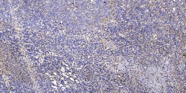
| Sample | Paraffin-embedded human tonsil tissue; fixative not specified (datasheet A01690-1) |
| Fixation | Image fixative and duration unreported (datasheet A01690-1); verify before use. |
| Sectioning | 4–5 µm sections on charged slides (standard) |
| Deparaffinisation | Xylene, graded ethanol series to water (standard) |
| Antigen retrieval | Heat retrieval: Tris-EDTA pH 9.0 (datasheet A01690-1); 20 min, 95–100 °C (standard) |
| Peroxidase block | 3% H2O2, 10 min, room temperature (standard) |
| Blocking | 10% normal serum of the secondary host, 30 min, room temperature (standard) |
| Primary antibody | Rabbit anti-FCGR2B, 1:100-1:300 (datasheet A01690-1) |
| Primary incubation | Overnight at 4 °C (standard) |
| Detection | HRP-polymer secondary, DAB chromogen 5–10 min (standard) |
| Counterstain | Hematoxylin, blue, dehydrate and mount (standard) |
| Expected result | FCGR2B-positive staining in endothelial cells of placenta (HPA tissue IHC: High). HPA tissue profile: Cytoplasmic expression in endothelial cells in placenta and subsets of lymphoid cells. No signal in the no-primary control. |
FCGR2B is a cell membrane receptor with an extracellular region at residues 43–217 and a cytoplasmic tail at 241–310 (UniProt P31994 topology). In paraffin tissue IHC, expect the strongest reported staining in placental endothelial cells, with weaker staining in selected hematopoietic and lymphoid cells (HPA tissue IHC). HPA describes the observed placental and lymphoid staining as cytoplasmic; its tissue profile is rated Enhanced but carries a cross-gene staining caution (HPA tissue IHC).
| Distinct staining in placental endothelial cells, with an apparent membrane edge or cytoplasmic signal. | This fits the strongest reported tissue result: High placental endothelial staining (HPA tissue IHC). A membrane edge fits receptor topology (UniProt P31994 topology), while cytoplasmic appearance fits HPA's description of its IHC images (HPA tissue IHC). Record the compartment actually seen; the two sources describe different kinds of evidence. |
| Moderate staining in selected bone marrow hematopoietic cells or lymph node, tonsil, or splenic white-pulp cells. | These are reported Medium staining populations, rather than uniformly positive compartments (HPA tissue IHC). Score the named cells or regions and compare them with adjacent unstained cells. HPA's tissue reliability is Enhanced, with medium consistency between staining and RNA data and a warning that staining may include protein from more than one gene (HPA tissue IHC). |
| Predominantly nuclear staining, especially without the expected placental endothelial signal. | A nuclear-only pattern does not match the cell membrane assignment (UniProt P31994 subcellular location) or HPA's cytoplasmic tissue description (HPA tissue IHC). Treat it as unconfirmed and inspect controls, tissue morphology, and detection background before interpreting it as FCGR2B; a nuclear signal alone cannot establish a new localisation. |
| Strong staining in adipocytes, brain glia, or glandular cells where HPA reports no detection. | HPA reports no detection in adipocytes from adipose tissue and breast, caudate glial cells, and duodenal glandular cells (HPA tissue IHC). Such staining warrants review for antibody cross-reactivity or nonspecific detection (general IHC practice). HPA also cautions that its antibody staining may represent protein from more than one gene (HPA tissue IHC). |
| Widespread, weak, featureless colour across tissue and blank areas. | A diffuse deposit cannot establish the reported cell-specific distribution (HPA tissue IHC). Compare with a no-primary control and inspect blocking, washes, and chromogen development (general IHC practice). If the deposit appears in the control, resolve the detection background before assigning any cellular staining to FCGR2B. |
| Cell compartment and assay | FCGR2B has one transmembrane segment at residues 218–240 (UniProt P31994 topology). HPA's tissue IHC description calls the observed signal cytoplasmic, whereas its ICC-IF summary says Membrane and supplies no cell-line images (HPA tissue IHC; HPA subcellular). Use tissue IHC evidence for this paraffin-section decision; the ICC-IF summary does not establish an IF result in a named cell line. |
| Tissue and cell selection | Placental endothelial cells are High; bone marrow hematopoietic cells and specified lymphoid regions are Medium (HPA tissue IHC). Lung macrophages are Low, so they offer a less conspicuous comparison (HPA tissue IHC). Specify the cell population when assessing a section: HPA also reports no detection in cerebral-cortex and colon endothelial cells (HPA tissue IHC). |
| Antibody-specific confidence | HPA lists CAB007796 as IHC Enhanced and HPA014730 as IHC Supported (HPA antibodies). Its tissue profile is also rated Enhanced, but HPA reports only medium agreement between antibody staining and RNA and warns of possible staining from more than one gene (HPA tissue IHC). Treat matching anatomy as supporting evidence, not proof of exclusive FCGR2B recognition. |
| Protein forms and assay limits | UniProt lists five FCGR2B isoforms, three N-glycosylation sites, and a signal peptide at residues 1–42 (UniProt P31994 isoforms, glycosylation, processing). These features identify potential differences among protein forms; the supplied sources do not identify the antibodies' epitopes or show how these features change paraffin IHC staining. Avoid assigning a compartment difference or failed stain to a specific isoform or modification. |
| Situation | Likely cause | Next action |
|---|---|---|
| No signal in placental endothelial cells. | The expected High population is absent (HPA tissue IHC); the available evidence does not establish a target-specific fixation or retrieval cause. | Confirm that endothelial cells are present, then check positive and no-primary controls, antibody application, detection reagents, and the documented retrieval procedure (general IHC practice). Record any retrieval change as an assay adjustment, not a demonstrated FCGR2B fixation effect. |
| Only faint signal in bone marrow or lymphoid tissue. | These populations are reported Medium, below the High placental endothelial result (HPA tissue IHC). | Identify the reported hematopoietic cells or lymphoid regions before scoring (HPA tissue IHC). Compare a placental positive control and examine exposure or chromogen development within the assay's validated settings (general IHC practice); do not require every cell to stain. |
| Strong nuclear-only staining. | A nuclear-only result conflicts with the assigned cell membrane location and HPA's tissue IHC description (UniProt P31994 subcellular location; HPA tissue IHC). | Compare with a no-primary control and assess morphology and detection background (general IHC practice). Repeat with an independently validated antibody if available; HPA lists distinct IHC validation statuses for CAB007796 and HPA014730 (HPA antibodies). |
| Placenta and many HPA-negative cells stain equally strongly. | A cell-independent pattern conflicts with High placental endothelial staining and reported nondetection in several named populations (HPA tissue IHC). Cross-reactivity or nonspecific detection is possible (general IHC practice). | Score endothelial cells separately, run a no-primary control, and compare reported negative cell populations (HPA tissue IHC; general IHC practice). Interpret persistent staining cautiously because HPA warns that antibody staining may include protein from more than one gene (HPA tissue IHC). |
| Brown deposit appears throughout the section or in the no-primary control. | A signal in the no-primary control implicates the detection workflow rather than primary-antibody recognition (general IHC practice). | Review endogenous activity blocking where relevant to the chosen chromogenic system, reagent carryover, wash quality, and development time (general IHC practice). Reassess cellular localisation after background is reduced; diffuse colour alone does not reproduce HPA's described distribution (HPA tissue IHC). |
| An IF result appears inconsistent with the paraffin IHC section. | HPA's ICC-IF summary says Membrane but provides no cell-line images; tissue IHC describes cytoplasmic staining in placental endothelial and selected lymphoid cells (HPA subcellular; HPA tissue IHC). | Evaluate IF with its own controls and guide. For this paraffin IHC section, use the tissue-specific IHC pattern and document whether staining appears membranous or cytoplasmic (HPA tissue IHC; UniProt P31994 topology). Do not infer an IF protocol or a named-cell IF pattern from the supplied summary. |
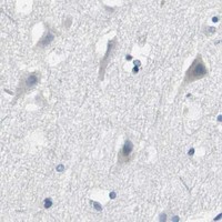
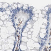

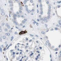
Comprehensive Human Protein Atlas IHC scoring per tissue (reliability: Enhanced — Medium consistency between antibody staining and RNA expression data. Caution, targets protein from more than one gene. Antibody staining in cells/structures not annotated, view images.). Rows are taken directly from the HPA tissue chart — click any row's HPA link to view the source.
| Tissue | Cell type | Level | Evidence | Source |
|---|---|---|---|---|
| Placenta | Endothelial cells | High | Protein (IHC) | HPA → |
| Bone marrow | Hematopoietic cells | Medium | Protein (IHC) | HPA → |
| Lymph node | Non-germinal center cells | Medium | Protein (IHC) | HPA → |
| Spleen | Cells in white pulp | Medium | Protein (IHC) | HPA → |
| Tonsil | Non-germinal center cells | Medium | Protein (IHC) | HPA → |
| Tissue | Cell type | Level | Evidence | Source |
|---|---|---|---|---|
| Adipose tissue | Adipocytes | Not detected | Protein (IHC) | HPA → |
| Breast | Adipocytes | Not detected | Protein (IHC) | HPA → |
| Caudate | Glial cells | Not detected | Protein (IHC) | HPA → |
| Cerebellum | Cells in granular layer | Not detected | Protein (IHC) | HPA → |
| Cerebral cortex | Endothelial cells | Not detected | Protein (IHC) | HPA → |
Troubleshoot FCGR2B staining in paraffin sections by checking retrieval, cell identity and subcellular pattern against the available IHC evidence.
A01690-1 has a human paraffin-section IHC image (catalog image caption). IF is listed without an IF image (catalog applications; image list); Human, Mouse and Rat reactivity is listed (catalog reactivity).
A01690-1 will render with its own IHC image of paraffin-embedded human tonsil, using a 1:200 primary dilution and Tris-EDTA retrieval at pH 9.0 (catalog image caption). The catalog lists IF and Human, Mouse and Rat reactivity, but provides no IF image; the IHC caption does not report the fixative (catalog applications, reactivity and image caption).
Which to pick: Choose A01690-1 for paraffin-section IHC because its own human tonsil image documents that preparation; the fixative is unreported (catalog image caption). For IF, A01690-1 lists a 1:50 dilution, but has no IF image, and ICC is not listed (catalog dilution, applications and image list). For cross-species planning, A01690-1 lists Human, Mouse and Rat reactivity; its host is Rabbit, and clonality is unreported (catalog reactivity, host and clone).