Click image to see more details
-

-
-
-
-
+4
Product Info Summary
| SKU: | A01690-1 |
|---|---|
| Size: | 100ul |
| Reactive Species: | Human, Mouse, Rat |
| Host: | Rabbit |
| Application: | ELISA, IF, IHC, WB |
Customers Who Bought This Also Bought

Product info
Product Name
Anti-CD32 FCGR2B Antibody
SKU/Catalog Number
A01690-1
Size
100ul
Form
Liquid
Description
Boster Bio Anti-CD32 FCGR2B Antibody catalog # A01690-1. Tested in ELISA, IHC, WB applications. This antibody reacts with Human, Mouse, Rat.
Storage & Handling
Store at -20°C for one year. For short term storage and frequent use, store at 4°C for up to one month. Avoid repeated freeze-thaw cycles.
Cite This Product
Anti-CD32 FCGR2B Antibody (Boster Biological Technology, Pleasanton CA, USA, Catalog # A01690-1)
Host
Rabbit
Contents
Liquid in PBS containing 50% glycerol, 0.5% stabilizing protein and 0.02% sodium azide.
*This antibody is supplied in a stabilized formulation.
Compatibility with conjugation reactions depends on the chemistry of the conjugation method used.
For conjugation methods that are not compatible with the stabilizing components present in this formulation, a carrier-free antibody format is required.
Clonality
Polyclonal
Isotype
IgG
Immunogen
The antiserum was produced against synthesized peptide derived from human CD32. AA range:277-326
Cross-reactivity
No cross reactivity with other proteins.
Reactive Species
A01690-1 is reactive to FCGR2B in Human, Mouse, Rat
Calculated molecular weight
34.0 kDa
Antibody Validation
Boster validates all antibodies on WB, IHC, ICC, Immunofluorescence, and ELISA with known positive control and negative samples to ensure specificity and high affinity, including thorough antibody incubations.
Application & Images
Applications
A01690-1 is guaranteed for ELISA, IF, IHC, WB Boster Guarantee
Recommend Dilution
WB 1:500-1:2000
IHC 1:100-1:300
ELISA 1:5000
IF 1:50-200
Validation Images & Assay Conditions
Click image to see more details
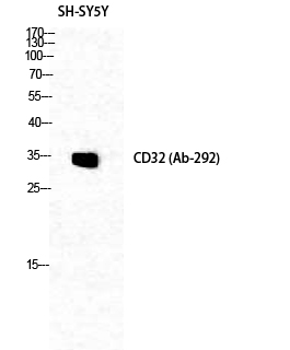
Western Blot analysis of SH-SY5Y cells using CD32 Polyclonal Antibody diluted at 1:1000

Click image to see more details
Western blot analysis of lysates from 293, HepG2, and HeLa cells, treated with PMA 125ng/ml 30', using CD32 Antibody. The lane on the right is blocked with the synthesized peptide.

Click image to see more details
Immunohistochemical analysis of paraffin-embedded human tonsil. 1, Antibody was diluted at 1:200 (4° overnight). 2, Tris-EDTA, pH9.0 was used for antigen retrieval. 3, Secondary antibody was diluted at 1:200 (room temperature, 30min).

Click image to see more details
FCGR2B were up-regulated in hippocampus of DM mice. A qRT-PCR was performed to detect the expression of ALB, AREG and FCGR2B mRNA expression in hippocampus of mice. B Western blot was conducted to detect the ALB, AREG and FCGR2B protein expression in hippocampus of mice. C IHC assay was employed to examine the ALB, AREG and FCGR2B protein expression in hippocampus of mice. D IF staining was utilized to detect the expression of FCGR2B and NeuN in hippocampus of mice. E Western blot was performed to detect the SHC1 protein expression in hippocampus of mice. F IF staining was performed to detect the expression of SHC1 and NeuN in hippocampus of mice. G Western blot was used to detect the p-PI3K and p-AKT protein expression in hippocampus of mice. *** P < 0.001 Full size image
Index in PubMed under a CC BY license. PMID: 40537751

Click image to see more details
Knockdown FCGR2B could promote the PI3K/AKT signaling pathway in vivo. A The work flow chart of how to construct a DM mouse model. B The mRNA levels of FCGR2B and SHC1 were assessed by qRT-PCR. C The protein levels of FCGR2B and SHC1 were evaluated by Western blot. D The protein level of SHC1 was determined by Western blot. E IF staining was used to detect the expression of FCGR2B and NeuN in hippocampus of mice. F IF staining was performed to detect the expression of SHC1 and NeuN in hippocampus of mice. G The expressions of p-PI3K and p-AKT in hippocampus of mice were evaluated by Western blot. ** P < 0.01, *** P < 0.001 Full size image
Index in PubMed under a CC BY license. PMID: 40537751

Click image to see more details
FCGR2B regulates the PI3K/AKT signaling pathway via downstream SHC1 in vitro. A qRT-PCR was employed to detect the expression of FCGR2B and SHC1 mRNA expression in HT22 cells after transfection of FCGR2B shRNA or FCGR2B plasmid. B Western blot was used to examine the expression of FCGR2B and SHC1protein expression in HT22 cells after transfection of FCGR2B shRNA or FCGR2B plasmid. C qRT-PCR was performed to detect the expression of FCGR2B and SHC1 mRNA expression in HT22 cells after transfection of SHC1 shRNA or SHC1 plasmid. D Western blot was used to examine the expression of FCGR2B and SHC1 protein expression in HT22 cells after transfection of SHC1 shRNA or SHC1 plasmid. E Western blot was conducted to examine the expression of p-PI3K and p-AKT expression in HT22 cells after transfection of SHC1 shRNA or SHC1 plasmid. F Western blot was used to examine the expression of p-PI3K and p-AKT expression in HT22 cells after transfection of SHC1 plasmid alone or combined with FCGR2B plasmid. G Western blot was performed to examine the expression of p-PI3K and p-AKT expression in HT22 cells after transfection of FCGR2B plasmid alone or combined with SHC1 shRNA. H Western blot was performed to examine the expression of p-PI3K and p-AKT expression in HT22 cells after transfection of FCGR2BshRNA alone or combined with SHC1 plasmid. I Western blot was performed to examine the expression of p-PI3K and p-AKT expression in HT22 cells after transfection of FCGR2B shRNA alone or combined with SHC1 shRNA. * P < 0.05, ** P < 0.01, *** P < 0.001 Full size image
Index in PubMed under a CC BY license. PMID: 40537751

Click image to see more details
Knockdown FCGR2B improved hippocampal neuronal excitability. A Representative images of Golgi staining of the hippocampal neuronal spines from the mice. B IHC assay was performed to examine the expression of NeuN in hippocampus of mice. C IHC assay was used to examine the expression of c-fos and GABAA in hippocampus of mice. D The expressions of c-fos, CaMKII, GABAA, and GABAARAP in hippocampus of mice were detected by Western blot. E TUNEL staining was conducted to assess cell apoptosis in hippocampus. F IF was used to detect BrdU and Ki67 positive cells in hippocampaltissue to analyzecell proliferation * P < 0.05, ** P < 0.01, *** P < 0.001 Full size image
Index in PubMed under a CC BY license. PMID: 40537751

Click image to see more details
Knockdown FCGR2B alleviated DM-induced cognition impairment in vivo. A H&E staining evaluated the pathological changes of hippocampus. B Neuronal damage of the hippocampal region was assessed by Nissl staining. C - D Morris water maze test was evaluated the learning and memory ability of mice by the escape latency time, number of crossing platform and swimming time in quadrant. E The recognition index among 4 groups in the novel object recognition test. * P < 0.05,** P < 0.01 Full size image
Index in PubMed under a CC BY license. PMID: 40537751
Specific Publications For Anti-CD32 FCGR2B Antibody (A01690-1)
Loading publications
Recommended Resources
Here are featured tools and databases that you might find useful.
- Boster's Pathways Library
- Protein Databases
- Bioscience Research Protocol Resources
- Data Processing & Analysis Software
- Photo Editing Software
- Scientific Literature Resources
- Research Paper Management Tools
- Molecular Biology Software
- Primer Design Tools
- Bioinformatics Tools
- Phylogenetic Tree Analysis
Customer Reviews
Have you used Anti-CD32 FCGR2B Antibody?
Share your experimental results or join a short interview to earn up to $1,000 in product credits or other rewards.
0 Reviews For Anti-CD32 FCGR2B Antibody
Customer Q&As
Have a question?
Find answers in Q&As, reviews.
Can't find your answer?
Submit your question
