This website uses cookies to ensure you get the best experience on our website.
- Table of Contents
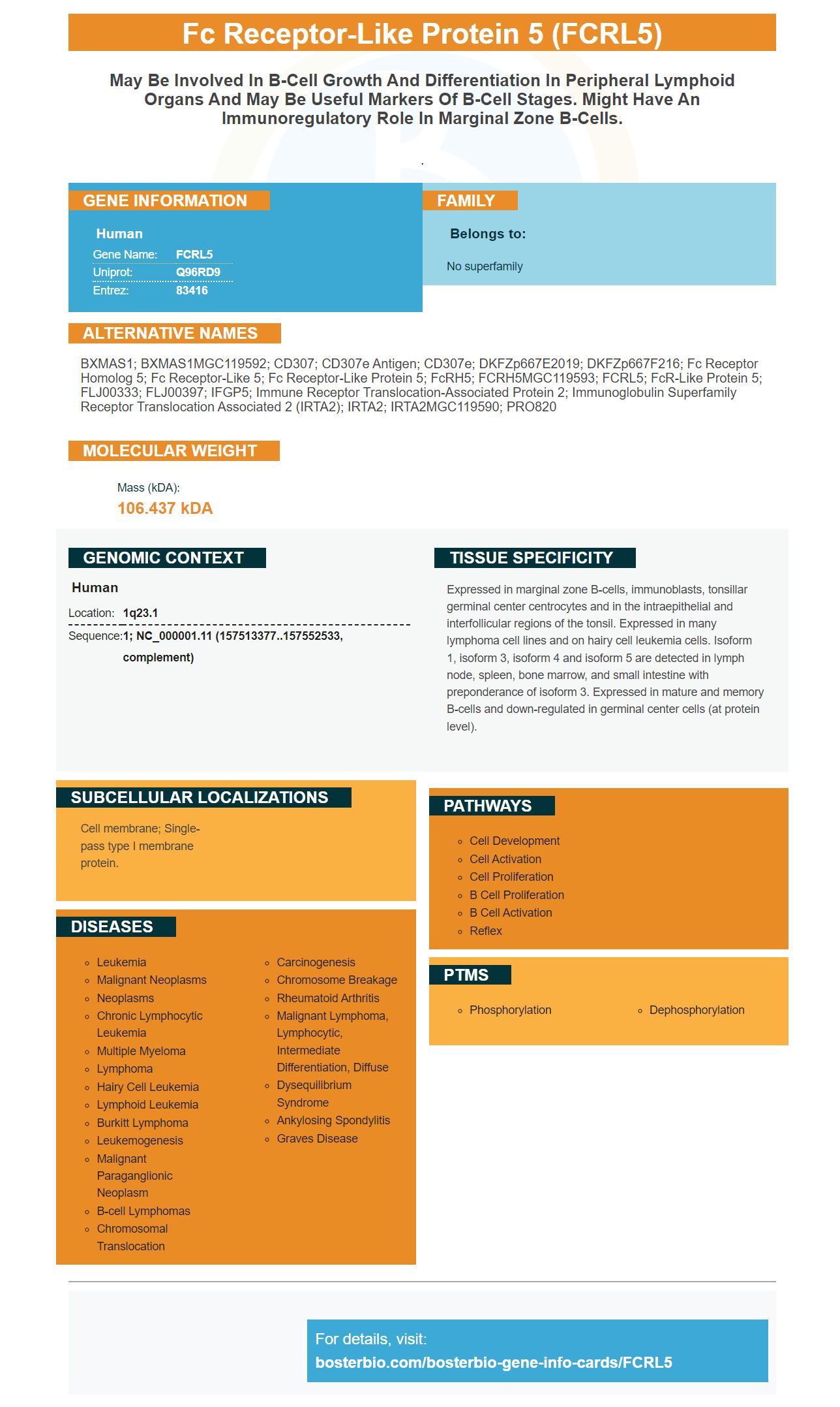
Facts about Fc receptor-like protein 5.
.
Human | |
---|---|
Gene Name: | FCRL5 |
Uniprot: | Q96RD9 |
Entrez: | 83416 |
Belongs to: |
---|
No superfamily |
BXMAS1; BXMAS1MGC119592; CD307; CD307e antigen; CD307e; DKFZp667E2019; DKFZp667F216; Fc receptor homolog 5; Fc receptor-like 5; Fc receptor-like protein 5; FcRH5; FCRH5MGC119593; FCRL5; FcR-like protein 5; FLJ00333; FLJ00397; IFGP5; Immune receptor translocation-associated protein 2; immunoglobulin superfamily receptor translocation associated 2 (IRTA2); IRTA2; IRTA2MGC119590; PRO820
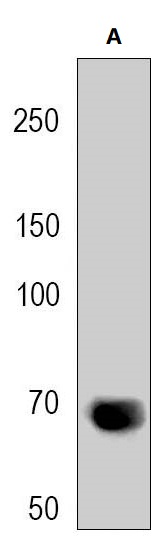
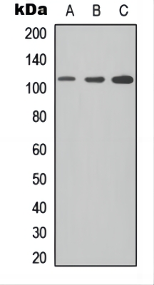
Mass (kDA):
106.437 kDA
Human | |
---|---|
Location: | 1q23.1 |
Sequence: | 1; NC_000001.11 (157513377..157552533, complement) |
Expressed in marginal zone B-cells, immunoblasts, tonsillar germinal center centrocytes and in the intraepithelial and interfollicular regions of the tonsil. Expressed in many lymphoma cell lines and on hairy cell leukemia cells. Isoform 1, isoform 3, isoform 4 and isoform 5 are detected in lymph node, spleen, bone marrow, and small intestine with preponderance of isoform 3. Expressed in mature and memory B-cells and down-regulated in germinal center cells (at protein level).
Cell membrane; Single-pass type I membrane protein.
When optimizing your experiment using the Anti Fc receptorlike protein 5 FCRL5Marker, there are many factors to consider. This article will describe the application, optimization, and safety of this agent. This article will explain the benefits and applications of this reagent to those who are not familiar with it. You'll be glad you did! This article also includes optimization tips and guides.
The FCRL5 gene encodes the identity of a novel member in the immunoglobulin Superfamily. It is expressed predominantly by B cells. This gene has been implicated as a cause of AIDs in a genome-wide study. This gene is associated with a lower activation level in peripheral blood mononuclear cell, while it is significantly greater in peripheral blood blood B cells. A Boster Bio antibody to FCRL5 is available to test this receptor's function.
The antibody targets the Fc receptor-like protein, Y281, in the anti-FcR receptor subunit of B cells. The antibody targets a variety BCR-derived protein under a variety conditions. It is highly sensitive, specific and highly accurate. The antibody can detect mRNA and DNA as well as protein based upon the BCR derived peptide.
The HA -FcRL1 (WT), mCherry antigen is a common monoclonal Tetramer. It can detect both mutant and wild-type FcRL1 tyrosine tyrosine residues. Although not fully investigated, this antibody is useful in studying B cell activation.
The mRNA coding for the moFcRL1 spliced variant of FCRL5 mRNA was purchased from American Type Culture Collection. The mRNA was kept in complete RPMI 1640 containing 10% fetal bovine serum. The yeast expression of the FCRL5 variant was compared to GAPDH, a housekeeping gene. The qPCR data was analyzed using BioMark Gene Expression Data Analysis Software from Fluidigm Corporation. The relative abundance was calculated using formula 2-DCt and normalized with a reference gene GAPDH.
FCRL molecules regulate activation of PTPN11, PTPN6 in addition to BCR signaling. Interestingly, BONITA revealed that the CD79A and CD79B components of the BCR are required for downstream signaling. These studies indicate that FCRL molecules could act through the LYN to activate BCR signaling pathways.
Boster Bio's optimization for the FCRL5 gene marker is an exciting method to identify immune system B cells. MVA reduced the critical pathway for B cell signaling in a clinical study. Protein boosters containing FCRL5 gene also inhibited critical B cell signals. This study has implications on future vaccine development and diagnostics.
In vaccine design, it is crucial to determine whether the FCRL5 marker works well in identifying B cell types that are unable and unwilling to recognize an antigen. FCRL5 is expressed in antigen-specific MBCs but is not found in plasma cells or blasts. This study addresses this question by identifying a population of B cells that express FCRL5.
Plasmablasts are more likely to develop from MBCs that express FCRL5 than those that don't. These cells are also more likely to express CD11b and CD11c, which is a good indicator for the atypical phenotype. This is consistent also with previous findings that FCRL5+ MBCs produce more antibodies in comparison to FCRL5+.
Mice were infected as P. chabaudi AS. FCRL5 expressing B cells were analysed using flow cytometry. FCRL5 expression was found on B cells from the blood, spleen, and marginal zone. CD138+ cells also expressed FCRL5. FCRL5 expression was detected in B cells from one mouse at the time, as well in mice that were not contaminated with P. chabaudi.
The FCRL5 mark is a powerful tool to determine FcgR's status. This marker controls activation of B cells as well as the antibody response. Furthermore, it helps in identifying T cells that are able to recognize antigens. Researchers can identify the protein in immune cells to identify the best antibody target. Boster Bio uses this marker to tag antibodies.
The phenotypic characteristics and behavior of FCRL5+ murine MBCs are similar to those of human atMBCs. They exhibit an expansion of blood cells following Plasmodium infection, and contract when the infection is cleared. This is similar the function of FCRL5+MBCs in chronic diseases. FCRL5+ cells also have similar immune-related gene expressions to the human atMBCs such as T-bet. Other ABC markers are also expressed in FCRL5+ cells, such as CD73 or CD80.
Patients with chronic diseases are likely to have FCRL5+ MBCs in their peripheral blood. These cells exhibit high levels antigen-specificity. They are long-lived antigen specific and have a high number of FCRL5+ cells. Boster Bio provides a valuable tool for detecting long-lived antigen-specific, antigen-specific MBCs.
The immunoglobulin superfamily also includes the FCRL5 gene. It is also known as IFGP5, CD307 and BXMAS1, and CCRL1. Its function is unclear. It is expressed in plasma cells, and it increases in multiple myeloma tissues. FCRL5 is widely used in diagnostic and research applications despite the fact that it has no known function.
In a recent study FCRL1-human Fc Fusion proteins were used for hybridoma screen. These fusion proteins were added onto microtiter plates, and stained with a specific fluorescent method. These experiments were published in BioRxiv. The authors concluded that FCRL1 human Fc fusion proteins could be safely used in humans. Further research is necessary to confirm their safety.
The FCRL1 gene has been implicated in multiple hematologic malignancies, such as leukemia and multiple sclerosis. Its expression may be a target for treatment. E9Fv)-PE38 may also have cytotoxic properties on FCRL1+ cells. This could make it a potential new therapy to treat FCRL1+ malignancies. This article has been marked as an advertisement and compensated in part with page charges.
PMID: 11453668 by Nakayama Y., et al. BXMAS1 identifies a cluster of homologous genes differentially expressed in B cells.
PMID: 11290337 by Hatzivassiliou G., et al. IRTA1 and IRTA2, novel immunoglobulin superfamily receptors expressed in B cells and involved in chromosome 1q21 abnormalities in B cell malignancy.