This website uses cookies to ensure you get the best experience on our website.


- Table of Contents
Plan chromogenic paraffin IHC for FDCSP using tonsil squamous epithelium as a positive reference (HPA tissue IHC). This guide covers the IHC-validated antibody at 0.5–1 μg/mL (datasheet A13093-1), fixation consistency, detection, and interpretation of a secreted target (UniProt).

Expected localisation, validated protocols, controls and antibodies — the at-a-glance facts below, then the full design guide.
| Expected localisation | Secreted; tissue compartment unresolved (UniProt; HPA tissue IHC) | |
| Staining pattern | Tonsil squamous; salivary glandular; compartment unclear (HPA tissue IHC) | |
| Antigen retrieval | Citrate pH 6 HIER, heat-mediated (datasheet A13093-1) | |
| Positive control | Tonsil+1 more · see all | |
| Negative control | Adipose tissue+4 more · see all |
| Fixation | Keep fixation consistent across paraffin sections. (standard IHC practice; not target-specific) | |
| Caveat | Secretion can separate protein staining from RNA location (HPA tissue IHC) | |
| Regulation | Tissue-specific expression (UniProt) | |
| Isoform / epitope | 0 isoforms; signal peptide cleaved, no cytoplasmic tail (UniProt) |
The catalog antibody’s IHC-P protocol (datasheet A13093-1) is accompanied by a published breast tissue IHC protocol (PMC12507714).
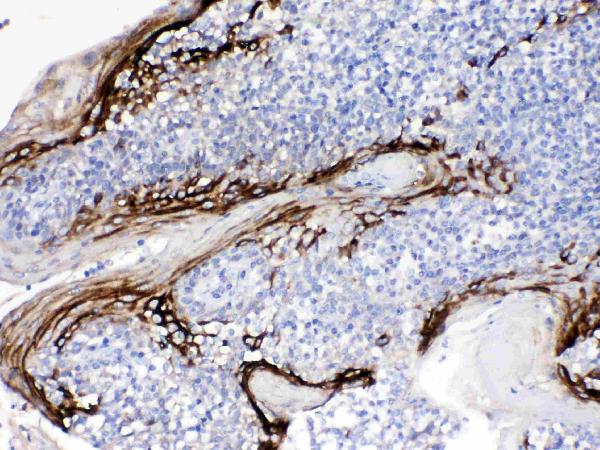
| Sample | Paraffin-embedded human tonsil tissues; fixative not specified (datasheet A13093-1) |
| Fixation | Image fixative and duration unreported (datasheet A13093-1); verify before use. |
| Sectioning | 4–5 µm sections on charged slides (standard) |
| Deparaffinisation | Xylene, graded ethanol series to water (standard) |
| Antigen retrieval | Heat retrieval: Citrate pH 6, 20 min (datasheet A13093-1) |
| Peroxidase block | 3% H2O2, 10 min, room temperature (standard) |
| Blocking | 10% goat serum (datasheet A13093-1) |
| Primary antibody | Rabbit anti-FDCSP, 0.5-1μg/ml (datasheet A13093-1) |
| Primary incubation | Overnight at 4 °C (datasheet A13093-1) |
| Detection | Streptavidin-biotin complex (SABC), DAB chromogen (datasheet A13093-1) |
| Counterstain | Hematoxylin, blue, dehydrate and mount (standard) |
| Expected result | FDCSP-positive staining in squamous epithelial cells of tonsil (HPA tissue IHC: High). HPA tissue profile: Distinct expression was observed in the squamous epithelium of tonsil and salivary gland. No signal in the no-primary control. |
FDCSP is a secreted peptide with no transmembrane segment (UniProt Q8NFU4 topology). In paraffin-section IHC, expect the clearest staining in tonsil squamous epithelial cells and weaker staining in salivary gland glandular cells (HPA: High; Medium). Its secreted status permits protein to appear beyond its producing cells (UniProt Q8NFU4; HPA reliability note). HPA rates the tissue pattern Enhanced, with medium consistency against RNA data and external verification pending (HPA reliability).
| Strong staining in tonsil squamous epithelium, with moderate staining in salivary gland glandular cells. | This matches the reported cell types and relative levels (HPA: tonsil High; salivary gland Medium). Assess the stained cells as well as intensity: surrounding deposited secreted protein may complicate assignment to its source (UniProt Q8NFU4 secreted; HPA reliability note). |
| Predominantly nuclear staining, or a sharply defined membrane pattern presented as the only convincing signal. | Neither is an established FDCSP location here: HPA lists no ICC-IF main location, and UniProt describes a secreted peptide without a transmembrane segment (HPA subcellular; UniProt Q8NFU4 topology). Treat the pattern as suspect and check controls before scoring it as specific. |
| Strong staining in adipocytes, marrow hematopoietic cells, or bronchial respiratory epithelium. | Those cell populations were not detected in the supplied tissue survey (HPA: adipose tissue, bone marrow, bronchus). Check antibody-dependent staining and, for chromogenic IHC, endogenous detection activity using appropriate controls (standard IHC practice). |
| Broad, low-contrast colour across cells and surrounding tissue, obscuring epithelial boundaries. | Diffuse background prevents a reliable comparison with the reported tonsil and salivary gland pattern (HPA tissue IHC). Review blocking, washing, primary-antibody concentration and chromogen development with matched controls (standard IHC practice); do not assign every extracellular deposit to FDCSP merely because it is secreted (UniProt Q8NFU4). |
| No staining in tonsil squamous epithelial cells on the test section. | Tonsil squamous epithelium is the strongest supplied positive reference (HPA: High). First verify tissue identity and that a known-positive control worked, then review retrieval, detection and antibody conditions as general IHC checks (standard IHC practice). The supplied sources do not identify an FDCSP-specific retrieval condition. |
| Secretion and topology (UniProt Q8NFU4). | FDCSP has a signal peptide and no transmembrane segment (UniProt Q8NFU4 topology). Cell-associated staining can support the expected pattern, but nearby extracellular signal need not mark the cell that produced it (UniProt Q8NFU4 secreted; HPA reliability note). |
| Processing (UniProt Q8NFU4). | The annotated signal peptide spans residues 1–17 and the mature chain spans 18–85 (UniProt Q8NFU4 processing). Epitope position is not supplied, so do not infer whether the catalog antibody recognizes precursor, mature peptide, or both. |
| Tissue contrast (HPA tissue IHC). | Use tonsil squamous epithelium as the strongest supplied positive reference and salivary gland glandular cells as a second, moderate reference (HPA: High; Medium). Lymph-node germinal-center cells are reported Low, so faint staining there is less decisive (HPA: Low). |
| Evidence strength (HPA tissue IHC). | The tissue profile is rated Enhanced, and both listed antibodies have Enhanced IHC status (HPA reliability; HPA antibodies: HPA014326, CAB014890). RNA and antibody staining have medium consistency, and external verification is pending (HPA reliability); interpret discordant fields cautiously. |
| IF localisation evidence (HPA subcellular). | HPA calls FDCSP secreted but provides no main ICC-IF location or cell-line images (HPA subcellular). The tissue IHC profile therefore cannot specify an IF puncta, membrane, or nuclear pattern; evaluate IF/ICC on its separate guide page. |
| Situation | Likely cause | Next action |
|---|---|---|
| Tonsil positive control is blank or very weak. | The run has not reproduced the reported High signal in tonsil squamous epithelial cells (HPA: High); the record does not establish an FDCSP-specific technical cause. | Confirm the squamous epithelium is present, then check antibody use, retrieval, detection reagents and run controls under the IHC protocol (standard IHC practice). Avoid declaring the tissue negative until the control works. |
| Salivary gland stains more strongly than tonsil. | That reverses the supplied relative tissue levels, although section and run differences can affect apparent intensity (HPA: tonsil High; salivary gland Medium; standard IHC practice). | Compare the stated cell populations under matched staining and scoring conditions, with a tonsil control in the same run (HPA tissue IHC; standard IHC practice). |
| Unexpected staining appears in a reported negative cell population. | Cross-reactivity or endogenous chromogenic detection activity is possible (standard IHC practice); the supplied HPA survey reports several such populations as Not detected (HPA tissue IHC). | Inspect a no-primary control and the relevant endogenous-activity control; compare the unexpected signal with the tonsil pattern before assigning specificity (standard IHC practice; HPA: tonsil High). |
| Colour is widespread, faint and hard to localise. | Nonspecific background or excessive development can obscure interpretable cell staining (standard IHC practice). FDCSP secretion alone does not validate an indiscriminate diffuse pattern (UniProt Q8NFU4 secreted). | Review primary-antibody dilution, blocking, washes and development against controls (standard IHC practice); score only signal that can be assigned reliably to the reported cells (HPA tissue IHC). |
| Lymph node is faint while tonsil is clearly positive. | This can agree with the survey: germinal-center cells in lymph node are Low, versus High tonsil squamous epithelial staining (HPA tissue IHC). | Use tonsil for the primary positive check, and describe lymph-node staining by its observed cell population and intensity rather than requiring tonsil-level signal (HPA: tonsil High; lymph node Low). |
| Can the IHC result define the expected IF/ICC pattern? | No ICC-IF main location or image-bearing cell lines are supplied, despite the secreted summary (HPA subcellular). | Use the separate IF/ICC guide for that application; do not transfer a chromogenic tissue pattern into a required IF compartment or invent an IF protocol (HPA subcellular; HPA tissue IHC). |


Comprehensive Human Protein Atlas IHC scoring per tissue (reliability: Enhanced — Medium consistency between antibody staining and RNA expression data. Pending external verification. Secreted protein, tissue location of RNA and protein is expected to differ.). Rows are taken directly from the HPA tissue chart — click any row's HPA link to view the source.
| Tissue | Cell type | Level | Evidence | Source |
|---|---|---|---|---|
| Tonsil | Squamous epithelial cells | High | Protein (IHC) | HPA → |
| Salivary gland | Glandular cells | Medium | Protein (IHC) | HPA → |
| Tissue | Cell type | Level | Evidence | Source |
|---|---|---|---|---|
| Adipose tissue | Adipocytes | Not detected | Protein (IHC) | HPA → |
| Adrenal gland | Glandular cells | Not detected | Protein (IHC) | HPA → |
| Bone marrow | Hematopoietic cells | Not detected | Protein (IHC) | HPA → |
| Breast | Adipocytes | Not detected | Protein (IHC) | HPA → |
| Bronchus | Respiratory epithelial cells | Not detected | Protein (IHC) | HPA → |
Troubleshoot FDCSP staining in paraffin sections using the catalog antibody’s tonsil IHC result and the reported tissue expression pattern.
Anti-FDCSP antibody A13093-1 has IHC and IF images from human paraffin-embedded tonsil sections (catalog IHC and IF captions).
A13093-1 was tested by chromogenic IHC on a human paraffin-embedded tonsil section (catalog IHC caption). A13093-1 was also tested by IF on a human paraffin-embedded tonsil section (catalog IF caption).
Which to pick: For tissue IHC, choose A13093-1 for human paraffin sections (catalog IHC caption; catalog reactivity: Human); the IHC caption does not report the fixative. For IF, A13093-1 has a human paraffin-section image, while ICC is not listed among its applications (catalog IF caption; catalog applications: IF, IHC). No cross-species choice is supported because A13093-1 lists human reactivity only (catalog reactivity: Human).