FGB / Fibrinogen beta chain · IHC design guide

Design Immunohistochemistry for FGB

Plan chromogenic FGB IHC on paraffin sections with the catalog antibody at 2–5 μg/mL (datasheet A01204-1). Expect strong plasma and extracellular staining, with medium staining in kidney proximal tubule cell bodies (HPA tissue IHC).

Evidence assembled Oct 2026 · For research use; verify linked source records and product datasheet before use
Immunohistochemistry protocol sheet for FGB (IHC for FGB): expected localisation Plasma and extracellular spaces (HPA tissue IHC), antibody A01204-1, validated IHC image, and IHC protocol steps
Printable FGB IHC protocol sheet — expected localisation Plasma and extracellular spaces (HPA tissue IHC), antibody A01204-1, controls and protocol steps. Open the full FGB IHC guide →

FGB Immunohistochemistry Experimental Design Guide

Expected localisation, validated protocols, controls and antibodies — the at-a-glance facts below, then the full design guide.

Must know before staining
Expected localisation Plasma and extracellular spaces (HPA tissue IHC)
Staining pattern Strong plasma and extracellular staining (HPA tissue IHC)
Antigen retrieval EDTA pH 8.0 HIER, heat-mediated (datasheet A01204-1)
Positive control ⓘ Kidney
Negative control ⓘ Adipose tissue+4 more · see all
Important caveats
Reasons your staining may differ from the expected pattern.
Fixation Keep fixation consistent across sections. (standard IHC practice; not target-specific)
Caveat Circulating FGB can stain outside its site of synthesis (UniProt; HPA tissue IHC)
Regulation Liver-enriched RNA (HPA tissue RNA)
Isoform / epitope 0 isoforms listed; mature chain spans aa 45–491 (UniProt)
Section 1

Recommended FGB IHC & IF Protocols

Start with the catalog antibody’s IHC-P protocol, then compare the 3 published IHC protocols below (datasheet: IHC-P; PMC12679381; PMC12506357; PMC10401294).

Recommended immunohistochemistry (IHC-P) protocol parameters
SampleParaffin-embedded human liver cancer tissue; fixative not specified (datasheet A01204-1)
FixationImage fixative and duration unreported (datasheet A01204-1); verify before use.
Sectioning4–5 µm sections on charged slides (standard)
DeparaffinisationXylene, graded ethanol series to water (standard)
Antigen retrievalHeat retrieval: EDTA pH 8.0 (datasheet A01204-1); 20 min, 95–100 °C (standard)
Peroxidase block3% H2O2, 10 min, room temperature (standard)
Blocking10% goat serum (datasheet A01204-1)
Primary antibodyRabbit anti-FGB, 2-5μg/ml (datasheet A01204-1)
Primary incubationOvernight at 4 °C (datasheet A01204-1)
DetectionHRP-conjugated secondary, DAB chromogen (datasheet A01204-1)
CounterstainHematoxylin, blue, dehydrate and mount (standard)
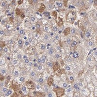
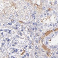
Expected resultFGB-positive staining in proximal tubules (cell body) of kidney (HPA tissue IHC: Medium). HPA tissue profile: Strong positivity in plasma and extracellular compartments. No signal in the no-primary control.
💡Decision noteTry heat-mediated EDTA retrieval at pH 8.0 first for the catalog antibody (datasheet: EDTA pH 8.0 HIER). Check each article’s antibody and sample before adopting its dilution.
Section 2

What Is the Expected FGB Staining Pattern?

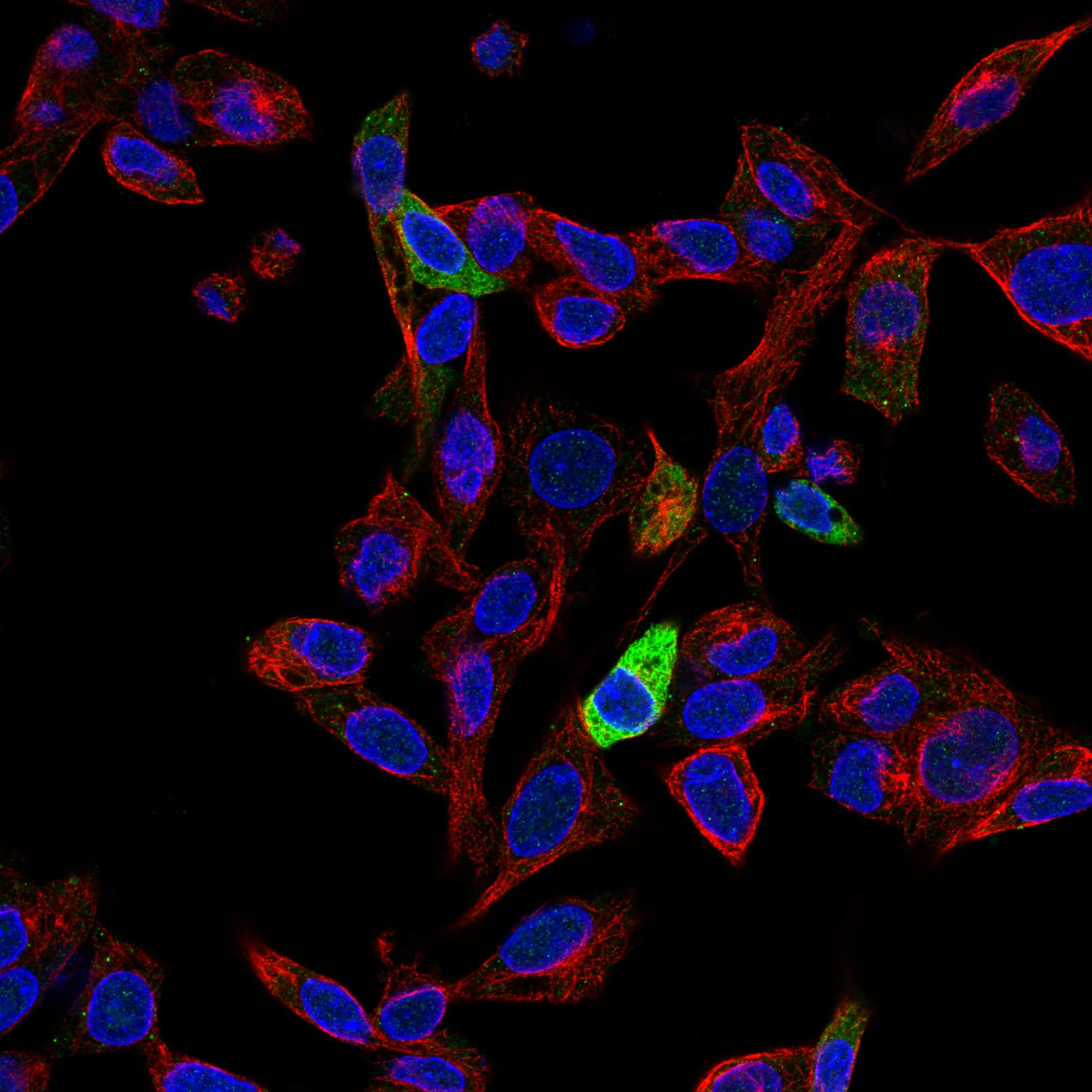
FGB is secreted and has no transmembrane segment (UniProt P02675 topology). In paraffin-section IHC, expect strong plasma and extracellular staining, with medium cell-body staining reported in kidney proximal tubules (HPA tissue IHC; reliability: Enhanced). HPA also reports endoplasmic-reticulum localisation by ICC-IF; that intracellular result should be interpreted in its own assay context (HPA subcellular ICC-IF).

What am I looking at on my slide?
Strong staining in plasma and extracellular spaces.This matches the dominant tissue pattern for a secreted protein (HPA tissue IHC; UniProt P02675 topology). Judge cellular staining separately from material around cells.
Medium cell-body staining in kidney proximal tubules.This matches the reported kidney observation (HPA tissue IHC: Medium). It does not, by itself, establish whether the detected protein was made or taken up by those cells.
Predominantly nuclear or membrane-outlined staining, without the expected extracellular signal.That distribution conflicts with FGB's secreted, non-transmembrane topology (UniProt P02675 topology) and warrants an artefact or antibody-specificity check.
Prominent cellular staining in a listed HPA-negative cell population.Adipocytes and adrenal glandular cells are reported as not detected (HPA tissue IHC). Check cross-reactivity and endogenous detection activity before calling such staining FGB.
Diffuse haze, or no signal where a positive pattern is expected.Haze obscures the plasma and extracellular pattern; absent signal prevents its assessment (HPA tissue IHC). Review controls, detection background and the IHC workflow (standard IHC practice).
💡Expected FGB appearanceCall positive when strong plasma or extracellular staining is evident, with possible medium kidney proximal-tubule cell-body staining (HPA tissue IHC); isolated nuclear staining is discordant with secreted FGB (UniProt P02675 topology).
How each factor affects the staining
Secretion and tissue compartment (UniProt P02675 topology; HPA tissue IHC)FGB can appear outside cells; extracellular positivity should not automatically be assigned to adjacent cells as their own expression.
Kidney cellular observation (HPA tissue IHC: Medium)The proximal-tubule cell-body result supports a local staining call, but the supplied evidence does not identify the source of protein within those cells.
RNA versus protein location (HPA tissue IHC)FGB RNA is tissue enriched in liver, while the tissue IHC profile emphasizes plasma and extracellular compartments. Do not infer liver IHC intensity from RNA enrichment.
ICC-IF compartment (HPA subcellular ICC-IF: enhanced)Endoplasmic-reticulum localisation is reported in ICC-IF. It informs an IF question, but does not replace the plasma and extracellular expectation for paraffin IHC.
Antibody validation (HPA antibodies)HPA001900 and HPA001901 have Enhanced IHC status; CAB008624 is Supported. These statuses describe validation, not a guaranteed result in every specimen.
Processing and modification (UniProt P02675)FGB has a signal peptide at residues 1–30 and a glycosylation site at 394. Epitope-dependent effects cannot be predicted without the antibody's epitope information.
Why is my staining missing, weak or wrong?
SituationLikely causeNext action
Expected extracellular signal is absent.The run may have insufficient detectable signal; the supplied sources give no FGB-specific retrieval condition.Check a known-positive control, antibody dilution and detection steps; assess retrieval using the antibody's IHC instructions (standard IHC practice).
The slide has widespread brown haze.Nonspecific staining or endogenous chromogenic activity can obscure the extracellular pattern (standard IHC practice).Review no-primary and detection controls, blocking and washes; compare the result with the HPA plasma and extracellular pattern (HPA tissue IHC).
Nuclei or cell borders dominate the result.That pattern is discordant with secreted FGB and its lack of a transmembrane segment (UniProt P02675 topology).Check control sections and antibody specificity; do not score the discordant compartment alone as an expected positive.
Adipocytes or adrenal glandular cells stain strongly.Both cell populations are listed as not detected, so cross-reactivity or endogenous activity needs consideration (HPA tissue IHC).Compare with a no-primary control and a second validated antibody, if available (standard IHC practice; HPA antibodies).
Kidney proximal tubules show cell-body staining.Medium staining is reported there, although its cellular source is unresolved by the supplied evidence (HPA tissue IHC).Score the observed compartment and intensity; avoid interpreting cell-body staining alone as proof of local FGB production.
An ICC-IF image shows intracellular reticular signal.Endoplasmic-reticulum localisation is reported for ICC-IF (HPA subcellular ICC-IF: enhanced).Interpret it within the IF assay and its controls; use the plasma and extracellular profile when judging paraffin-section IHC (HPA tissue IHC).

Sample controls for FGB IHC & IF

🧪Run kidney first and score proximal tubule cell bodies for staining (HPA: Medium in kidney proximal tubules). Run adipose tissue as a negative comparator and score adipocytes for absent staining (HPA: Not detected in adipocytes); on the kidney slide, use cells outside the scored proximal tubules to assess background without assuming they are FGB-negative.
Positive control tissue: Kidney (Proximal tubules (cell body), HPA Medium)
Negative control tissue: Adipose tissue (HPA Not detected)
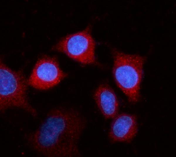
ICC-IF cell lines (HPA subcellular resource): HPA ICC-IF images show FGB in Hep-G2, U2OS, with annotated localisation: Endoplasmic reticulum (enhanced) (HPA subcellular).
Technical controls: Include no-primary (secondary-only) and host- and immunoglobulin-class-matched isotype controls, plus an FGB knockout sample if available to assess specificity (standard IHC practice). Quench endogenous peroxidase in kidney sections before HRP/DAB detection (caption: HRP/DAB; standard IHC practice).
⚠️Feasibility: A target-specific fixation window or fixation effect is unreported, and the selected A01204-1 paraffin-section caption does not state a fixative (caption: fixative not stated). The caption uses heat retrieval in EDTA at pH 8.0, but does not establish that retrieval is required; the supplied evidence does not establish whether frozen sections or IF are easier (caption: EDTA retrieval). Because FGB is secreted and detected in plasma, interpret staining near blood-containing spaces cautiously when scoring kidney cells (UniProt P02675: secreted; detected in blood plasma).

HPA tissue IHC evidence for FGB

Comprehensive Human Protein Atlas IHC scoring per tissue (reliability: Enhanced — At least one protein variant secreted, tissue location of RNA and protein might differ and correlation is complex.). Rows are taken directly from the HPA tissue chart — click any row's HPA link to view the source.

Positive expression · recommended positive controls

TissueCell typeLevelEvidenceSource
Kidney Proximal tubules (cell body) Medium Protein (IHC) HPA →

Undetected expression · recommended negative controls

TissueCell typeLevelEvidenceSource
Adipose tissue Adipocytes Not detected Protein (IHC) HPA →
Adrenal gland Glandular cells Not detected Protein (IHC) HPA →
Appendix Glandular cells Not detected Protein (IHC) HPA →
Bone marrow Hematopoietic cells Not detected Protein (IHC) HPA →
Breast Adipocytes Not detected Protein (IHC) HPA →
Section 3

Advanced FGB IHC Tips

Troubleshoot FGB staining by evaluating extracellular signal, cell body staining, and processing controls separately (HPA tissue IHC; UniProt P02675).

Which retrieval conditions should I use for FGB in paraffin sections?
Start with heat-mediated retrieval in EDTA at pH 8.0 for paraffin sections (datasheet A01204-1). Keep heating and cooling conditions identical across comparison sections so retrieval variation does not change apparent staining (standard IHC practice). The selected tissue image used 10% goat serum blocking and 2 μg/ml primary antibody overnight at 4°C after this retrieval (datasheet A01204-1). If staining is weak, compare modest changes in heating duration on adjacent sections while keeping antibody and detection conditions fixed (standard IHC practice). Score plasma and extracellular staining separately from cell body staining when judging the result (HPA tissue IHC).
How can I assess whether fixation caused weak FGB staining?
FGB-specific sensitivity to fixation is unknown: the selected paraffin-section caption does not state its fixative or fixation time (datasheet A01204-1). Record the fixative, fixation interval, processing schedule, and section thickness for every comparison specimen (standard IHC practice). Compare matched sections using the documented EDTA retrieval at pH 8.0 and 2 μg/ml primary antibody overnight at 4°C (datasheet A01204-1). Include a section with expected plasma or extracellular signal to check whether the staining run worked (HPA tissue IHC). If that control stains but the specimen does not, assess sampling and tissue preservation before attributing the difference to fixation (standard IHC practice).
Where should FGB staining appear, and how should intracellular signal be judged?
Expect strong plasma and extracellular staining in tissue IHC because FGB is secreted and the reference profile highlights those compartments (UniProt P02675; HPA tissue IHC). Kidney proximal tubule cell bodies show medium staining in the reference profile, so assess them separately from surrounding extracellular material (HPA tissue IHC). FGB has no transmembrane segment, and an ICC/IF reference reports endoplasmic reticulum localisation (UniProt P02675; HPA subcellular). Intracellular staining therefore needs careful compartment assessment; a diffuse nuclear pattern does not match those reference locations (UniProt P02675; HPA subcellular). Use the same compartment definitions across sections and document any vessel-associated signal separately (standard IHC practice).
Could processing or epitope position change what the FGB antibody detects?
The record lists 0 isoforms, a signal peptide at residues 1–30, and a fibrinogen beta chain spanning residues 45–491 (UniProt P02675). It also lists a glycosylation site at residue 394, a modified residue at 31, and a C-terminal domain spanning 232–488 (UniProt P02675). The antibody epitope is unspecified in the supplied caption, so its accessibility after retrieval cannot be assigned to any of these regions (datasheet A01204-1). Thrombin cleavage contributes to fibrin formation, but staining alone cannot distinguish circulating fibrinogen from deposited fibrin without epitope-specific evidence (UniProt P02675). Interpret a changed pattern alongside tissue morphology and staining controls (standard IHC practice).
How should I plan complementary IF staining for FGB?
For complementary IF, pair FGB with a marker identifying proximal tubules when examining kidney, where the reference IHC profile reports medium cell body staining (HPA tissue IHC). Choose a spectrally separated, preferably far-red fluorophore when tissue autofluorescence overlaps shorter wavelengths, and include single-color controls for multiplexing (standard IF practice). FGB is secreted and lacks a transmembrane segment; compare staining without detergent for extracellular antigen with mild permeabilisation when testing intracellular endoplasmic reticulum signal (UniProt P02675; HPA subcellular; standard IF practice). Include secondary-only controls and keep imaging settings consistent across samples (standard IF practice). The paraffin-section IHC caption supplies no IF validation for this antibody (datasheet A01204-1).
How do I reduce background without losing genuine extracellular FGB signal?
Begin with consistent section washing and a matched negative control, then inspect where unwanted chromogen accumulates (standard IHC practice). The selected IHC image used 10% goat serum blocking and 2 μg/ml primary antibody overnight at 4°C (datasheet A01204-1). For peroxidase detection, include an endogenous peroxidase block and assess a no-primary control before interpreting DAB staining (standard IHC practice). Strong plasma and extracellular positivity can be genuine for this secreted protein, so record it apart from diffuse staining over unrelated tissue structures (HPA tissue IHC; UniProt P02675). If background persists, compare shorter primary incubation or lower concentration on matched sections (standard IHC practice).
How should I quantify FGB across sections with different plasma content? ⚠ ANSWER MARKED FOR VERIFICATION
Define cell body and extracellular compartments before scoring because the reference profile reports strong plasma and extracellular staining alongside medium kidney proximal tubule cell body staining (HPA tissue IHC). For cell body signal, report the percentage of positive cells and an intensity-weighted H-score using the same thresholds across sections (standard IHC practice). For extracellular signal, report positive area or chromogen optical density per mm² of evaluable tissue, with vessel and plasma-rich regions measured separately (standard IHC practice). Normalise each compartment to its corresponding tissue area or cell count, and apply consistent exclusion rules for folds and necrosis (standard IHC practice). Hold retrieval and detection settings constant (standard IHC practice).
What distinguishes true FGB staining from artefact in chromogenic IHC?
A credible pattern includes plasma or extracellular positivity, consistent with the reference tissue profile and FGB secretion (HPA tissue IHC; UniProt P02675). Kidney proximal tubule cell body staining is also reported at medium intensity, while adipocytes in adipose tissue are reported as undetected (HPA tissue IHC). Treat staining confined to cut edges, folds, or necrotic debris cautiously and compare it with intact tissue on the same section (standard IHC practice). Diffuse nuclear staining conflicts with the supplied secreted and endoplasmic reticulum localisation evidence (UniProt P02675; HPA subcellular). Check a no-primary control for endogenous enzyme or detection-system signal before calling weak DAB deposits positive (standard IHC practice).
Boster reagents

Best FGB / Fibrinogen beta chain IHC Antibodies

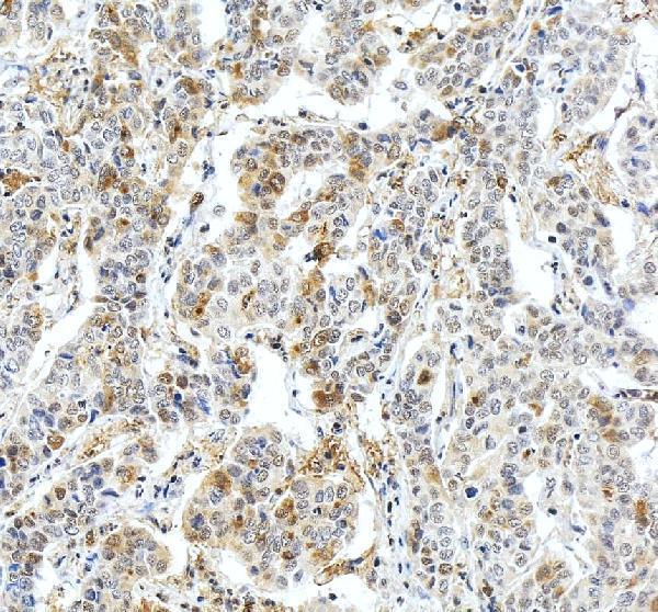
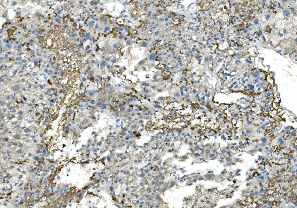
Both anti-FGB antibodies have IHC images from paraffin sections of human liver cancer tissue (A01204-1 and M01204-1 IHC captions). A01204-1 also has IF data from A549 cells (A01204-1 IF caption).

Real IHC data IHC analysis of Fibrinogen Beta Chain/FGB using anti-Fibrinogen Beta Chain/FGB antibody (A01204-1). Fibrinogen Beta Chain/FGB was detected in a paraffin-embedded section of human liver cancer tissue. Heat mediated antigen retrieval was performed in EDTA buffer (pH 8.0, epitope retrieval solution). The tissue section was blocked with 10% goat serum. The tissue section was then incubated with 2 μg/ml rabbit anti-Fibrinogen Beta Chain/FGB Antibody (A01204-1) overnight at 4°C. Peroxidase Conjugated Goat Anti-rabbit IgG was used as secondary antibody and incubated for 30 minutes at 37°C. The tissue section was developed using HRP Conjugated Rabbit IgG Super Vision Assay Kit (Catalog # SV0002) with DAB as the chromogen.
Anti-Fibrinogen beta chain/FGB Antibody ®
Cat # A01204-1
Real IHC data IHC analysis of Fibrinogen beta chain/FGB using anti-Fibrinogen beta chain/FGB antibody (M01204-1). Fibrinogen beta chain/FGB was detected in paraffin-embedded section of human liver cancer tissue. Heat mediated antigen retrieval was performed in EDTA buffer (pH8.0, epitope retrieval solution). The tissue section was blocked with 10% goat serum. The tissue section was then incubated with 2μg/ml mouse anti-Fibrinogen beta chain/FGB Antibody (M01204-1) overnight at 4°C. Biotinylated goat anti-mouse IgG was used as secondary antibody and incubated for 30 minutes at 37°C. The tissue section was developed using Strepavidin-Biotin-Complex (SABC) (Catalog # SA1021) with DAB as the chromogen.
Anti-Fibrinogen beta chain/FGB Antibody ® (monoclonal, 6D12)
Cat # M01204-1

A01204-1 will render with its human liver cancer tissue IHC image; its application list includes IHC, IF and ICC, and its IF image shows A549 cells (A01204-1 IHC/IF captions; catalog applications). M01204-1 will render with its human liver cancer tissue IHC image and lists Human reactivity (M01204-1 IHC caption; catalog reactivity).

Which to pick: For tissue IHC, either SKU has a paraffin-section example using EDTA retrieval at pH 8.0; M01204-1 is a mouse monoclonal, clone 6D12, while A01204-1 is rabbit (each SKU’s IHC caption; catalog host/clone). For IF/ICC, choose A01204-1, which lists both applications and has an A549 IF image; it also lists Human, Mouse and Rat reactivity, while M01204-1 lists Human reactivity and no IF/ICC application (catalog applications/reactivity; A01204-1 IF caption). The fixative is unreported for both paraffin-section IHC examples (each SKU’s IHC caption).

Each figure is that product's own IHC / IF validation image from its datasheet.

References

  1. UniProt Consortium. UniProt entry P02675 (FIBB_HUMAN, Fibrinogen beta chain).
  2. Human Protein Atlas. FGB tissue IHC expression (reliability: Enhanced).
  3. Human Protein Atlas. FGB subcellular location (ICC-IF): Localized to the endoplasmic reticulum..
  4. Human Protein Atlas. FGB antibody validation summary (3 antibodies).
  5. Histological analysis of thrombus composition in acute ischemic stroke with large vessel occlusion: impact of r-tPA treatment. Frontiers in neuroscience 2025 — PMC12679381.
  6. Pleural thickening induced by Glaesserella parasuis infection was linked to increased collagen and elastin. Frontiers in cellular and infection microbiology 2022 — PMC9409878.
  7. Bioinformatics analysis of the proteome in the pathway of complement and coagulation cascades in COVID-19: discovering potential biomarkers of FGB and SERPINA5. BMC infectious diseases 2025 — PMC12506357.
  8. Bioprosthetic Valve Deterioration: Accumulation of Circulating Proteins and Macrophages in the Valve Interstitium. JACC. Basic to translational science 2023 — PMC10401294.
  9. PubMed PMID:6688356 — UniProt-cited evidence.
  10. PubMed PMID:2102623 — UniProt-cited evidence.
  11. PubMed PMID:14702039 — UniProt-cited evidence.