This website uses cookies to ensure you get the best experience on our website.


- Table of Contents
Plan FGF19 chromogenic IHC on paraffin sections using the catalog antibody’s human IHC evidence (datasheet). Compare cytoplasmic staining and extracellular deposits with the tissue profile, allowing for differences between RNA and protein location in this secreted protein (HPA tissue IHC; UniProt).

Expected localisation, validated protocols, controls and antibodies — the at-a-glance facts below, then the full design guide.
| Expected localisation | Extracellular deposits and cytoplasm (HPA tissue IHC) | |
| Staining pattern | Cytoplasmic staining and extracellular deposits across tissues (HPA tissue IHC) | |
| Antigen retrieval | Citrate pH 6.0 HIER, 95–98 °C, 20 min (rule: cytoplasmic / membrane antigen) | |
| Positive control | Caudate+4 more · see all | |
| Negative control | Bone marrow+4 more · see all |
| Fixation | Keep fixation consistent across paraffin sections. (standard IHC practice; not target-specific) | |
| Caveat | Secretion can separate protein staining from RNA expression (UniProt; HPA tissue IHC) | |
| Regulation | Expression control not specified (UniProt) | |
| Isoform / epitope | No isoforms annotated; mature chain 25–216, with no cytoplasmic tail (UniProt) |
The catalog antibody’s IHC-P protocol is followed by three published FGF19 IHC protocols for prostate, nasopharyngeal, and liver samples (PMC3630260; PMC10899426; PMC6054564).
| Sample | Tissue sections; selected-image fixative not specified (standard IHC workflow) |
| Fixation | Image fixative and duration unreported (datasheet PA1476); verify before use. |
| Sectioning | 4–5 µm sections on charged slides (standard) |
| Deparaffinisation | Xylene, graded ethanol series to water (standard) |
| Antigen retrieval | Heat-induced epitope retrieval in citrate buffer, pH 6.0, 20 min at 95–98 °C (standard rule: cytoplasmic / membrane antigen) |
| Peroxidase block | 3% H2O2, 10 min, room temperature (standard) |
| Blocking | 10% normal serum of the secondary host, 30 min, room temperature (standard) |
| Primary antibody | Rabbit anti-FGF19, 0.5-1μg/ml (datasheet PA1476) |
| Primary incubation | Overnight at 4 °C (standard) |
| Detection | HRP-polymer secondary, DAB chromogen 5–10 min (standard) |
| Counterstain | Hematoxylin, blue, dehydrate and mount (standard) |
| Expected result | FGF19-positive staining in glial cells of caudate (HPA tissue IHC: High). HPA tissue profile: Extracellular deposits and cytoplasmic expression in all tissues, including gallbladder. No signal in the no-primary control. |
FGF19 is secreted and has no transmembrane segment (UniProt O95750 topology). In tissue IHC, expect cytoplasmic staining and extracellular deposits, including in gallbladder (HPA tissue IHC: Approved). Gallbladder and duodenal glandular cells, cerebellar Purkinje cells, caudate glial cells, kidney tubule cells, and small-intestinal glandular cells have high reported staining (HPA tissue IHC). HPA notes that its tissue pattern awaits external verification (HPA reliability description).
| Cytoplasmic staining in gallbladder glandular cells, with extracellular deposits (HPA tissue IHC). | This fits the reported high gallbladder signal and the distribution of a secreted protein (HPA tissue IHC; UniProt O95750: secreted). Compare cell-associated signal with deposits so that extracellular material is not scored as a new positive cell population (standard IHC interpretation). |
| A strong, exclusively nuclear or sharply membrane-confined pattern (HPA tissue IHC: expected cytoplasmic and extracellular pattern). | This does not match the reported tissue pattern or a protein without a transmembrane segment (HPA tissue IHC; UniProt O95750 topology). Check staining specificity and morphology before calling those cells FGF19 positive (standard IHC practice). |
| Signal in liver cholangiocytes or bone-marrow hematopoietic cells (HPA tissue IHC: not detected in these cell populations). | Treat an unexpected positive result as a specificity or endogenous-detection concern; inspect matched negative controls and the staining distribution (standard IHC practice). The HPA finding applies to the named cell populations, not every cell in either tissue (HPA tissue IHC). |
| Uniform haze across tissue and surrounding space, with little cell-to-cell distinction (standard IHC interpretation). | Diffuse background can obscure the cytoplasmic and extracellular pattern reported for FGF19 (HPA tissue IHC). Check a negative control and review blocking, washing, antibody concentration, and detection conditions (standard IHC practice). |
| No staining in gallbladder glandular cells or another reported high-staining population (HPA tissue IHC). | A failed positive control leaves a negative study sample uninterpretable (standard IHC practice). Check tissue preservation and morphology, primary-antibody application, retrieval, detection reagents, and counterstain before inferring absent FGF19 (standard IHC practice). |
| Secretion and topology (UniProt O95750) | FGF19 has a signal peptide at residues 1–24, a mature chain at 25–216, and no transmembrane segment (UniProt O95750). Interpret deposits alongside cytoplasmic staining; a membrane-only pattern needs a specificity check (HPA tissue IHC; standard IHC practice). |
| Tissue and cell selection (HPA tissue IHC) | Gallbladder, duodenum, and small-intestinal glandular cells are reported high, as are Purkinje cells, caudate glial cells, and kidney tubule cells (HPA tissue IHC). Appendix glandular and bronchial respiratory epithelial cells are medium; their weaker expected staining can complicate control assessment (HPA tissue IHC; standard IHC interpretation). |
| RNA versus protein (HPA tissue IHC) | Gallbladder RNA is tissue enriched, while HPA reports extracellular deposits and cytoplasmic protein staining across tissues (HPA tissue IHC). Because FGF19 is secreted, RNA location and protein location may differ; avoid assigning a deposit to its producing cell from IHC alone (HPA reliability description; UniProt O95750). |
| Strength of validation (HPA tissue IHC; HPA antibodies) | HPA labels its tissue profile Approved and says external verification is pending (HPA reliability description). Antibody HPA036082 is IHC Approved, with no ICC validation listed; these labels do not make an unexpected compartment or cell type automatically specific (HPA antibodies; standard IHC interpretation). |
| IF/ICC Q&A (HPA subcellular; HPA antibodies) | Q: What IF/ICC pattern is established? A: HPA summarizes FGF19 as secreted but supplies no main subcellular location, ICC-IF images, or ICC validation for HPA036082 (HPA subcellular; HPA antibodies). Treat an IF/ICC compartment call as unverified here (HPA subcellular). |
| Situation | Likely cause | Next action |
|---|---|---|
| Reported high-staining gallbladder glandular cells appear negative (HPA tissue IHC). | An assay failure or an unsuitable section can produce an uninformative negative result (standard IHC practice). | Check morphology, primary-antibody application, retrieval, detection reagents, and a positive-control section before interpreting sample negatives (standard IHC practice). |
| The slide shows strong nuclear or membrane-only staining (HPA tissue IHC: cytoplasmic and extracellular pattern). | The compartment conflicts with the observed tissue profile and with the lack of a transmembrane segment (HPA tissue IHC; UniProt O95750 topology). | Inspect morphology and controls, then reassess antibody specificity and detection conditions before assigning FGF19 positivity (standard IHC practice). |
| Liver cholangiocytes stain strongly (HPA tissue IHC: not detected). | Cross-reactivity or endogenous detection activity is possible when a reported negative cell population stains (HPA tissue IHC; standard IHC interpretation). | Compare a no-primary control and a reported positive population on the same staining run; restrict conclusions to identifiable cell types (standard IHC practice; HPA tissue IHC). |
| Broad haze obscures cells and extracellular deposits (standard IHC interpretation). | Excess nonspecific staining or detection background can blur the reported FGF19 pattern (standard IHC practice; HPA tissue IHC). | Review negative controls, blocking, washing, antibody concentration, and detection development; judge the result only after cell boundaries are readable (standard IHC practice). |
| Extracellular material stains while nearby cells appear weak (HPA tissue IHC: extracellular deposits and cytoplasmic expression). | A secreted protein can be seen away from its producing cell; signal location alone does not establish cellular origin (UniProt O95750: secreted; HPA reliability description). | Record deposits separately from cell-associated staining and use cell morphology when scoring named populations (standard IHC practice; HPA tissue IHC). |
| A weak or negative sample disagrees with gallbladder RNA enrichment (HPA tissue IHC). | RNA location need not match protein location for secreted FGF19, and HPA's tissue pattern awaits external verification (HPA reliability description). | Compare the sample with a reported high-staining cell population and assay controls; report the observed IHC result without treating RNA enrichment as proof of protein staining (HPA tissue IHC; standard IHC practice). |

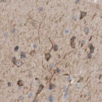


Comprehensive Human Protein Atlas IHC scoring per tissue (reliability: Approved — Secreted protein, tissue location of RNA and protein is expected to differ. Pending external verification.). Rows are taken directly from the HPA tissue chart — click any row's HPA link to view the source.
| Tissue | Cell type | Level | Evidence | Source |
|---|---|---|---|---|
| Caudate | Glial cells | High | Protein (IHC) | HPA → |
| Cerebellum | Purkinje cells | High | Protein (IHC) | HPA → |
| Duodenum | Glandular cells | High | Protein (IHC) | HPA → |
| Gallbladder | Glandular cells | High | Protein (IHC) | HPA → |
| Kidney | Cells in tubules | High | Protein (IHC) | HPA → |
| Tissue | Cell type | Level | Evidence | Source |
|---|---|---|---|---|
| Bone marrow | Hematopoietic cells | Not detected | Protein (IHC) | HPA → |
| Liver | Cholangiocytes | Not detected | Protein (IHC) | HPA → |
| Lymph node | Germinal center cells | Not detected | Protein (IHC) | HPA → |
| Oral mucosa | Squamous epithelial cells | Not detected | Protein (IHC) | HPA → |
| Ovary | Ovarian stroma cells | Not detected | Protein (IHC) | HPA → |
Use matched controls and compartment-aware scoring to troubleshoot FGF19 staining in paraffin sections (UniProt O95750; HPA tissue IHC profile).
The catalog contains one human-reactive anti-FGF19 antibody with an IHC(P) image from human gallbladder cancer tissue (catalog: reactivity; PA1476 image caption). No IF image is supplied (catalog: IF images).
PA1476 is listed for human IHC (catalog: applications and reactivity). Its IHC(P) image shows human gallbladder cancer tissue (PA1476 image caption).
Which to pick: Choose PA1476 for paraffin-section tissue IHC because its own image caption documents IHC(P) in human gallbladder cancer tissue; the fixative is unreported (PA1476 image caption). No SKU is documented for IF/ICC: PA1476 lists IHC and WB, with no IF image or dilution (catalog: applications, IF images and IF dilution). No cross-species option is documented because PA1476 lists human reactivity only; its host is rabbit, and its clone is unreported (catalog: reactivity, host and clone).