FGF4 · Western blot design guide

Design a Western Blot for FGF4

Real validated FGF4 Western blot protocols, expected-band and isoform facts, troubleshooting for weak or shifted signal, and recommended anti-FGF4 WB antibodies. Everything you need to plan the experiment before you commit precious samples.
Last reviewed: May 2026 · Scientific review: Boster Bio technical team
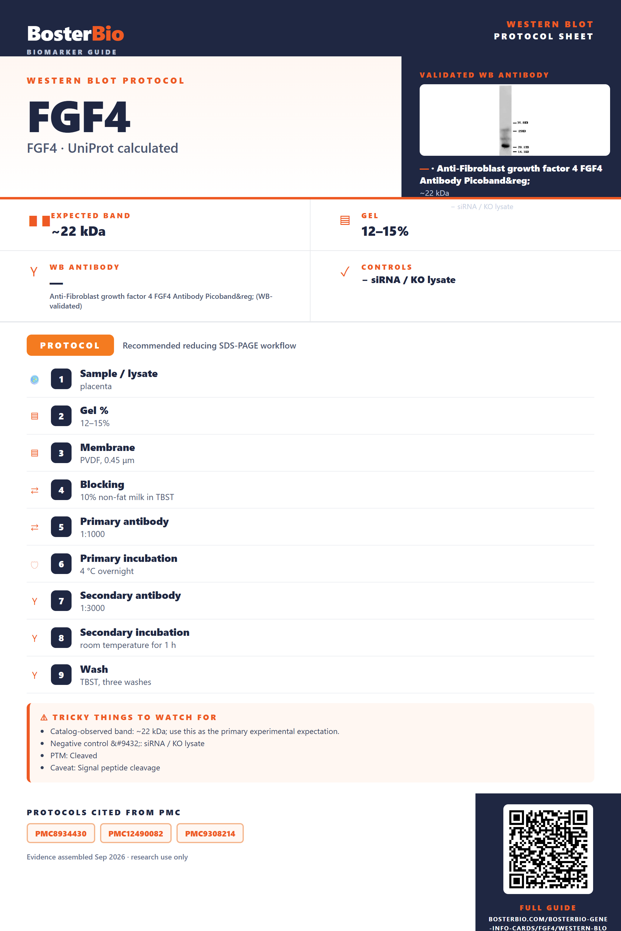
Western blot protocol sheet for FGF4: expected band ~22 kDa, antibody , and PMC-cited SDS-PAGE protocol steps
FGF4 Western blot protocol sheet — expected band ~22 kDa, controls and PMC citations. Open the full FGF4 WB guide →

FGF4 Western Blot Experimental Design Guide

Expected bands, validated protocols, controls and antibodies — the at-a-glance facts below, then the full design guide.

Must know before running
Expected band ~22 kDa
Gel 12–15%
Negative control ⓘ siRNA / KO lysate
Important caveats
Reasons your observed band may differ from the expected size.
PTM Cleaved
Caveat Signal peptide cleavage
Regulation LPS-induced
Isoform 2 isoform(s)
Section 1

Real Curated FGF4 Western Blot Protocols

Literature-validated Western blot parameters for FGF4 — gel percentage, transfer, blocking, antibody incubation and detection, extracted from published methods.

Recommended Western blot protocol parameters
Sample / lysateplacenta
Gel %12–15%
MembranePVDF, 0.45 μm
Blocking10% non-fat milk in TBST
Primary antibody1:1000
Primary incubation4 °C overnight
Secondary antibody1:3000
Secondary incubationroom temperature for 1 h
WashTBST, three washes
DetectionEasySee Western Blot Kit; enhanced chemiluminescence (ECL) reagents
Section 2

What Is the Expected FGF4 Western Blot Band Size?

FGF4 is a 22 kDa secreted precursor that loses its 30-residue signal peptide during secretion, giving a smaller, unglycosylated monomeric mature band.

What am I looking at on my blot?
single band smaller than the 22 kDa full-length predictionthe 30-residue signal peptide is cleaved during secretion, producing a smaller mature FGF4 species than the unprocessed precursor
little or no band in standard whole-cell lysateFGF4 is a secreted protein, so it accumulates in conditioned medium/extracellular space rather than inside the cell
two distinct bands at different apparent massesFGF4 has two annotated splice isoforms that can migrate at different positions
single sharp, non-smeared bandFGF4 has no annotated glycosylation sites, so there is no glycoform heterogeneity to broaden the band
no shift in band position between reducing and non-reducing conditionsFGF4 has no annotated inter-chain disulfide bonds, so it runs as a monomer regardless of reduction
💡Expected FGF4 appearanceExpect a single secreted mature FGF4 band below the 22 kDa full-length prediction after cleavage of the 30-residue signal peptide, seen mainly in conditioned medium rather than whole-cell lysate.
How each factor affects band size
predicted full-length mass from UniProtsets the 22 kDa reference point for the unprocessed FGF4 precursor
signal peptide cleavage (residues 1-30)removes the N-terminal signal sequence during secretion, shifting the mature band below the 22 kDa precursor mass
named splice isoforms (isoform 1, isoform 2)can produce an extra band at a different relative mass depending on which isoform is expressed or detected
secreted subcellular localizationshifts where the band is detectable, favoring conditioned media/concentrated supernatant over standard whole-cell lysate
Why is my band missing or off?
SituationLikely causeNext action
No band in lysateFGF4 is secreted and does not accumulate intracellularlyprobe concentrated conditioned medium or a heparin-affinity-enriched fraction instead of whole-cell lysate
Band higher than expectedincomplete cleavage of the 30-residue signal peptide leaves an unprocessed precursor formcompare against the 22 kDa precursor prediction and confirm processing with a signal-peptide-specific control
Band lower than expectedthe small secreted mature protein is prone to proteolytic clipping during collection or handlingadd protease inhibitors to collection media, keep samples cold, and minimize freeze-thaw cycles
Multiple bandsthe two annotated splice isoforms of FGF4 differ in massconfirm isoform identity by sequence or isoform-specific detection rather than assuming degradation
Weak or no signallow levels of secreted FGF4 are diluted in culture medium relative to intracellular proteinsconcentrate conditioned medium before loading and increase total protein loaded per lane

Sample controls for FGF4 Western blot

🧪For positive controls for FGF4 in Western blot, you can use lysate or conditioned medium from cells transiently transfected with an FGF4 expression construct, or recombinant FGF4 protein, since no tissue or cell line with confirmed high endogenous expression is listed in the available evidence.
Positive control: FGF4-transfected cell lysate / conditioned medium
Negative control: untransfected parental cell lysate (no confirmed endogenous expression data)
Loading controls: Run GAPDH and β-actin antibody blots alongside a total-protein stain (stain-free gel, Ponceau S, or REVERT) to confirm equal loading.
⚠️Feasibility: As a secreted growth factor with no confirmed endogenous tissue expression data, FGF4 is unlikely to be detectable in standard whole-cell lysates, so rely on conditioned medium or an overexpression system for a reliable positive signal.

Comprehensive Human Protein Atlas IHC scoring per tissue. Rows are taken directly from the HPA tissue chart — click any row's HPA link to view the source.

Positive expression · recommended positive controls

TissueCell typeLevelEvidenceSource

Undetected expression · recommended negative controls

TissueCell typeLevelEvidenceSource
Section 3

Advanced FGF4 Western Blot Tips

Deeper troubleshooting and optimisation questions for FGF4, answered from its protein features.

Why does FGF4 run smaller than its 22 kDa prediction?
The predicted mass includes a signal peptide (residues 1-30) that is cleaved during secretion. The mature secreted protein detected in conditioned media or extracellular fractions will migrate below the 22 kDa full-length prediction, so compare lysate versus secreted-fraction samples when assessing band size.
How many FGF4 isoforms exist and will they resolve?
UniProt lists two isoforms (1 and 2) generated by alternative splicing. Depending on the size difference between them, they may appear as a doublet or single band on standard SDS-PAGE; use a high-percentage gel with extended run time to separate closely spaced isoform bands if both are expressed.
Does FGF4 carry glycosylation or other modifications?
No glycosylation sites or modified residues are annotated for FGF4, and no disulfide bonds are listed. This means a single unmodified species is expected per isoform, and reducing versus non-reducing conditions should give equivalent migration, ruling out modification-driven band shifts as an explanation for anomalies.
How to optimize blocking for FGF4 detection?
FGF4 is a heparin-binding secreted growth factor, so use BSA-based blocking buffer rather than milk to avoid nonspecific glycoprotein interactions that can mask a low-abundance mitogen. Since it is secreted, ensure conditioned media samples are concentrated before loading to bring signal above background.
What transfer method to use for FGF4 Western blot?
FGF4 is a small (~22 kDa) secreted protein, so use a low-molecular-weight-optimized transfer: semi-dry or wet transfer with a 0.2 micron pore PVDF or nitrocellulose membrane, reduced methanol in the transfer buffer, and a shortened transfer time to prevent blow-through of the small mature protein.
How should FGF4 be normalized for quantitation?
Because FGF4 is secreted, standard intracellular housekeeping controls like actin or GAPDH are not appropriate for conditioned media samples. Normalize to total protein loaded using a stain such as Ponceau S or a stain-free gel system, and keep collection volume and concentration steps consistent across samples.
What explains extra or unexpected bands on FGF4 blots?
Extra bands can reflect the uncleaved signal-peptide-containing precursor versus the mature secreted form, isoform-specific products from alternative splicing, or heparin-mediated self-association given FGF4's heparin-binding activity. Confirm identity by comparing intracellular versus secreted fractions and checking migration against the mature protein's expected reduced size.
When is FGF4 expression induced or upregulated?
FGF4 is annotated as a developmental protein involved in differentiation and is a known proto-oncogene. Expect induction during developmental or differentiation signaling contexts and potential overexpression in tumor samples where FGF4 acts as an oncogenic driver; include relevant developmental or tumor-derived lysates as positive controls.
Boster reagents

Best FGF4 Western Blot Antibodies

BosterBio's FGF4 antibodies are among the best-performing WB antibodies on the market — well cited, thoroughly validated, and orthogonally cross-validated against negative tissues and complementary methods.

Real WB data Anti-FGF4 antibody, PA1033, Western blotting WB: HELA Cell Lysate
Anti-Fibroblast growth factor 4 FGF4 Antibody Picoband®
Cat # PA1033

Our recommended anti-FGF4 antibody is Boster's best-performing option, extensively cited and thoroughly validated by Western blot, with specificity orthogonally confirmed through negative-tissue controls and complementary detection methods, delivering dependable, publication-ready results for confident FGF4 detection.

Which to pick: Only one FGF4 antibody is catalogued, PA1033, which includes an actual Western blot validation image (HeLa cell lysate), making it the clear and only choice since no alternative SKU is available for comparison.

Source: BosterBio FGF4 gene-info card — filtered to Western-blot-capable antibodies; each card shows that product's actual WB validation figure.

References

  1. UniProt Consortium. UniProt entry P08620.
  2. Human Protein Atlas. FGF4 tissue expression.

