FGFR2 / Fibroblast growth factor receptor 2 · IHC design guide

Design Immunohistochemistry for FGFR2

Plan FGFR2 chromogenic IHC on paraffin sections using 2 μg/ml catalog antibody as a documented starting condition (datasheet A00231-2). Compare general cytoplasmic tissue staining (HPA tissue IHC) with membrane and vesicle localization (UniProt), while accounting for uncertain tissue staining reliability (HPA tissue IHC).

Evidence assembled Oct 2026 · For research use; verify linked source records and product datasheet before use
Immunohistochemistry protocol sheet for FGFR2 (IHC for FGFR2): expected localisation General cytoplasmic tissue staining (HPA tissue IHC), antibody A00231-2, validated IHC image, and IHC protocol steps
Printable FGFR2 IHC protocol sheet — expected localisation General cytoplasmic tissue staining (HPA tissue IHC), antibody A00231-2, controls and protocol steps. Open the full FGFR2 IHC guide →

FGFR2 Immunohistochemistry Experimental Design Guide

Expected localisation, validated protocols, controls and antibodies — the at-a-glance facts below, then the full design guide.

Must know before staining
Expected localisation General cytoplasmic tissue staining (HPA tissue IHC)
Staining pattern High epithelial staining; generally cytoplasmic (HPA tissue IHC)
Antigen retrieval EDTA pH 8.0 HIER, heat-mediated (datasheet A00231-2)
Positive control ⓘ Bronchus+4 more · see all
Negative control ⓘ Adipose tissue+4 more · see all
Important caveats
Reasons your staining may differ from the expected pattern.
Fixation Keep fixation consistent across paraffin sections (standard IHC practice; not target-specific)
Caveat A secreted variant may alter the tissue staining pattern (HPA tissue IHC)
Regulation Ligand-induced internalization and degradation (UniProt)
Isoform / epitope 17 isoforms; map extracellular versus cytoplasmic epitopes (UniProt)
Section 1

Recommended FGFR2 IHC & IF Protocols

The catalog antibody’s IHC-P protocol (datasheet: A00231-2) is accompanied by published FGFR2 staining protocols for gastric cancer, breast carcinoma, and corneal tissue (PMC8846517; PMC7564845; PMC5996875).

Recommended immunohistochemistry (IHC-P) protocol parameters
SampleParaffin-embedded human liver cancer tissue; fixative not specified (datasheet A00231-2)
FixationImage fixative and duration unreported (datasheet A00231-2); verify before use.
Sectioning4–5 µm sections on charged slides (standard)
DeparaffinisationXylene, graded ethanol series to water (standard)
Antigen retrievalHeat retrieval: EDTA pH 8.0 (datasheet A00231-2); 20 min, 95–100 °C (standard)
Peroxidase block3% H2O2, 10 min, room temperature (standard)
Blocking10% goat serum (datasheet A00231-2)
Primary antibodyRabbit anti-FGFR2, 2-5 μg/ml (datasheet A00231-2)
Primary incubationOvernight at 4 °C (datasheet A00231-2)
DetectionHRP-conjugated secondary, DAB chromogen (datasheet A00231-2)
CounterstainHematoxylin, blue, dehydrate and mount (standard)
Expected resultFGFR2-positive staining in respiratory epithelial cells of bronchus (HPA tissue IHC: High). HPA tissue profile: General cytoplasmic expression. No signal in the no-primary control.
💡Decision noteStart with heat-mediated EDTA retrieval at pH 8.0 for the catalog antibody (datasheet: A00231-2); citrate retrieval at pH 6.0 is a published alternative (PMC8846517).
Section 2

What Is the Expected FGFR2 Staining Pattern?

FGFR2 spans the cell membrane, with an extracellular domain and a cytoplasmic kinase region; it is also reported in Golgi and cytoplasmic vesicles (UniProt P21802 topology and subcellular location). In tissue IHC, expect staining mainly in the cytoplasm of selected epithelial cells and keratinocytes (HPA tissue IHC). Treat that expectation as provisional: HPA rates the tissue IHC pattern Uncertain.

What am I looking at on my slide?
Strong staining in bronchial respiratory epithelium or skin keratinocytes, with cytoplasmic signal and possible membrane accentuation.These are reported high staining cell types (HPA tissue IHC); cytoplasmic staining matches the reported tissue profile (HPA tissue IHC), while membrane staining is consistent with receptor topology (UniProt P21802 topology). Record the compartment and cell type separately because HPA rates tissue IHC reliability Uncertain.
Predominantly nuclear staining in a paraffin section, without the expected epithelial cytoplasmic pattern.An isolated nuclear IHC pattern is unsupported by the reported general cytoplasmic tissue profile (HPA tissue IHC). Check controls and detection artefacts before assigning it to FGFR2. HPA reports additional nucleoplasmic localisation in ICC-IF (HPA subcellular), so compartment judgments should remain application specific.
Strong signal in adipocytes or soft tissue fibroblasts while the selected positive epithelium is unstained.HPA reports FGFR2 as not detected in those cell types (HPA tissue IHC). This reversal raises concern for antibody cross-reactivity or endogenous detection activity (general IHC practice); it does not establish either cause without reagent controls, particularly given the Uncertain HPA tissue IHC rating.
Diffuse chromogen over cells and surrounding section, without a clear cell or compartment pattern.This is difficult to score as FGFR2 because the reported IHC profile is cellular and generally cytoplasmic (HPA tissue IHC). Review the no-primary control, blocking, washing and detection conditions for background (general IHC practice). A broad haze alone does not identify its source.
No staining in bronchial respiratory epithelial cells or skin keratinocytes.Both are reported high staining cell types (HPA tissue IHC), so absence of signal warrants checking section quality, retrieval and detection controls (general IHC practice). It is not proof that FGFR2 is absent: HPA rates tissue IHC reliability Uncertain, and staining can vary with the antibody and specimen.
💡Expected FGFR2 appearanceCall a section positive when selected epithelial cells or keratinocytes show clear, relatively strong cytoplasmic staining, with possible membrane accentuation (HPA tissue IHC; UniProt P21802 topology); diffuse haze or dominant staining in HPA not-detected cell types is suspect (HPA tissue IHC; general IHC practice).
How each factor affects the staining
Compartment and epitope locationFGFR2 has extracellular residues 22–377, a transmembrane segment at 378–398 and a cytoplasmic region at 399–821 (UniProt P21802 topology). Interpret membrane and cytoplasmic signal in light of the antibody's stated epitope if available; the supplied record gives no epitope for the catalog antibody.
Isoforms and secreted proteinUniProt lists 17 isoforms and a secreted location (UniProt P21802 isoforms and subcellular location). HPA notes that at least one protein variant is secreted and that RNA and protein locations may differ (HPA tissue IHC reliability description). Do not infer the detected isoform from staining alone.
InternalisationThe activated receptor is rapidly internalised and degraded after ligand binding (UniProt P21802 subcellular location). Vesicular or cytoplasmic signal can therefore fit receptor trafficking, but IHC appearance alone cannot establish ligand exposure or activation (UniProt P21802; general IHC interpretation).
Antibody validationHPA marks IHC results for HPA035305 and CAB010886 Uncertain and rates the tissue profile Uncertain (HPA antibodies; HPA tissue IHC). Use the reported positive and not-detected cell types as comparative controls, rather than treating one stained section as definitive antibody specificity.
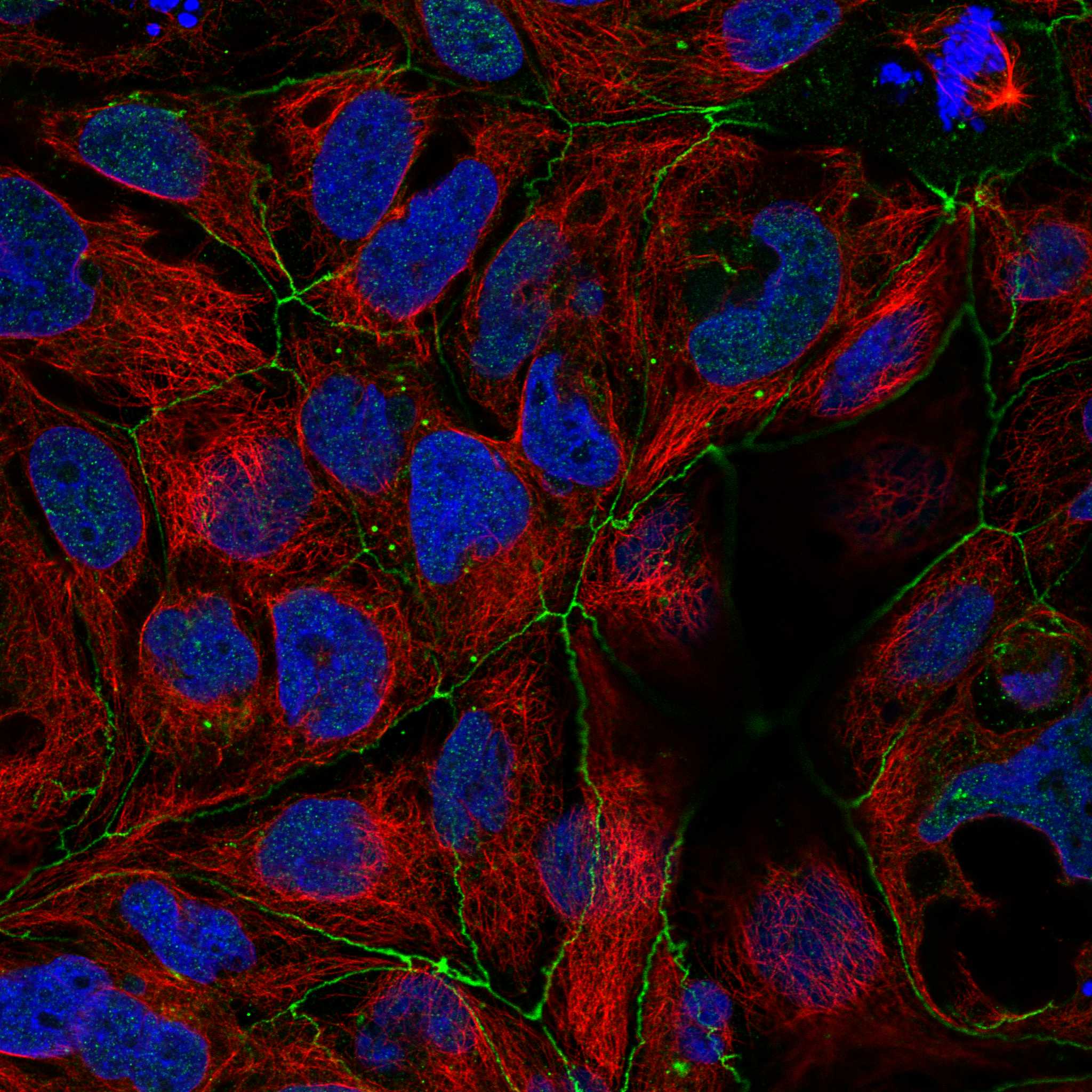
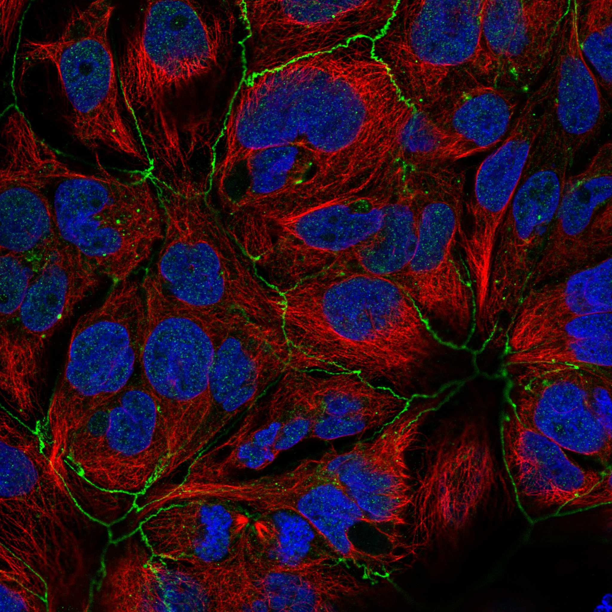
IF/ICC Q&A: where should signal appear?HPA reports cell junctions as the main approved ICC-IF location, with additional approved nucleoplasmic and vesicular locations (HPA subcellular). These IF observations answer the localisation question; they do not supply an IF/ICC protocol for this IHC-P guide.
Antigen retrieval and endogenous detectionRetrieval and blocking are general IHC workflow variables (general IHC practice). No target-specific fixation or retrieval sensitivity is supplied for FGFR2, so compare conditions with controls rather than predicting which condition will increase FGFR2 staining.
Why is my staining missing, weak or wrong?
SituationLikely causeNext action
Positive epithelium and the assay control are both blank.A shared retrieval, primary antibody or chromogenic detection failure is possible (general IHC practice).Check reagent preparation and the detection run; repeat with a reported high staining tissue such as bronchus, assessing respiratory epithelium (HPA tissue IHC; general IHC practice).
The assay control stains, but bronchial respiratory epithelium is blank.Specimen variation or antibody performance in this section is possible; HPA tissue IHC reliability is Uncertain (HPA tissue IHC).Review section integrity and compare another reported high staining cell type, such as skin keratinocytes, under the same IHC conditions (HPA tissue IHC; general IHC practice).
Adipocytes or soft tissue fibroblasts stain strongly.Those cell types are reported as not detected (HPA tissue IHC); cross-reactivity or endogenous detection activity is possible (general IHC practice).Compare a no-primary control and a positive epithelial control, then assess whether signal follows the antibody or detection system (general IHC practice; HPA tissue IHC).
Diffuse chromogen obscures cell borders and compartments.Background from insufficient blocking, washing or detection control is possible (general IHC practice).Review the no-primary control and optimise blocking, washing or detection conditions before scoring cell-specific signal (general IHC practice).
Only nuclear IHC signal is visible.This differs from the general cytoplasmic tissue IHC profile (HPA tissue IHC), although HPA reports additional nucleoplasmic signal in ICC-IF (HPA subcellular).Check controls and review the matching cell type and compartment before calling the IHC pattern FGFR2-positive (general IHC practice; HPA tissue IHC).
Membrane and cytoplasmic staining differ between otherwise comparable sections.FGFR2 occupies membrane and intracellular locations, and activated receptor can internalise (UniProt P21802 subcellular location); staining alone cannot identify the mechanism.Score membrane and cytoplasmic compartments separately, confirm the intended cell type and compare controls before interpreting the difference (general IHC practice; HPA tissue IHC).

Sample controls for FGFR2 IHC & IF

🧪Run bronchus first and look for staining in respiratory epithelial cells (HPA: bronchus respiratory epithelial cells High); use adipose tissue as the negative comparator, focusing on adipocytes (HPA: adipocytes Not detected). On the bronchus slide, cells outside the stained respiratory epithelium should show only background signal, but HPA does not identify a specific bronchial cell type as negative (HPA: bronchus respiratory epithelial cells High).
Positive control tissue: Bronchus (Respiratory epithelial cells, HPA High)
Negative control tissue: Adipose tissue (HPA Not detected)
ICC-IF cell lines (HPA subcellular resource): HPA ICC-IF images show FGFR2 in CACO-2, HEK293, U2OS, hTERT-RPE1 (serum starved), with annotated localisation: Cell Junctions (approved) (HPA subcellular).
Technical controls: Include a no-primary, secondary-only control; a host-, class-, and clonality-matched rabbit immunoglobulin control; and an FGFR2-knockout specimen if available (selected-SKU caption: rabbit anti-FGFR2 antibody). Block endogenous peroxidase and check the bronchus section for pigment or nonspecific DAB signal before scoring (selected-SKU caption: peroxidase detection with DAB).
⚠️Feasibility: A target-specific fixation window or fixation effect is unreported, and the selected-SKU paraffin-section caption does not report the fixative (selected-SKU caption: fixative not stated). That caption used heat-mediated retrieval in EDTA at pH 8.0, but it does not establish whether retrieval is essential (selected-SKU caption: EDTA retrieval). The supplied evidence does not establish that frozen sections or IF are easier for FGFR2; in bronchus, distinguish epithelial staining from pigment or nonspecific DAB signal with the controls (HPA: bronchus respiratory epithelial cells High; selected-SKU caption: DAB detection).

HPA tissue IHC evidence for FGFR2

Comprehensive Human Protein Atlas IHC scoring per tissue (reliability: Uncertain — Low consistency between antibody staining and RNA expression data. At least one protein variant secreted, tissue location of RNA and protein might differ and correlation is complex.). Rows are taken directly from the HPA tissue chart — click any row's HPA link to view the source.

Positive expression · recommended positive controls

TissueCell typeLevelEvidenceSource
Bronchus Respiratory epithelial cells High Protein (IHC) HPA →
Cervix Squamous epithelial cells High Protein (IHC) HPA →
Epididymis Glandular cells High Protein (IHC) HPA →
Esophagus Squamous epithelial cells High Protein (IHC) HPA →
Nasopharynx Respiratory epithelial cells High Protein (IHC) HPA →

Undetected expression · recommended negative controls

TissueCell typeLevelEvidenceSource
Adipose tissue Adipocytes Not detected Protein (IHC) HPA →
Fallopian tube Glandular cells Not detected Protein (IHC) HPA →
Ovary Ovarian stroma cells Not detected Protein (IHC) HPA →
Prostate Glandular cells Not detected Protein (IHC) HPA →
Salivary gland Glandular cells Not detected Protein (IHC) HPA →
Section 3

Advanced FGFR2 IHC Tips

Troubleshoot FGFR2 staining in paraffin sections by checking retrieval, antibody access, cellular location and controls before interpreting chromogenic signal.

Which antigen retrieval conditions should I start with for FGFR2 paraffin IHC?
Start with heat-mediated retrieval in EDTA at pH 8.0 for the catalog antibody (datasheet A00231-2). Its paraffin-section example used that retrieval before 10% goat-serum blocking and 2 μg/ml primary antibody overnight at 4°C (datasheet A00231-2). If staining is weak, compare a second retrieval condition on adjacent sections while keeping detection and imaging conditions constant, and inspect tissue integrity after heating (standard IHC practice). Record membrane-associated and cytoplasmic staining separately because FGFR2 spans the membrane and can enter vesicles after ligand binding (UniProt P21802 topology and subcellular annotation).
How should I assess fixation when FGFR2 staining is weak or uneven?
The selected paraffin-section caption does not state a fixative, so target-specific fixation sensitivity is unknown for this antibody (datasheet A00231-2). Check processing records and compare sections with documented fixation and similar thickness, retrieval and detection conditions (standard IHC practice). Inspect whether weak staining tracks tissue folds, poor preservation or section edges before attributing it to FGFR2 biology (standard IHC practice). Use the documented EDTA retrieval at pH 8.0 as the starting condition, then change only one preparation variable at a time (datasheet A00231-2; standard IHC practice). Do not infer fixation tolerance from FGFR2 topology or tissue staining patterns (UniProt P21802 topology; HPA tissue IHC).
How do I evaluate membrane versus cytoplasmic FGFR2 staining?
Score membrane-associated and cytoplasmic signal separately: FGFR2 has one transmembrane segment at residues 378–398 and a cytoplasmic region at 399–821 (UniProt P21802 topology). Cytoplasmic puncta can be plausible because FGFR2 is annotated in vesicles and the Golgi, and activated receptor is internalized after ligand binding (UniProt P21802 subcellular annotation). HPA describes general cytoplasmic tissue staining but rates its tissue IHC reliability uncertain, so that pattern alone cannot validate an individual section (HPA tissue IHC). Compare staining with a no-primary control and inspect whether membrane signal follows cell outlines rather than folds or section edges (standard IHC practice).
Could FGFR2 isoforms or epitope location explain discordant staining?
Map the antibody immunogen or stated epitope to the expressed FGFR2 sequence before comparing samples; 17 isoforms are listed, but this payload does not identify the catalog antibody’s epitope (UniProt P21802 isoforms; datasheet A00231-2). The third extracellular Ig-like domain occupies residues 256–358, while the kinase domain occupies 481–770 (UniProt P21802 domains). Isoform differences in the third Ig-like domain affect ligand specificity, so an epitope there warrants particular scrutiny when interpreting variant coverage (UniProt P21802 subunit annotation). Compare antibody documentation and appropriate controls before calling discrepant staining an isoform-specific result (standard IHC practice).
How should I investigate FGFR2 localisation with multiplex IF?
For a separate IF experiment, pair FGFR2 with a marker of the expected cell type, such as an epithelial marker when examining HPA-reported keratinocyte staining (HPA: high in skin keratinocytes; standard IF practice). Choose a fluorophore channel after checking tissue autofluorescence, and use single-stain controls to assess channel bleed-through (standard IF practice). Match permeabilisation to the antibody epitope: access to the cytoplasmic region at residues 399–821 generally requires permeabilisation, whereas an accessible extracellular epitope may be assessed without it (UniProt P21802 topology; standard IF practice). The epitope is unspecified here, so verify antibody documentation before assigning either approach (datasheet A00231-2).
What should I check when FGFR2 DAB staining is diffuse?
Run a no-primary control to assess secondary-reagent and detection background, and include a peroxidase block in the chromogenic workflow (standard IHC practice). The catalog example used 10% goat serum, 2 μg/ml primary antibody overnight at 4°C, and DAB detection (datasheet A00231-2). If background rises across the section, compare primary-antibody concentration and wash conditions on adjacent sections while holding development conditions constant (standard IHC practice). Check whether apparent cytoplasmic staining has cellular boundaries or puncta; HPA reports general cytoplasmic expression but assigns uncertain reliability to its tissue IHC pattern (HPA tissue IHC).
How should I quantify FGFR2 staining across paraffin sections? ⚠ ANSWER MARKED FOR VERIFICATION
Define the scoring compartment and cell population before measuring FGFR2, then report the percentage of positive cells and an intensity-based H-score within the same region (standard IHC practice). If spatial distribution matters, report positive-cell density per mm² of viable tissue and normalize counts to the annotated tissue area (standard IHC practice). Keep retrieval, antibody concentration, DAB development, counterstain and image analysis settings consistent across compared sections (standard IHC practice). Record membrane-associated and cytoplasmic scores separately because FGFR2 is a membrane receptor that can localize to vesicles, while HPA tissue IHC chiefly describes cytoplasmic staining with uncertain reliability (UniProt P21802 subcellular annotation; HPA tissue IHC).
What evidence separates true FGFR2 staining from tissue artefact?
Look for reproducible cellular staining in the expected compartment and evaluate it against a no-primary control and adjacent tissue morphology (standard IHC practice). FGFR2 can occur at the cell membrane, Golgi and cytoplasmic vesicles, but isolated diffuse signal over necrosis or section edges warrants scrutiny (UniProt P21802 subcellular annotation; standard IHC practice). Compare the stained cell population with documented patterns, such as high staining in skin keratinocytes and undetected staining in adipocytes, without treating either as universal ground truth (HPA tissue IHC). Exclude endogenous peroxidase signal with an appropriate block and control, and interpret HPA concordance cautiously because its tissue IHC reliability is uncertain (standard IHC practice; HPA tissue IHC).
Boster reagents

Best FGFR2 / Fibroblast growth factor receptor 2 IHC Antibodies

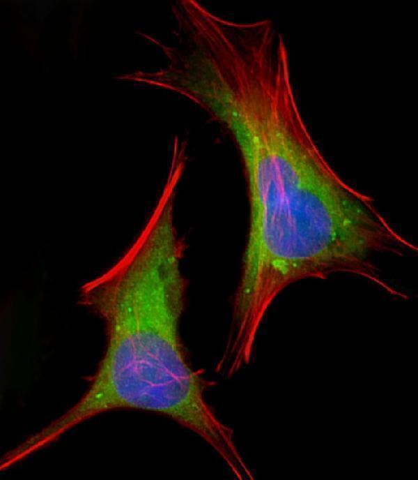
Two anti-FGFR2 antibodies have human paraffin-section IHC images (catalog IHC captions); A00231 also has IF images from HeLa cells (A00231 IF caption) and lists mouse reactivity (A00231 catalog).

Real IHC data IHC analysis of FGFR2 using anti-FGFR2 antibody (A00231-2). FGFR2 was detected in a paraffin-embedded section of human liver cancer tissue. Heat mediated antigen retrieval was performed in EDTA buffer (pH 8.0, epitope retrieval solution). The tissue section was blocked with 10% goat serum. The tissue section was then incubated with 2 μg/ml rabbit anti-FGFR2 Antibody (A00231-2) overnight at 4°C. Peroxidase Conjugated Goat Anti-rabbit IgG was used as secondary antibody and incubated for 30 minutes at 37°C. The tissue section was developed using HRP Conjugated Rabbit IgG Super Vision Assay Kit (Catalog # SV0002) with DAB as the chromogen.
Anti-FGFR2 Antibody ®
Cat # A00231-2
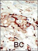
Real IHC data Formalin-fixed and paraffin-embedded human cancer tissue reacted with the primary antibody, which was peroxidase-conjugated to the secondary antibody, followed by AEC staining. This data demonstrates the use of this antibody for immunohistochemistry; clinical relevance has not been evaluated. BC = breast carcinoma; HC = hepatocarcinoma.
Anti-FGFR2 Antibody (N-term)
Cat # A00231

A00231-2 will render with human liver cancer tissue IHC and is listed for human reactivity (A00231-2 IHC caption; catalog). A00231 will render with formalin-fixed, paraffin-embedded human cancer tissue IHC; its separate IF images show HeLa cells (A00231 IHC and IF captions).

Which to pick: For human tissue IHC, choose A00231-2 if you want a documented starting protocol: its paraffin-section caption specifies EDTA retrieval and 2 μg/ml primary antibody, but does not report the fixative (A00231-2 IHC caption). For IF/ICC, choose A00231: its IF caption shows paraformaldehyde-fixed, permeabilized HeLa cells at 1:25 (A00231 IF caption). For human and mouse work, A00231 is the polyclonal option listed for both species, though its pictured IHC example is human tissue (A00231 catalog; IHC caption).

Each figure is that product's own IHC / IF validation image from its datasheet.

References

  1. UniProt Consortium. UniProt entry P21802 (FGFR2_HUMAN, Fibroblast growth factor receptor 2).
  2. Human Protein Atlas. FGFR2 tissue IHC expression (reliability: Uncertain).
  3. Human Protein Atlas. FGFR2 subcellular location (ICC-IF): Mainly localized to the cell junctions. In addition localized to the nucleoplasm and vesicles..
  4. Human Protein Atlas. FGFR2 antibody validation summary (2 antibodies).
  5. Cancer-related FGFR2 overexpression and gene amplification in Japanese patients with gastric cancer. Japanese journal of clinical oncology 2021 — PMC8491535.
  6. FGFR2 overexpression and compromised survival in diffuse-type gastric cancer in a large central European cohort. PloS one 2022 — PMC8846517.
  7. Hormonal Receptor Status Determines Prognostic Significance of FGFR2 in Invasive Breast Carcinoma. Cancers 2020 — PMC7564845.
  8. Modeling Keratoconus Using Induced Pluripotent Stem Cells. Investigative ophthalmology & visual science 2016 — PMC5996875.
  9. PubMed PMID:1697263 — UniProt-cited evidence.
  10. PubMed PMID:2172978 — UniProt-cited evidence.
  11. PubMed PMID:1647213 — UniProt-cited evidence.